Regulating ErbB Signaling in the Zebrafish Epidermis
Sven Reischauer, Mitchell P. Levesque, Christiane Nu¨sslein-Volhard, Mahendra Sonawane¤*
Max-Planck Institut fu¨r Entwicklungsbiologie, Department of Genetics, Tuebingen, Germany
Abstract
Changes in tissue homeostasis, acquisition of invasive cell characteristics, and tumor formation can often be linked to the loss of epithelial cell polarity. In carcinogenesis, the grade of neoplasia correlates with impaired cell polarity. In Drosophila,
lethal giant larvae (lgl),discs large (dlg), and scribble, which are components of the epithelial apico-basal cell polarity machinery, act as tumor suppressors, and orthologs of this evolutionary conserved pathway are lost in human carcinoma with high frequency. However, a mechanistic link between neoplasia and vertebrate orthologs of these tumor-suppressor genes remains to be fully explored at the organismal level. Here, we show that thepen/lgl2mutant phenotype shares two key cellular and molecular features of mammalian malignancy: cell autonomous epidermal neoplasia and epithelial-to-mesenchymal-transition (EMT) of basal epidermal cells including the differential expression of several regulators of EMT. Further, we found that epidermal neoplasia and EMT inpen/lgl2mutant epidermal cells is promoted by ErbB signalling, a pathway of high significance in human carcinomas. Intriguingly, EMT in thepen/lgl2 mutant is facilitated specifically by ErbB2 mediated E-cadherin mislocalization and not via canonical snail–dependent down-regulation of E-cadherin
expression. Our data reveal that pen/lgl2 functions as a tumor suppressor gene in vertebrates, establishing zebrafish
pen/lgl2mutants as a valuable cancer model.
Citation:Reischauer S, Levesque MP, Nu¨sslein-Volhard C, Sonawane M (2009) Lgl2 Executes Its Function as a Tumor Suppressor by Regulating ErbB Signaling in the Zebrafish Epidermis. PLoS Genet 5(11): e1000720. doi:10.1371/journal.pgen.1000720
Editor:Mary C. Mullins, University of Pennsylvania School of Medicine, United States of America
ReceivedJune 17, 2009;AcceptedOctober 15, 2009;PublishedNovember 13, 2009
Copyright:ß2009 Reischauer et al. This is an open-access article distributed under the terms of the Creative Commons Attribution License, which permits unrestricted use, distribution, and reproduction in any medium, provided the original author and source are credited.
Funding:This research was funded by the Max-Planck Society, Germany (http://www.mpg.de). The funders had no role in study design, data collection and analysis, decision to publish, or preparation of the manuscript.
Competing Interests:The authors have declared that no competing interests exist.
* E-mail: mahendra.sonawane@tuebingen.mpg.de
¤ Current address: Tata Institute of Fundamental Research, Department of Biological Sciences, Colaba, Mumbai, India
Introduction
Tumor suppression is a concept first formulated inDrosophilaafter emerging evidence that recessive mutations can lead to the formation of cellular overgrowth [1,2]. To date, more than 50 tumor suppressor genes have been identified inDrosophila[3]. Their deficiencies result in benign hyperplasias to malignant neoplasms. Amongst these tumor suppressors, mutations inlethal giant larvae(lgl), cause malignant neoplasias in imaginal discs and the brain when transplanted into wild type adult host flies [4,5]. In Drosophila
neuroblasts,lglfunction is essential for localization of the cell fate determinant Numb, mislocalization of which inlglmutant larvae prevents the neuroblasts from dividing asymmetrically and therefore causes neuroblastoma [6–9]. Furthermore, it has been proposed thatlglprevents tumor formation by antagonizing the activation of Dpp signaling by semaphorin 5c in the brain [10]. Although epithelial overgrowth phenotypes have been reported inlglmutant larvae inDrosophila[1], the mechanism by whichlgl manifests its effects on epithelial growth remains to be understood. Nevertheless, it is known that along withlgl, two other tumor suppressor genes, discs large (dlg) and scribble (scrib), primarily act in the maintenance of apico-basal cell polarity in epithelial cells [11].
The establishment as well as the maintenance of apico-basal cell polarity and eventually the depolarization of a cell is a complex process, involving several factors. In recent years, a conserved mechanism for the establishment and maintenance of apico-basal
cell polarization has emerged, which mainly involves two pathways. Accordingly, the formation of the apical domain is controlled by the Par (partitioning defective) pathway, which consists of the PDZ domain containing proteins Par3, Par6, and atypical protein kinase C (aPKC). In contrast, a pathway consisting ofdisc-large(dlg),scribble
(scrib) and lethal giant larvae (lgl) regulates the formation and maintenance of the baso-lateral domain [12,13]. Intriguingly, only mutations in genes that act in the baso-lateral pathway (e.g.lgl, dlg, scrib) lead to a neoplastic growth phenotype inDrosophila[2,14–16]. The two vertebrate orthologs of theDrosophila lethal giant larvaegene have conserved functions in the maintenance of cell polarity and tissue homeostasis. Disruption oflgl1function results in the loss of apical junctional complex in neuroblasts and hyperplasia of the brain in mouse [17]. Furthermore, it has been shown for human melanoma cell lines, that a human homolog oflgl,hugl1is significantly down-regulated. Artificial induction ofhugl1in these cell lines reduces their migratory potential with concomitant transcriptional up-regulation of the cell adhesion molecule E-cadherin (E-cad) and a down-regulation of matrix-metalloproteinases (mmps), both of which are known to be involved in suppression of epithelial-to-mesenchymal transition (EMT), a process which enables an epithelial cell to gain mesenchymal or migratory properties [18]. Recently, a significant correlation between the loss ofhugl1and a poor clinical prognosis for cancer patients has been shown [19].
epidermal integrity, which is a stratified epithelium. Thepen/lgl2
deficiency primarily results in the loss of hemidesmosomes, cellular junctions that mediate cell-matrix adhesion [20]. It has been shown that Lgl2 localizes to the lateral domain of the epidermal cells and regulates hemidesmosome formation by mediating the targeting of ITGa6, a component of hemidesmosomes, to the membrane [21]. Furthermore, epithelial cells in the pen/lgl2
mutant exhibit altered epidermal cell morphology as well as cell polarity and enhanced epidermal growth [20,21]. Recently, it has
been shown that in colorectal and breast carcinoma cell lines, a member of the ZFH family of repressors ZEB1 regulates the levels of Lgl2. The loss of ZEB1 function restores Lgl2 levels and the epithelial phenotypes in tumor cells, which suggests that Lgl2 acts as an effector of ZEB1 in tumor suppression [22].
From the analysis of several cancer models it is evident that autocrine self-stimulation with growth factors is one of the hallmarks of tumorigenicity [23,24]. However, whether activation of growth factor signaling is a consequence of the loss of baso-lateral pathway components remains unclear to date. Moreover, whetherlgl1andlgl2deficient clonal cell populations can promote tumor formation in vertebrate tissues, a typical characteristic of tumor suppressor genes, remains unresolved. Here, we show that
lgl2 deficient clones indeed promote tumor formation in the zebrafish epidermis. Moreover, pen/lgl2 mutant basal epidermal cells undergo EMT. Using biochemical analysis, chemical inhibitors and genetic interaction studies, we demonstrate that these phenotypes are a consequence of an over-activation oferbB
signaling involving at least oneerbBfamily member,erbB2. Our microarray and immuno-histological analysis reveal that activation oferbBsignaling facilitates EMT by transcriptional up-regulation of key EMT regulators and a reduction in the membrane localization of E-cad, a known suppressor of EMT.
Results
lgl2functions as a tumor suppressor gene in the zebrafish epidermis
We have previously shown that zebrafishpen/lgl2 larvae show overgrowth of epidermal cells (Figure 1A and 1B; and [20]). As
pen/lgl2mutant larvae die at 4–5 days post fertilization (dpf), it was not clear whether these hyper-proliferating cells would be able to form tumor like structures. To test this, we transplanted cells from
pen/lgl2 homozygous mutant donor individuals, into wild type Author Summary
In metazoans, the body surface and linings of several organs are formed from membranous tissue called epithelia. The functions of epithelia include secretion, absorption, and protection. Epithelial cells exhibit polar-ized distribution of several proteins, which is essential for their function. In carcinomas, which are cancers of epithelial origin, this epithelial cell polarity is impaired. Intriguingly, defects in cell polarization can also lead to tumorigenesis in some animal model systems. It is thus important to understand how cell polarization and epithelial growth control are linked so as to treat the carcinomas better by identifying new drug targets. Here we show that in zebrafish a gene namedlethal giant larvae 2 (lgl2), which is essential for the establishment of epithelial cell polarity, also acts as a suppressor of malignant growth properties in the epidermis, an epithelial component of the skin. We further show that in absence of
lgl2function increased epidermal growth factor receptor activity imparts malignant properties to the epidermal cells. Thus, we report here a mechanism by which epithelial cells acquire malignant characteristics when cell polarity is impaired in absence oflgl2function.
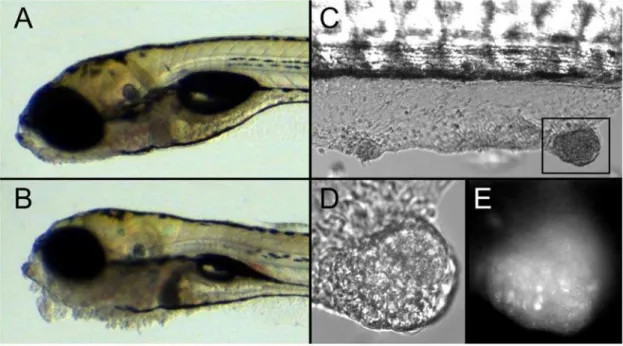
Figure 1.pen/lgl2deficient epidermal cells form tumors in a cell autonomous fashion.DIC images of 5-day-old wild-type (A) andpen/lgl2 mutant larvae (B). DIC image of wt host withpen/lgl2mutant skin clones (C). Close-up of a tumor in DIC (D) and GFP channel (E). In comparison to 5 day wild-type larvae (A) thepen/lgl2 mutant larvae exhibit neoplasias, most prominently in the ventral jaw region (B). Seven days after the transplantation ofpen/lgl2mutant cells at blastula stage, recipients develop tumor like structures in the skin (C). These tumor like structures (D) contain GFP labelled cells (E) indicating that they are derived from mutant clones.
hosts during blastula stage. In order to trace the donor cells in the wild type recipients, donor embryos were transgenic for ubiqui-tously expressed GFP. The recipient larvae with epidermal cells from mutants or wild type siblings were monitored for potential tumor development. On the 7th day after the transplantation, 17 of the 24 (71%) larvae that had received pen/lgl2deficient cells (GFP marked) in the epidermis developed epidermal tumors (Figure 1C and 1D). In contrast, none of the larvae that had received cells from wild type siblings developed epidermal tumors (n = 77). Fluorescence microscopic analysis of these tumors revealed that they contain GFP positive cells, indicating that the
lgl22/2 cells are inducing the formation of epidermal tumors (Figure 1E). Interestingly,lgl22/2cells in other tissues, including brain, did not show any visible hyperplasia at this stage. We conclude that pen/lgl2 mutant cells are capable of inducing epidermal tumor formation, even if surrounded by wild type tissue. The levels of GFP expression varied in the tumor cells (Figure 1E). This may reflect a variegated expression of GFP. However, we cannot rule out the possibility that some wild type cells also contribute to the neoplastic tissue. Our results support the notion that lgl2 acts as a tumor suppressor gene specifically in the epidermis.
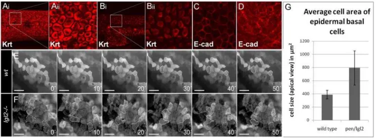
Loss ofpen/lgl2function results in EMT of basal epidermal cells
Along with uncontrolled cell proliferation, migratory behavior mediated by EMT is another hallmark of cancer cells [25]. Inpen/ lgl2mutant larvae, epidermal cells not only hyper proliferate but also exhibit different morphological shape as evident from the changes in the keratin organization from basal polygonal in wild type to peri-nuclear and spindle shaped in pen/lgl2 mutants (Figure 2A and 2B, [20]). Such alterations in cell morphology are indicative of EMT. Since E-cad membrane levels are inversely correlated with acquisition of EMT [26–28], we investigated the
localization of E-cad in pen/lgl2 mutants. We found that the membrane localization of E-cad was severely reduced in the mutant epidermis, while the cytoplasmic fraction appeared to be increased compared to the wild type siblings (Figure 2C and 2D). To observe the behavior of basal epidermal cells of pen/lgl2
mutants, we generated a transgenic line driving the expression of GFP in basal epidermal cells under the control of DN-p63
promoter [29]. Accordingly, tg(DN-p63::Gal4,UAS::GFP) zebrafish larvae show GFP expression exclusively in this cell type (Figure S1). To test whether pen/lgl2 mutant cells acquire migratory properties, we performed time lapse studies of tg(DN-p63:: Gal4,UAS::GFP);lgl22/2larvae. These revealed that, in contrast to their wild type siblings, basal epidermal cells inpen/lgl2mutant larvae dramatically alter their shape, form numerous lamellipodia like structures and exhibit net displacement over time (Figure 2 and Videos S1, S2, S3, S4). From an apical perspective, the observed changes in cell morphology also lead to an increase in average cell area (Figure 2G). The mutant epidermal cells appear larger when compared to wild type cells as they exhibit a highly flattened morphology and develop lamellipodia like cell protrusions.
We conclude that in absence of pen/lgl2 function the basal epidermal cells lose their epithelial morphology and acquire the morphology of migratory (mesenchymal) cells indicating that these cells undergo the morphological changes associated with EMT.
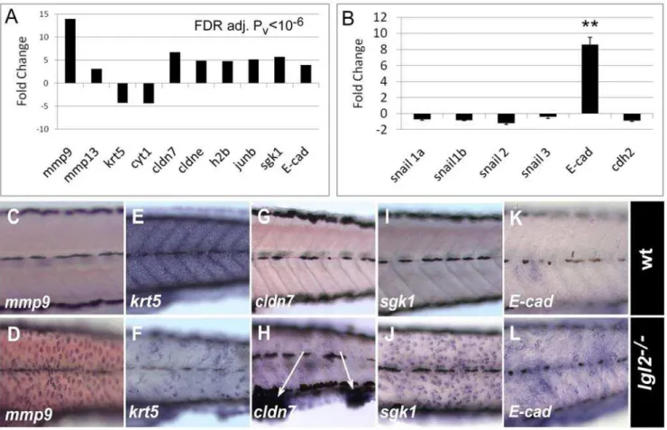
Expression profile ofpen/lgl2mutant larvae reveals differential transcriptional regulation of known molecular regulators of EMT
To understand if pen/lgl2 mutants also show molecular signatures of EMT and if so, which of the known EMT related genes are active in pen/lgl2 mutant basal epidermal cells, we performed a genome wide expression profiling ofpen/lgl2mutant larvae showing neoplastic overgrowth at 108hpf using the Agilent
Figure 2. Epidermal cells undergo EMT inpen/lgl2mutant larvae.Basal epidermal cells of wild type andpen/lgl2mutant 5dpf stained using pan 1–8 Cytokeratin antibody (Ai - Bii) and E-cad antibody (C, D). Time lapse analysis of tg(DNp63::Gal4,UAS::GFP) labelled cells in wild-type (E) and pen/lgl2mutant larvae (F) at 5dpf. Analysis of cell area in wild-type andpen/lgl2mutant larvae (G). In contrast to wild-type larvae (Ai, Aii), inpen/lgl2 mutant larvae the cells appear spindle shaped with keratin accumulation around the nucleus (Bi, Bii). Furthermore, in wild-type basal epidermal cells (C), E-cad localizes to the cell membrane; cells exhibit perfect polygonal shapes. Inpen/lgl2mutant larvae (D) membrane localization of E-cad is strongly reduced with concomitant increase in the cytoplasmic fraction. Time-lapse analysis reveals (E, F) that there is no change in the shapes of wild-type epidermal cells (E). However, in pen/lgl2 mutants (F) the shape of epidermal cells dramatically changes over time, indicating their metastable cell fate. Cells develop lamellipodia like structures, a classic trait exhibited by mesenchymal cell types (see Videos S1, S2, S3, S4 in addition). As epidermal basal cells flatten and develop lamellipodia like cell protrusions inpen/lgl2, the cell area in apical view is increased in these larvae (G). However, average area of epidermal basal cells in these mutants is highly variable compared to wild types.
microarray platform. Subsequent statistical analysis revealed 117 genes to be significantly differentially regulated inpen/lgl2mutant larvae (FDR; P#1026) (Figure 3A and Table S1). Amongst these
differentially regulated genes, we found a very strong transcrip-tional induction ofmmps such asmmp9(11.1 fold) andmmp13(3.1 fold), which are known regulators of EMT, mainly in the context of malignancy [30–32]. Further, we found a set ofcytokeratins,krt5,
ckrt1,ckrt2, and collagens to be down-regulated within a range of 3.5 to 4.4 fold, which is consistent with the previous analysis of the role of cytokeratins in EMT [28]. Moreover, genes involved in cell cycle regulation (e.g. histone-b, jun-b, N-ras), cell survival (e.g.
sgk1) and tight-junction formation (e.g.cldn-7,cldn-e, cldn-c,cldn-i) were also up-regulated from 3.6 to 5.6 fold (Figure 3A).
The canonical way to achieve EMT is to down-regulateE-cadat the transcriptional level. The transcriptional repressors, mainly those of the snail family, play an important role in this process [27,33–35]. Intriguingly, we observed robust up-regulation of E-cad expression at the mRNA level (Figure 3A). By performing quantitative RT-PCR in pen/lgl2 mutant larvae, we detected 8 times higherE-cad RNA levels in mutants compared to the wild type sibling controls (Figure 3B). Further, examination of E-cad by western blot analysis revealed increase in protein levels inpen/lgl2
mutant larvae compared to their wild type siblings (data not
shown). Thus, although the membrane localization is drastically perturbed (Figure 2C and 2D), E-cad protein levels are higher in
pen/lgl2mutants. We further estimated the expression levels ofsnail
family members. Consistent with up-regulation of E-cad levels, none of thesnail family members shows increased expression in
pen/lgl2mutants. These data indicate that inpen/lgl2mutant larvae EMT of basal epidermal cells is facilitated by mis-localizing E-cad rather than its snail mediated repression.
We confirmed the tissue specificity of differential expression of the genesmmp9,sgk1,cldn7,krt5andE-cadby in-situ hybridization (ISH) (Figure 3C–3L). We verified the up-regulation ofmmp9as well as
sgk1and down-regulation ofkrt5specifically in the basal epidermal cells. Interestingly, the up-regulation of the tight-junction genecldn7
was observed mainly in the cells that form epidermal cell aggregates in the ventral jaw region and in the fin-fold (Figure 3H).
Our analyses indicate that basal epidermal cells in pen/lgl2
larvae exhibit both the morphological and transcriptional characteristics of cells undergoing EMT. We conclude thatpen/ lgl2function is essential to suppress epidermal neoplasia and EMT. Thus, pen/lgl2 acts as a recessive tumor suppressor gene in vertebrates. Furthermore, canonical snail mediated repression of E-cad is not involved in EMT inpen/lgl2mutant. Instead, EMT is facilitated by removal of E-cad from the plasma membrane.
Figure 3.pen/lgl2mutant epidermal cells differentially express EMT regulators.Graphical representation of expression data of some of the relevant genes obtained by microarray (A) and quantitative RT-PCR (B). RNA In situ hybridization analysis in wild-type (C, E, G, I, K) andpen/lgl2mutant larvae (D, F, H, J, L) at 5dpf. Gene expression data obtained by microarray analysis reveals strong transcriptional activation of EMT associated matrix metalloproteinases likemmp9andmmp13, cell-cycle related genes like proto-oncogenejun-band histone 2b, whereas expression of cytokeratins like krt5 or cyt1 is decreased. Interestingly, tight junction proteins cldn7, cldne as well as adherens junction component E-cad and serum- and glucocorticoid-induced kinase-1(sgk1) were also up-regulated inpen/lgl2 mutant. RT-PCR analysis (B) revealed thatE-cad expression is indeed significantly up-regulated in mutant larvae with marginal decrease in snail expression levels. In situ hybridization analysis (C–L) reveals that the relevant genes are differentially expressed in the epidermis. Note thatcldn7is highly up-regulated in the cellular clumps in the finfold (arrows in H). ** = P,0.005
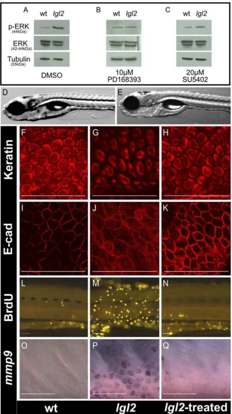
Inhibitors of ErbB signaling suppress cell proliferation and EMT in basal epidermal cells of pen/lgl2mutant larvae
As human carcinomas are often a consequence of massive over activation of growth factor signaling [36], we assayed pen/lgl2
mutant larvae for the phosphorylation level of the mitogen activated protein kinase Erk, a common member of growth factor signaling cascades, by western blot [37]. We found elevated levels of phosphorylated Erk in the mutants compared to their wild type siblings (Figure 4A). To identify which growth factor signaling is activated, we treated larvae from heterozygous pen/lgl2 carriers with inhibitors for three different receptor tyrosine kinases (RTKs), FGFR, IGFR and EGFR, starting at 96 hpf just prior to the appearance of the pen/lgl2 mutant phenotype. These treatments revealed that an inhibitor of ErbB (PD168393) reduced the levels of phosphorylated Erk (Figure 4B) and rescued the epidermal neoplasia phenotype as well (Figure 4D and 4E). Similar rescue in the epidermal phenotype was observed with another ErbB inhibitor AG1478 (data not shown). In contrast, Inhibition of FGFR (SU5402) neither affected the levels of phosphorylated Erk (Figure 4C) nor did it rescue the epidermal overgrowth phenotype (data not shown). Similarly, there was no rescue in the epidermal overgrowth phenotype when IGFR signaling was inhibited using AG1024 (data not shown). Genotyping of an entire clutch treated with ErbB inhibitor (PD168393) revealed the expected Mendelian proportion (27 out of 120) of pen/lgl2 homozygous larvae. We further asked whether the EMT and over-proliferation phenotypes were suppressed after treatment with PD168393. Indeed, cytokeratin and E-cad antibody stainings of inhibitor treated
pen/lgl2 mutant larvae were indistinguishable from wild type siblings (Figure 4H and 4K). Consistently, epidermal cell proliferation and mmp9 transcript levels were strongly reduced (Figure 4L–4N, Figure 4O–4Q). However, hemidesmosomes did not form in PD168393 treated pen/lgl2 mutant larvae as revealed by electron microscopic analysis (Figure S4).
Our data indicate that loss of functionalpen/lgl2results in over activation of ErbB signaling which promotes over proliferation of basal epidermal cells as well as cellular EMT by transcriptional modulation of EMT regulators. We further conclude that over-activation of ErbB signaling is not the cause for the absence of hemidesmosomes in thepen/lgl2mutant. This observation further indicates that disruption ofpen/lgl2primarily affects hemidesmo-some formation, which is consistent with the previous analysis [20,21].
erbB2promotes EMT but not the cell proliferation in the basal epidermal cells ofpen/lgl2 mutant larvae
In mammals four ErbB receptors are known, ErbB1 to ErbB4, which get activated upon the binding of ligands such as EGF, HB-EGF, neuregulins, betacellulin [38]. Ligand binding leads to the formation of homo- or heterodimers amongst these receptors, resulting in signal transduction [38]). Our bioinformatic and phylogenetic analysis coupled with the previous analysis of some of the family members [39,40] revealed that with the exception of
erbB2, all other members of this family exist in duplicates in the zebrafish (Figure 5A).erbB2as well aspcs/erbB3bzebrafish mutants exhibit defects in glia development and regeneration [39–42]. We founderbB2to have an additional epidermal phenotype in the fin-fold (Figure 5C) and it is also expressed in the epidermis (Figure 5E). To determine which of the ErbB receptors is activated in pen/lgl2, we performed loss of function studies in a pen/lgl2
mutant background. We knocked down erbB1awith a splice site antisense morpholino. Injections of erbB1a morpholino did not
interfere with the pen/lgl2 phenotype but reproduced the cardiovascular phenotype published earlier [43] (data not shown). However, we cannot exclude the involvement of erbB1a in promoting the EMT and growth phenotype in pen/lgl2 mutant larvae as we found the effect of the morpholino to decrease beyond 48hpf, possibly due to dilution effects (Figure S2). Double mutants ofpcs/erbB3b2/2,pen/lgl22/2genotype did not show suppression of the pen/lgl2 neoplastic phenotype and immuno-histological analysis using the pan 1–8 Cytokeratin antibody did not reveal any reduction in strength and initiation of EMT phenotype (data not shown, Figure 5I). Taken together, these results demonstrate that
erbB3bfunction is not essential for neoplasia and EMT phenotype inpen/lgl2mutants.
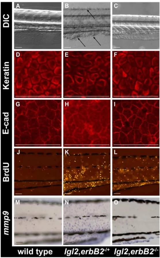
In zebrafish,lgl2anderbB2are both located on chromosome 12 (23.6mb distance). To study double mutants, chromosomal recombinants were made (see Materials and Methods). We investigated progeny fromlgl22/+
,erbB22/+
double heterozygous fish (3 crosses; n = 296), which were sorted for the morphological epidermal lgl2 phenotype at 108hpf as well as 132hpf and subsequently genotyped. We identified 40 larvae that were homozygous for bothlglanderbB2. In 37 of these, no epidermal phenotype was detected up to 132hpf (Figure 6C). In contrast, all
lgl2 larvae with erbB2+/2 or erbB2+/+ genotype showed strong
phenotypes or lethality at the same stage (Figure 6B, Table S2). In a separate experiment, we analyzed Cytokeratin and E-cadherin localization,mmp9expression and BrdU incorporation inlgl22/2,
erbB22/2 double mutants at 108hpf when the lgl2 phenotype becomes apparent. The morphology of the epidermal cells in the double mutant larvae, as revealed by Keratin staining, appeared completely normal at 108hpf Figure 6D–6F). Occasionally, epidermal cells inpen/lgl2; erbB2double mutant larvae exhibited milder changes in Keratin organization, indicative of altered cellular morphology at 132 hpf (data not shown). E-cadherin localization appeared normal in the epidermis of the double mutants (Figure 6G–6I) and mmp9 expression levels in the epidermis of double mutant larvae were comparable to those in wild type (Figure 6M–6O). Interestingly, however, the BrdU incorporation analysis revealed that (Figure 6J–6L) there is no significant decrease (t-test, p.0.05) in the epidermal cell proliferation in a predefined area in lgl22/2,erbB22/2 double mutants (2961.0, n = 3) as compared tolgl22/2,erbB22/+
(3269, n = 3) larvae.
Our data suggest thaterbB2but noterbB3bmediated signaling is responsible mainly for the EMT phenotype in pen/lgl2 mutant basal epidermis. We did not observe rescue in the epidermal cell-proliferation phenotype in pen/lgl2; erbB2 double mutants and epideramal cell morphology was altered as well at later stages indicating that erbB2 deficiency doesn’t completely rescue the phenotype. Since the inhibitors completely rescue both the proliferation as well as EMT phenotypes, we propose that other ErbB receptors might be involved in promoting epidermal cell-proliferation inpen/lgl2mutants.
Discussion
Figure 4. Inhibition of ErbB signaling restores the epidermal morphology and cell cycle inpen/lgl2mutant larvae.Western blot analysis of Erk1/2 phosphorylation in untreated wild-type andpen/lgl2larvae (A), larvae treated with PD168393 (B) and larvae treated with SU5402 (C). DIC images of wild-type (D) andpen/lgl2mutant (E) treated with the ErbB inhibitor PD168393 and genotyped. The pan 1–8 cytokeratin antibody staining (F–H), anti E-cad staining (I–K), anti BrdU antibody staining (L–N) and in situ hybridization staining formmp9(O–Q) of the epidermis of wild-type (F, I, L, O)pen/lgl2mutant (G, J, M, P) and mutant larvae treated with PD168393 (H, K, N, Q). In comparison to wild-type larvae (A, left lane, 44kD) Erk shows higher level of phosphorylation inpen/lgl2mutant larvae (A, right lane 44kD). The levels of Erk phosphorylation are equal in the mutants treated with PD168393 (B) but not with SU5402 (C). The a-Tubulin levels (55kD) are indicative of equal protein loading. The epidermal cell morphology, E-cad localization, cell proliferation andmmp9expression inpen/lgl2larvae, treated with PD168393 (H, K, N, Q) appears similar to wild-type larvae (F, I, L, O) than the untreated mutant larvae (G, J, M, P). Note that PD168393 treated (rescued) larvae were genowild-typed to confirm their genotypes.
this pathway,lglanddlg, have multiple orthologues [45]. Loss of
lgl1 function results in brain hyperplasia in the mouse [17]. In zebrafish loss of lgl2 function leads to over-proliferation of epidermal cells [20]. Here, we demonstrate that transplantation ofpen/lgl2homozygous mutant cells during blastula stage into wild type embryos results in the formation of tumor like structures in the epidermis after 7 days. This analysis clearly shows that lgl2
behaves as a tumor suppressor gene in vertebrates.
The basal epidermal cells in pen/lgl2 mutants do not form hemidesmosomes and display an altered cell morphology instead of the polygonal cell shape found in wild type larvae [20]. E-cad localization at the plasma membrane is reduced in the mutant epidermal cells indicating that epidermal cells undergo EMT. Indeed, GFP labeling of basal epidermal cells revealed that inpen/ lgl2 mutant larvae these cells show continuous changes in their morphology, project lamellipodia and move as a sheet. This sheet like movement has been described as a metastable cell state in the context of EMT [28]. Consistent with the cellular analysis, the expression profiling oflgl2mutant basal epidermal cells revealed differential expression of the known EMT markers and regulators such asmatrix-metalloproteinasesandkeratins. Intriguingly, the levels of
E-cadRNA are high in thepen/lgl2mutant. This is in contrast to known developmental scenarios of EMT such as the delamination of neural crest cells or gastrulation where E-cad expression is downregulated at the transcriptional level by a transcriptional repressor, snail [34,35]. In addition, structural components of tight junctions, such as severalclaudins, are also up-regulated inpen/lgl2
mutants. Thus, the expression profiles of cell adhesion molecules in pen/lgl2 larvae resemble several human breast carcinomas including an inflammatory breast cancer (IBC), a highly aggressive subtype of human breast cancer, which has been characterized by E-cad and erbB2 over-expression [46–51]. The up-regulation of
claudin genesis intriguing. However, it is not clear whether Claudins localize properly in the epidermal cells. Even if the Claudins do localize properly, the in-situ expression analysis suggests that
claudins are up-regulated mostly in the cellular clumps that are formed in the median finfold or in the ventral jaw region. It is thus
clear that claudins are not expressed during the process or EMT but rather when the mesenchymal cells re-acquire partial epithelial phenotypes while forming tumor like aggregation. However, further analysis is required to test this notion.
How does deficiency in components of the baso-lateral pathway lead to epithelial cell proliferation and EMT? It has been suggested that the protein Scrib stabilizes the coupling between E-cad and the Catenins and thus behaves as a regulator of epithelial cell adhesion and migration [52]. Our data show that for Lgl2 the mechanism to suppress EMT and tumor formation is fundamentally different. Lgl2 may not manifest its function by stabilizing the coupling between E-cad and Catenins. Previously, we have shown that neither the loss of maternal nor zygoticlgl2
function primarily affect E-cad localization [20,21]. Consistent with mammalian data, knockout ofE-cadin the mouse or zebrafish epidermis does not result in EMT [21,53–55]. This indicates that although the loss of E-cad facilitates EMT [56], it may not be sufficient to induce it in animal models.
We show that inhibition oferbB2signaling, either genetically or by small chemical inhibitors, leads to suppression of EMT and a neoplastic phenotype inpen/lgl2mutant larvae. Intriguingly, E-cad membrane localization is restored in these larvae, indicating that the loss of E-cad is a consequence of activation of ErbB signaling rather than a cause of it. Thus, our analyses presented here suggest that Lgl2 acts as a tumor suppressor by regulating the amplitude of ErbB signaling in the epidermis. Since we did not observesnail -mediated down-regulation of E-cad, we propose that an erbB2
dependent pathway inpen/lgl2mutants leads to the destabilization of E-cad at adherens junctions. Indeed in pen/lgl2 mutant epidermal cells, known modifiers of E-cad function, such as
mmp9 and sgk1 are up-regulated (Figure 3). While Mmps are known to be involved in ecto-domain shedding of E-cad [57,58], Sgk1 functions in the phosphorylation of Ndrg1, a protein involved in vesicular recycling of E-cad [59,60]. Additionally, recently published data from cell culture suggests an involvement of RTK signaling in the destabilization of adherens junctions via Numb [61]. Here, Numb functions as an adapter protein coupling Figure 5. Phylogenetic analysis oferbBfamily members and zebrafish mutants oferbBparalogs.(A) Phylogenetic analysis oferbBfamily members in the zebrafish genome (zv7) with the human orthologs (Minimum evolution algorithm, 100 replicates). With the exception oferbB2, all othererbBfamily members are duplicated in zebrafish. (B–D) DIC Images of wild-type (B),erbB22/2(C) anderbB3b2/2(D) larvae at 132hpf. (E) In-situ hybridization analysis oferbB2at 48hpf. Keratin staining inerbB3bmutant larva (F)erbB3b/lgl2double mutant larva (G) andpen/lgl2mutant larva (H) at 132 hpf. Note that inerbB3B;lgl2double mutants (G) epidermal cells appear spindle shaped as inpen/lgl2mutant larvae (H).erbB3b(F) mutations alone do not affect the morphology of basal epidermal cells.
Figure 6.erbB2promotes EMT in the epidermis ofpen/lgl2mutant larvae.DIC images of the morphology of wild-type (A),lgl2,erbB2+/2(B) and lgl2,erbB22/2(C) larvae at 108hpf. Keratin (D–F), E-cadherin (G–I), BrdU (J–L) andmmp9(M–O) staining in wild-type (D, G, J, M),lgl2,erbB2+/2(E, H, K, N) andlgl2,erbB22/2(F, I, L, O) larvae at 108hpf. The DIC images of the morphology reveals that cellular clumps, a typical characteristic ofpen/lgl2 mutant larvae are present inlgl2,erbB2+/2(B) but are absent inlgl2,erbB22/2(C). The keratin, E-cadherin andmmp9staining inlgl2,erbB22/2(F, I, O) appear similar to wild-type (D, G, M) indicatingerbb2promotes the EMT phenotype inpen/lgl2mutant larvae. Note that, inlgl2,erbB22/2larvae (L) BrdU incorporation does not decrease to wild-type levels (J).
E-cadherin to the Par polarity-complex. This interaction was shown to be sensitive to elevated levels of RTK signaling, leading to mislocalization of E-cadherin [61]. Further analysis involving loss of function of mmps and sgk1 as well as studies of Numb localization in the pen/lgl2 mutant larvae would be necessary to clarify their contributions to the EMT phenotype.
The primary function of Lgl2 in hemidesmosome formation [20,21] is not dependent on ErbB signaling as hemidesmosomes do not form inpen/lgl2mutants even after the inhibition of ErbB signaling (Figure S4). This suggests that the loss of hemidesmo-somes might be the cause but not a consequence of the activation of ErbB signaling. This hypothesis is supported by the fact that components of hemidesmosomes (e.g. Itga6/Itgb4), physically interact with the ErbB2 receptor tyrosine kinase [62–64]. Thus, it is possible that mislocalization of hemidesmosomal components in
pen/lgl2mutants lead to an activation of ErbB2.
ErbB signaling plays an important role in the development of human carcinomas, as it is able to induce proliferation and EMT and further is able to suppress apoptosis [36,65]. This is in particular true for ErbB2 which is over-expressed in more than 25% of all breast carcinoma [66]. A causal link between ErbB activation and loss of cell polarity, which is one of the hallmarks of carcinomas, has recently been established. It has been shown that activated ErbB2 associates with Par6-aPKC leading to disruption of the apico-basal polarity [67]. While ErbB2 regulates apico-basal cell polarity by interacting with apical polarity components like Par6-aPKC, our analyses presented here suggest that one of the basolateral pathway components, Lgl2, regulates the activation of ErbB2. Thus, there seems to be a reciprocal interaction between cell polarity regulators and ErbB2 signaling. Interestingly, although ErbB2 activation induces cell proliferation, the associa-tion of ErbB2 with Par6-aPKC was not essential for regulating cell proliferation indicating that ErbB2 affect the cell proliferation independent of disruption of apico-basal cell polarity [67]. Our data suggest that at the organismal level loss of ErbB2 does not affect the epidermal cell proliferation phenotype in lgl2mutants but only prevents EMT. Since inhibitors, which do not discriminate between various ErbB family members rescue both, proliferation as well as EMT phenotypes, it appears that activation of more than one ErbB family member might be involved in promoting epidermal neoplasia in pen/lgl2 mutants. Thus, cell proliferation and EMT phenotypes are not coupled in pen/lgl2
mutant larvae.
The ErbB signaling pathway includes multiple ligands and receptors in vertebrates. ErbB1 and ErbB4 represent discrete RTKs, as they contain ligand binding sites as well as a kinase activity, essential for auto-phosphorylation and signal transduc-tion. In contrast, ErbB2 and ErbB3 do not independently transduce extracellular signals as homodimers since ErbB2 has no ligand-binding domain and ErbB3 has no kinase domain. Thus, ErbB2 and ErbB3 must interact with each other or with other ErbB receptors to transduce signals [38]. Analysis of the
Danio reriogenome revealed that genome duplication events have led to the duplication of erbB1, erbB3 and erbB4 making the situation even more complex. We found that homozygous mutations inerbB2but noterbB3bdramatically rescued the EMT phenotype, establishing ErbB2 signaling to be responsible for the onset of EMT. However, the direct activator of ErbB2, in promoting the EMT phenotype in lgl2 mutants remains unidentified at this point.
To summarize,lgl2function is essential for tumor suppression and EMT in the vertebrate epidermis. pen/lgl2 mutant cells are able to induce the formation of epidermal tumors when surrounded by wild type cells. In the absence of Lgl2,
hemidesmosomes do not form and ErbB signaling is activated, which results in induction of pathways involved in promoting EMT via mislocalization of E-cad as well as proliferation. Amongst severalerbBparalogues,erbB2is more directly involved in transducing the signal. Thus, thepen/lgl2mutant would serve as a very good model to perform chemical screens for compounds that are able to suppresserbBsignaling or important downstream effectors and thus have high potential for cancer therapeutic research.
Materials and Methods
Fish strains
The morphological and immuno-histological analysis ofpen/lgl2
mutant was carried out in Tuebingen (TUE) and WIK background. For transplantations, embryos frompen/lgl2mutants in the background of anb-actin::GFP transgenic line were used as donors and from albino fish as recipients. Live imaging was performed in transgenicDNp63::Gal4, UAS::GFP line. TheerbB2
mutant allele used in this work is st61 [39]. TheerbB3bmutant allele is HJ036, which contains a C to A point mutation at position 156 leading to a premature stop after 50 amino acids [41]. Aslgl2
and erbB2 are located on the same linkage group, to generate double mutants, we crossedpen/lgl2+/2fish witherbB2+/2fish and out-crossed the obtained trans-heterozygote F1 fish to albino fish. The F2 generation were then screened by genotyping for both, the
pen/lgl2anderbB2mutations.
Phylogenetic analysis
Protein sequences for ErbB zebrafish paralogues and human orthologues were obtained from Genebank (http://www.ncbi.nlm. nih.gov) and through Ensembl (http://www.ensembl.org/Danio_ rerio) databases. Alignments were performed using ClustalW. Phylogenetic analysis was run using neighbor joining, maximum parsimony and minimum evolution algorithms, (MEGA4, http:// www.megasoftware.net/). All analysis methods showed similar tree morphology with comparable bootstrap values (1000 replicates). Human orthologues (Genebank ID): ERBB1: 1956, ERBB2: 2064, ERBB3: 2065, ERBB4: 2066. Zebrafish paralogues (Ensembl transcript ID): erbB1a: ENSDART00000027219, erbB1b: EN-SDART00000031151, erbB2: ENSDART00000003932, erbB3a:
ENSDART00000014892,erbB3b:ENSDART00000049893,erbB4a:
ENSDART00000092114,erbB4b:ENSDART00000100398
GeneratingDNp63::Gal4, UAS::GFP transgenic fish The DNp63 promoter was cloned by enzymatic restriction of BAC dkey-13d19 with HhaI (NEB) and subsequent blunt-end cloning of a resulting 4.96 kb fragment into a plasmid containing a Gal4,UAS::GFP expression cassette and mini-TOL2 sites (based on [68,69]). Transgenesis was achieved by simultaneous injection of plasmid DNA and transposase RNA at 1-cell stage using WPI PV830 pneumatic injection system followed by F1 screen for GFP positive larvae.
BrdU labelling
5-day-old wild type andpen/lgl2mutant larvae were incubated with 10 mM BrdU solution in 2% DMSO in embryonic medium (E3) for 2 hours. After treatment, larvae were washed several times in E3 fixed overnight in 4% PFA in PBS at 4uC. Staining was performed as described below.
Immunohistochemistry
1–8 (Progen Biotechnik) and anti E-cad antibody (BD Transduc-tion Laboratory). Embryos were either fixed in 4% PFA (E-cad, BrdU) or in Dent’s fixative (cytokeratin). After downgrading the larvae to 0.1 M phosphate buffer (PB), they were washed with PBT (PB+0.8% Triton X-100) five times and blocked in 10% normal goat serum. For BrdU staining, larvae were treated with 4 N HCl for 20 min., washed in PB and blocked in 1% BSA for 1–3 hours. Antibodies were diluted as: Ks pan 1–8 (1:10), anti-E-cad (1:250), anti-BrdU (1:50) and samples were incubated at room temperature for 4 hours or overnight at 6–8uC. Afterwards, larvae were washed five times in PBT, incubated with Cy3 or Alexa 488 anti-mouse or anti-rat antibodies, post fixed in 4% PFA and upgraded in 70% glycerol for fluorescence/light microscopy.
In situ hybridization
DIG-labeled RNA probes forcldn7, krt5, mmp9 and sgk1were prepared from larval 5dpf total cDNA obtained frompen/lgl22/2
mutants. In situ hybridization was performed using Intavis in-situ robot (model: insituPro VSi).
Probe templates have been amplified with the following primer combinations by PCR reaction and subsequently cloned into pGEM-T Easy Vector (Promega). DIG-labeled probes were synthesized using T7 and Sp6 RNA polymerases (Roche).
E-cad: 59-TTACTTCTGCTATTGCTTGCT-39; 59 -TCATA-GTCTTGGTCGTTTCCT-39
cldn7: 59-TGGCACATAAAGGACTGCAA-39; 59 -CGATGA-AAATAGCTGCACCA-39
erbB2: 59-GTCATCCAGAACGAAGATCAG-39; 59 -CATCA-GTCTCCAGATCTCCA-39
krt5: 59-CAGGAGCTCAGTGTCCTTCC-39; 59 -CGGTT-GTTGAGGGTCTTGAT-39
mmp9: 59-GCTGCTCATGAGTTTGGAC-39; 59 -CCGAG-CTTCTCGATTTTACG-39
sgk1: 59-ATGGAACGACGTCAACCTTC-39; 59 -GCGTA-AGCTTCTTGGCATTC-39
Transplantations
Transplantations were carried out at blastula stage. After transplantations, donor and corresponding recipients embryos were cultured together (3–5 in number) in a 24 well plate. The
pen/lgl2mutant donors were identified at 4.5dpf by phenotype or by molecular genotyping at blastula stage (RFLP). The host larvae that received mutant or wild type clones (GFP-positive) in the skin were sorted and further raised up to 7–8 dpf and analyzed by microscopy for tumor phenotypes.
Inhibitor treatment
For screening, all inhibitors (SU5402; AG1024; PD168393; AG1478 all Calbiochem) were used at 10–50mM concentration in 1% DMSO in E3 Medium starting at 96hpf. For BrdU, IHC and ISH experiments, PD168393 and AG1478 were used at a concentration of 10mM starting at 96hpf.
Western blot and detection
For western blots, mutants were identified by the development of the characteristiclgl2mutant phenotype at 108hpf. Three times 40 mutants and equivalent number of wild type larvae were collected at 108hpf and than treated with either DMSO as a control, or inhibitors for ErbB signaling (10mM) or FGFR signaling (20mM) for 12 h. Subsequently, larval tails were collected by cutting posterior to the swimming bladder on ice. Proteins were extracted in DXB (25 mM Hepes, pH 6.8, 50 mM KCl, 1 mM MgCl2, 1 mM DTT, 250 mM sucrose) containing
Roche Complete protease inhibitors (Cat No. 11836153001) and Pierce Halt phosphatease inhibitors (Cat No. 78420). Protein concentration was determined using OD at 280 nm (NanoDrop). For PAGE, NuPage 4–12% gradient gels (Invitrogen) were used. After transfer, equal loading was re-checked by poinseau red staining. Antibodies used were: p-ERK (M9692; 1:600), Erk (M5670; 1:500), a-Tubulin (T9026; 1:10000) (all from Sigma). Secondary antibodies used were HRP conjugated anti-mouse (p-Erk, Tubulin) and anti-rabbit (Erk). Detection was performed using chemiluminiscence (ECL+; GE Healthcare).
Microarray analysis
The microarray experiment was conducted using tissues (tail) posterior of the anal opening of wild type (Tuebingen) and pen
larvae at 5 dpf. RNA for microarray analysis was extracted from four biological replicates using TRIZol Reagent (Gibco BRL, Eggenstein, Germany). Complementary RNA was prepared from 1mg total RNA from each replicate as described in the Agilent Low RNA Input Linear Amplification kit manual (Agilent Technologies, Palo Alto, CA, USA). Double-stranded cDNA was synthesized using the reagents from this kit, and Cy3- or Cy5-labeled cRNA was prepared by cDNA in vitro transcription in the presence of cyanine 5-CTP or cyanine 3-CTP dyes. Fluorescently labeled RNA was then purified with the Qiagen RNEasy spin columns, according to the manufacturer’s protocol (Qiagen, Hilden, Germany). After purification, cRNA was fragmented and used to hybridize to the zebrafish G2519F 4X44 microarray platform containing 4 duplicated arrays of the 22,000 probe-set design (Agilent Technologies, Palo Alto, CA, USA). Four biological replicates were conducted, including two dye swap experiments to minimize the effect of any potential dye bias. Hybridization, washing, and scanning were performed according to the manufacturer’s protocol. The microarrays were scanned on a Genepix Axon 4000B scanner (Molecular Devices, Union City, CA, USA) at five micron resolution with five-line averaging. Raw expression values from each probeset were extracted using the Genepix Pro 6.0 feature extraction software, and features were flagged manually for poor quality. The data were then analyzed in the R statistical programming environment using the Bioconduc-tor module Limma [70,71]. Duplicate probesets on each array were considered as technical replicates for the analysis in addition to the 4 biological replicates on separate arrays. A standard linear model for differential expression, with the Limma module in Bioconductor, was used to identify genes up and down regulated in the mutant versus wild type experiment. The resulting p-values from the hypothesis tests were adjusted for multiple testing with the false discovery approach (FDR) to control for false positives [72]. In addition, the empirical Bayes approach automatically adjusts raw p-values for multiple testing and generates a B-statistic that may also be used for ranking differentially expressed genes [70]. All microarray data may be accessed through the ArrayEx-press repository on the European Bioinformatics Institute database website (http://www.ebi.ac.uk/microarray-as/ae/).
Quantitative real-time PCR
(DDC(t) method) as described [74]. Statistical significance was determined using student’s t-test.
Genotyping of mutant larvae
Genotyping to indentify mutants was done by a PCR based restriction fragment length polymorphism (RFLP) method for
lgl2-anderbB2-or by DNA sequencing forerbB3bmutants. The RFLP analysis was done as follows- the mutations inerbB2cause a loss whereas in lgl2 a gain of a restriction site. PCR product from genomic DNA samples obtained by the primer pairs: 59 -ATGCATACCTTCCTGGAGTAG-39; 59 -TGTGGTTCTA-GTGGAGGAGGA-39 (for lgl2) or 59 -TGAAGAATGCTGG-TAGCTGG-39 and 59-GGACTCAGCAAAGG ACTTAC-39
(for erbB2) was digested with either SfcI (for lgl2 mutation) or BsrGI (for erbB2mutation) resulting in a genotype specific DNA band pattern (Figure S3). The erbB3b genotyping was done by scoring for the premature stop at position 156bp by DNA sequencing of a PCR product obtained from the genomic DNA using the primer pair 59-CGCTCTCCTGTTCCTCTGTG-39; 59-ACCCTCTTCCTCCATTGTCC-39 (Figure S3). In case of PFA fixation, individual genomic DNA samples were sampled before fixation (head tissue). The genotype of larvae used in ISH experiments was scored by the development of the characteristic
lgl2phenotype.
Movie processing and image analysis
Time-lapse movies were acquired using AxioVision 4.6 (Zeiss) and compressed in the Codec H264. For encoding in H264, the OSS Virtual Dub was used. Cell area (Figure 2G) was determined using the ‘‘outline’’ tool included in Zeiss AxioVision 4.6 (Zeiss) on three biological replicates each from wild type andpen/lgl2. Scale bars indicate equal sizes within one figure.
Supporting Information
Figure S1 Expression of GFP under the DNp63 promoter. Antibody staining of 5.5dpf tg(DNp63::Gal4,UAS::GFP) zebrafish larvae using anti Cytokeratin (red) and anti GFP antibody (green). The co-labeling of both antibodies reveals activity of the 4.96 kb upstream promoter element of DNp63 exclusively in basal epidermal cells in the skin.
Found at: doi:10.1371/journal.pgen.1000720.s001 (0.70 MB TIF)
Figure S2 Knockdown oferbB1ausing morpholinos. For knock-down oferbB1a, we used a morpholino targeted to a splice site. To test the activity of this morpholino over time, primers spanning the targeted intron-exon boundary were designed. (A) PCR performed on cDNA from morphant zebrafish larvae at different time points reveals morpholino efficiency. (B) A working morpholino causes splice events to fail at the targeted splice site (red) resulting in larger PCR product. Genomic DNA contamination of the cDNA causes a third, larger, product as the amplicon spans two introns on the genomic template. This analysis reveals that the used morpholino only prevents efficient splicing oferbB1aRNA before 48hpf, causing equal amounts of spliced vs. morphant RNA at later stages. Primers used for PCR: 59-CCACCAACA TCGACTCCTTT-39; 59-AAACCTTGAGGTCATCCGAG-39. Morpholino sequence: 59 -AAATGCTCTTCCTCACCCTCT-GAAT-39.
Found at: doi:10.1371/journal.pgen.1000720.s002 (0.54 MB TIF)
Figure S3 Genotyping oflgl2,erbB2, anderbB3b. The genotype of larvae presented in this work was scored using PCR-based restriction fragment length polymorphisms (RFLP) for erbB2 and lgl2or by sequencing for erbB3b. The mutation in lgl2 causes an artificial
restriction site for SfcI. The mutation inerbB2causes a loss of a BsrGI restriction site. Treatment of PCR products from individual genomic samples with either SfcI or BsrGI leads to a genotype specific DNA band pattern in agarose gel electrophoresis. TheerbB3bmutation was scored by sequencing of a PCR product spanning the site of lesion. The nature of the mutation is a cytosine to adenine transversion leading to a premature stop codon after 156bp. Found at: doi:10.1371/journal.pgen.1000720.s003 (0.66 MB TIF)
Figure S4 Inhibition of erbB signaling does not restore hemidesmosome formation in pen/lgl2 mutants. pen/lgl2 mutant basal cells are unable to form hemidesmosomes, even after inhibition of ErbB signaling. EM cross section through larval skin 5dpf reveals hemidesmosome formation at the basal membrane in wild types (A, arrows) whereas pen/lgl2 mutants (B) andpen/lgl2
mutants treated with ErbB inhibitor PD168393 (C) lack these structures.
Found at: doi:10.1371/journal.pgen.1000720.s004 (1.58 MB TIF)
Table S1 Expression profile ofpen/lgl2 compared to wild type. Using microarray technique the expression profile of wild-type versus mutant zebrafish larval tails, posterior to the anal opening was analysed. A significance threshold of adj. p-value of 1026 (FDR) resulted in 117 genes to be significantly differentially expressed. Within those, most prominent genes, involved in EMT and cell cycle, as well as cytoskeleton rearrangements, can be found. Additionally, genes involved in the formation of tight and adherens junctions are present. The down-regulated genes are indicated in red in this table whereas up-regulated genes are indicated in black.
Found at: doi:10.1371/journal.pgen.1000720.s005 (0.03 MB XLS)
Table S2 Comparison of phenotypes and genotypes inerbB2,lgl2
double mutant incrosses. Percental distribution of epidermal neoplasia inpen/lgl2 single- and lgl2,erbB2double mutants. Note that the loss of erbB2 strongly reduces the formation of the characteristic overgrowth phenotype in the pen/lgl2 mutant background, even at late time points.
Found at: doi:10.1371/journal.pgen.1000720.s006 (0.03 MB DOC)
Video S1 Phenotype of basal epidermal cells in the wild-type larvae. A 90-minute timelapse movie of tg(Np63::Gal4,UAS::GFP) wild-type zebrafish larva at 5dpf. Settings: 2061-min interval, 10 frames per second. The GFP labelled basal epidermal cells in the wild-type larvae remain static.
Found at: doi:10.1371/journal.pgen.1000720.s007 (1.81 MB MOV)
Video S2 Phenotype of basal epidermal cells in the pen/lgl2
mutants larvae. A 90-minute timelapse movie of tg(Np63:: Gal4,UAS::GFP) lgl22/2 zebrafish larva at 5dpf. Settings: 206 1-min interval, 10 frames per second. The GFP labelled basal epidermal cells exhibit migratory behavior in the mutant larvae. Cells show cell shape changes and development of lamellipodia like cell protrusions, indicating their mesenchymal character. Found at: doi:10.1371/journal.pgen.1000720.s008 (6.63 MB MOV)
Video S3 Phenotype of basal epidermal cells in wild-type larvae. A 240-minute timelapse movie of tg(DNp63::Gal4,UAS::GFP) wild-type zebrafish larva at 5 dpf. Settings: 2561-min interval, 10 frames per second. The GFP labelled basal epidermal cells remain static in the wild-type larvae.
Video S4 Phenotype of basal epidermal cells inpen/lgl2mutant larvae. A 240-minutes timelapse movie of tg(Np63::Gal4,UAS::GFP)
lgl22/2 zebrafish larva at 5dpf. Settings: 2561-min interval, 10 frames per second. The GFP labeled basal epidermal cells exhibit migratory properties in the mutant larva. Cells show cell shape changes and formation of lamellipodia like cell protrusions indicating their mesenchymal character.
Found at: doi:10.1371/journal.pgen.1000720.s010 (13.28 MB MOV)
Acknowledgments
We thank all members of our lab, in particular Nadine Wittkop, Jana Krauss, and Kellee Siegfried-Harris for the helpful discussions and comments on the manuscript. Our special thanks go to Matthew Harris, Martina Konantz, Hans-Martin Maischein, Heinz Schwarz, and So¨ren Alsheimer for help with some of the experiments. We further want to thank William Talbot and Matthew Voas for sending us the st61 allele oferbB2.
Author Contributions
Conceived and designed the experiments: SR MS. Performed the experiments: SR MPL. Analyzed the data: SR MPL MS. Wrote the paper: SR CNV MS.
References
1. Gateff E, Schneiderman HA (1967) Developmental studies of a new mutant of Drosophila melanogaster: Lethal malignant brain tumor (l(2)gl 4. American Zoologist 7: 760.
2. Gateff E SH (1974) Developmental Capacities of Benign and Malignant Neoplasms of Drosophila. Rouxs Arch Dev 176: 23–65.
3. Watson KL, Justice RW, Bryant PJ (1994) Drosophila in cancer research: the first fifty tumor suppressor genes. J Cell Sci Suppl 18: 19–33.
4. Woodhouse E, Hersperger E, Shearn A (1998) Growth, metastasis, and invasiveness of Drosophila tumors caused by mutations in specific tumor suppressor genes. Dev Genes Evol 207: 542–550.
5. Gateff E (1978) Malignant neoplasms of genetic origin in Drosophila melanogaster. Science 200: 1448–1459.
6. Gonzalez C (2007) Spindle orientation, asymmetric division and tumour suppression in Drosophila stem cells. Nat Rev Genet 8: 462–472.
7. Ohshiro T, Yagami T, Zhang C, Matsuzaki F (2000) Role of cortical tumour-suppressor proteins in asymmetric division of Drosophila neuroblast. Nature 408: 593–596.
8. Peng CY, Manning L, Albertson R, Doe CQ (2000) The tumour-suppressor genes lgl and dlg regulate basal protein targeting in Drosophila neuroblasts. Nature 408: 596–600.
9. Betschinger J, Mechtler K, Knoblich JA (2003) The Par complex directs asymmetric cell division by phosphorylating the cytoskeletal protein Lgl. Nature 422: 326–330.
10. Woodhouse EC, Fisher A, Bandle RW, Bryant-Greenwood B, Charboneau L, et al. (2003) Drosophila screening model for metastasis: Semaphorin 5c is required for l(2)gl cancer phenotype. Proc Natl Acad Sci U S A 100: 11463– 11468.
11. Bilder D, Li M, Perrimon N (2000) Cooperative regulation of cell polarity and growth by Drosophila tumor suppressors. Science 289: 113–116.
12. Tanentzapf G, Tepass U (2003) Interactions between the crumbs, lethal giant larvae and bazooka pathways in epithelial polarization. Nat Cell Biol 5: 46–52. 13. Bilder D, Schober M, Perrimon N (2003) Integrated activity of PDZ protein
complexes regulates epithelial polarity. Nat Cell Biol 5: 53–58.
14. Perrimon N (1988) The maternal effect of lethal(1)discs-large-1: a recessive oncogene of Drosophila melanogaster. Dev Biol 127: 392–407.
15. Mechler BM, McGinnis W, Gehring WJ (1985) Molecular cloning of lethal(2)giant larvae, a recessive oncogene of Drosophila melanogaster. Embo J 4: 1551–1557.
16. Bilder D, Perrimon N (2000) Localization of apical epithelial determinants by the basolateral PDZ protein Scribble. Nature 403: 676–680.
17. Klezovitch O, Fernandez TE, Tapscott SJ, Vasioukhin V (2004) Loss of cell polarity causes severe brain dysplasia in Lgl1 knockout mice. Genes Dev 18: 559–571.
18. Kuphal S, Wallner S, Schimanski CC, Bataille F, Hofer P, et al. (2006) Expression of Hugl-1 is strongly reduced in malignant melanoma. Oncogene 25: 103–110.
19. Tsuruga T, Nakagawa S, Watanabe M, Takizawa S, Matsumoto Y, et al. (2007) Loss of Hugl-1 expression associates with lymph node metastasis in endometrial cancer. Oncol Res 16: 431–435.
20. Sonawane M, Carpio Y, Geisler R, Schwarz H, Maischein HM, et al. (2005) Zebrafish penner/lethal giant larvae 2 functions in hemidesmosome formation, maintenance of cellular morphology and growth regulation in the developing basal epidermis. Development 132: 3255–3265.
21. Sonawane M, Martin-Maischein H, Schwarz H, Nusslein-Volhard C (2009) Lgl2 and E-cadherin act antagonistically to regulate hemidesmosome formation during epidermal development in zebrafish. Development.
22. Spaderna S, Schmalhofer O, Wahlbuhl M, Dimmler A, Bauer K, et al. (2008) The transcriptional repressor ZEB1 promotes metastasis and loss of cell polarity in cancer. Cancer Res 68: 537–544.
23. Hanahan D, Weinberg RA (2000) The hallmarks of cancer. Cell 100: 57–70. 24. Rajkumar T (2001) Growth factors and growth factor receptors in cancer.
Current Science 81: 535–541.
25. Lee M, Vasioukhin V (2008) Cell polarity and cancer–cell and tissue polarity as a non-canonical tumor suppressor. J Cell Sci 121: 1141–1150.
26. Hsu MY, Meier FE, Nesbit M, Hsu JY, Van Belle P, et al. (2000) E-cadherin expression in melanoma cells restores keratinocyte-mediated growth control and down-regulates expression of invasion-related adhesion receptors. Am J Pathol 156: 1515–1525.
27. Peinado H, Olmeda D, Cano A (2007) Snail, Zeb and bHLH factors in tumour progression: an alliance against the epithelial phenotype? Nat Rev Cancer 7: 415–428.
28. Lee JM, Dedhar S, Kalluri R, Thompson EW (2006) The epithelial-mesenchymal transition: new insights in signaling, development, and disease. J Cell Biol 172: 973–981.
29. Yang A, Kaghad M, Wang Y, Gillett E, Fleming MD, et al. (1998) p63, a p53 homolog at 3q27-29, encodes multiple products with transactivating, death-inducing, and dominant-negative activities. Mol Cell 2: 305–316.
30. Akgul B, Pfefferle R, Marcuzzi GP, Zigrino P, Krieg T, et al. (2006) Expression of matrix metalloproteinase (MMP)-2, MMP-9, MMP-13, and MT1-MMP in skin tumors of human papillomavirus type 8 transgenic mice. Exp Dermatol 15: 35–42.
31. Hua J, Muschel RJ (1996) Inhibition of matrix metalloproteinase 9 expression by a ribozyme blocks metastasis in a rat sarcoma model system. Cancer Res 56: 5279–5284.
32. Sehgal G, Hua J, Bernhard EJ, Sehgal I, Thompson TC, et al. (1998) Requirement for matrix metalloproteinase-9 (gelatinase B) expression in metastasis by murine prostate carcinoma. Am J Pathol 152: 591–596. 33. Batlle E, Sancho E, Franci C, Dominguez D, Monfar M, et al. (2000) The
transcription factor snail is a repressor of E-cadherin gene expression in epithelial tumour cells. Nat Cell Biol 2: 84–89.
34. Carl TF, Dufton C, Hanken J, Klymkowsky MW (1999) Inhibition of neural crest migration in Xenopus using antisense slug RNA. Dev Biol 213: 101–115. 35. Cano A, Perez-Moreno MA, Rodrigo I, Locascio A, Blanco MJ, et al. (2000) The transcription factor snail controls epithelial-mesenchymal transitions by repressing E-cadherin expression. Nat Cell Biol 2: 76–83.
36. Slamon DJ, Godolphin W, Jones LA, Holt JA, Wong SG, et al. (1989) Studies of the HER-2/neu proto-oncogene in human breast and ovarian cancer. Science 244: 707–712.
37. McKay MM, Morrison DK (2007) Integrating signals from RTKs to ERK/ MAPK. Oncogene 26: 3113–3121.
38. Citri A, Yarden Y (2006) EGF-ERBB signalling: towards the systems level. Nat Rev Mol Cell Biol 7: 505–516.
39. Lyons DA, Pogoda HM, Voas MG, Woods IG, Diamond B, et al. (2005) erbb3 and erbb2 are essential for schwann cell migration and myelination in zebrafish. Curr Biol 15: 513–524.
40. Rojas-Munoz A, Rajadhyksha S, Gilmour D, van Bebber F, Antos C, et al. (2009) ErbB2 and ErbB3 regulate amputation-induced proliferation and migration during vertebrate regeneration. Dev Biol 327: 177–190.
41. van Bebber F (2005) Neural crest mutants and the effect of glia during differentiation in the zebrafish. PhD Thesis, University of Tu¨bingen. 42. Budi EH, Patterson LB, Parichy DM (2008) Embryonic requirements for ErbB
signaling in neural crest development and adult pigment pattern formation. Development 135: 2603–2614.
43. Goishi K, Lee P, Davidson AJ, Nishi E, Zon LI, et al. (2003) Inhibition of zebrafish epidermal growth factor receptor activity results in cardiovascular defects. Mech Dev 120: 811–822.
44. Nolan ME, Aranda V, Lee S, Lakshmi B, Basu S, et al. (2008) The polarity protein Par6 induces cell proliferation and is overexpressed in breast cancer. Cancer Res 68: 8201–8209.
45. Humbert PO, Grzeschik NA, Brumby AM, Galea R, Elsum I, et al. (2008) Control of tumourigenesis by the Scribble/Dlg/Lgl polarity module. Oncogene 27: 6888–6907.
46. Alpaugh ML, Tomlinson JS, Shao ZM, Barsky SH (1999) A novel human xenograft model of inflammatory breast cancer. Cancer Res 59: 5079–5084. 47. Charafe-Jauffret E, Tarpin C, Bardou VJ, Bertucci F, Ginestier C, et al. (2004)
Immunophenotypic analysis of inflammatory breast cancers: identification of an ‘inflammatory signature’. J Pathol 202: 265–273.
49. Lanigan F, McKiernan E, Brennan DJ, Hegarty S, Millikan RC, et al. (2009) Increased claudin-4 expression is associated with poor prognosis and high tumour grade in breast cancer. Int J Cancer 124: 2088–2097.
50. Sobel G, Paska C, Szabo I, Kiss A, Kadar A, et al. (2005) Increased expression of claudins in cervical squamous intraepithelial neoplasia and invasive carcinoma. Hum Pathol 36: 162–169.
51. Tokes AM, Kulka J, Paku S, Szik A, Paska C, et al. (2005) Claudin-1, -3 and -4 proteins and mRNA expression in benign and malignant breast lesions: a research study. Breast Cancer Res 7: R296–305.
52. Qin Y, Capaldo C, Gumbiner BM, Macara IG (2005) The mammalian Scribble polarity protein regulates epithelial cell adhesion and migration through E-cadherin. J Cell Biol 171: 1061–1071.
53. Tunggal JA, Helfrich I, Schmitz A, Schwarz H, Gunzel D, et al. (2005) E-cadherin is essential for in vivo epidermal barrier function by regulating tight junctions. Embo J 24: 1146–1156.
54. Tinkle CL, Lechler T, Pasolli HA, Fuchs E (2004) Conditional targeting of E-cadherin in skin: insights into hyperproliferative and degenerative responses. Proc Natl Acad Sci U S A 101: 552–557.
55. Tinkle CL, Pasolli HA, Stokes N, Fuchs E (2008) New insights into cadherin function in epidermal sheet formation and maintenance of tissue integrity. Proc Natl Acad Sci U S A 105: 15405–15410.
56. Onder TT, Gupta PB, Mani SA, Yang J, Lander ES, et al. (2008) Loss of E-cadherin promotes metastasis via multiple downstream transcriptional pathways. Cancer Res 68: 3645–3654.
57. Sanceau J, Truchet S, Bauvois B (2003) Matrix metalloproteinase-9 silencing by RNA interference triggers the migratory-adhesive switch in Ewing’s sarcoma cells. J Biol Chem 278: 36537–36546.
58. Symowicz J, Adley BP, Gleason KJ, Johnson JJ, Ghosh S, et al. (2007) Engagement of collagen-binding integrins promotes matrix metalloproteinase-9-dependent E-cadherin ectodomain shedding in ovarian carcinoma cells. Cancer Res 67: 2030–2039.
59. Murray JT, Campbell DG, Morrice N, Auld GC, Shpiro N, et al. (2004) Exploitation of KESTREL to identify NDRG family members as physiological substrates for SGK1 and GSK3. Biochem J 384: 477–488.
60. Kachhap SK, Faith D, Qian DZ, Shabbeer S, Galloway NL, et al. (2007) The N-Myc down regulated Gene1 (NDRG1) Is a Rab4a effector involved in vesicular recycling of E-cadherin. PLoS ONE 2: e844. doi:10.1371/journal. pone.0000844.
61. Wang Z, Sandiford S, Wu C, Li SS (2009) Numb regulates cell-cell adhesion and polarity in response to tyrosine kinase signalling. Embo J 28: 2360–2373. 62. Falcioni R, Antonini A, Nistico P, Di Stefano S, Crescenzi M, et al. (1997) Alpha
6 beta 4 and alpha 6 beta 1 integrins associate with ErbB-2 in human carcinoma cell lines. Exp Cell Res 236: 76–85.
63. Folgiero V, Bachelder RE, Bon G, Sacchi A, Falcioni R, et al. (2007) The alpha6beta4 integrin can regulate ErbB-3 expression: implications for alpha6-beta4 signaling and function. Cancer Res 67: 1645–1652.
64. Gambaletta D, Marchetti A, Benedetti L, Mercurio AM, Sacchi A, et al. (2000) Cooperative signaling between alpha(6)beta(4) integrin and ErbB-2 receptor is required to promote phosphatidylinositol 3-kinase-dependent invasion. J Biol Chem 275: 10604–10610.
65. Slamon DJ, Clark GM, Wong SG, Levin WJ, Ullrich A, et al. (1987) Human breast cancer: correlation of relapse and survival with amplification of the HER-2/neu oncogene. Science 235: 177–182.
66. Hicks DG, Kulkarni S (2008) HER2+breast cancer: review of biologic relevance and optimal use of diagnostic tools. Am J Clin Pathol 129: 263–273. 67. Aranda V, Haire T, Nolan ME, Calarco JP, Rosenberg AZ, et al. (2006)
Par6-aPKC uncouples ErbB2 induced disruption of polarized epithelial organization from proliferation control. Nat Cell Biol 8: 1235–1245.
68. Balciunas D, Wangensteen KJ, Wilber A, Bell J, Geurts A, et al. (2006) Harnessing a High Cargo-Capacity Transposon for Genetic Applications in Vertebrates. PLoS Genet 2: e169. doi:10.1371/journal.pgen.0020169. 69. Koster RW, Fraser SE (2001) Tracing transgene expression in living zebrafish
embryos. Dev Biol 233: 329–346.
70. Smyth GK (2004) Linear models and empirical bayes methods for assessing differential expression in microarray experiments. Stat Appl Genet Mol Biol 3: Article3.
71. Gentleman RC, Carey VJ, Bates DM, Bolstad B, Dettling M, et al. (2004) Bioconductor: open software development for computational biology and bioinformatics. Genome Biol 5.
72. Benjamini Y, Hochberg Y (1995) Controlling the False Discovery Rate: A Practical and Powerful Approach to Multiple Testing.
73. Livak KJ, Schmittgen TD (2001) Analysis of relative gene expression data using real-time quantitative PCR and the 2(-Delta Delta C(T)) Method. Methods 25: 402–408.