Segmentation and Classification of Brain MRI Images Using
Improved Logismos-B Algorithm
S. Dilip kumar
1and P. Rupa Ezhil Arasi
2 1ME-CSE (Final year), Muthayammal Engineering College, Rasipuram
2
Assistant professor, Muthayammal Engineering College, Rasipuram
Abstract
Automated reconstruction and diagnosis of brain MRI images is one of the most challenging problems in medical imaging. Accurate segmentation of MRI images is a key step in contouring during radiotherapy analysis. Computed tomography (CT) and Magnetic resonance (MR) imaging are the most widely used radiographic techniques in diagnosis and treatment planning. Segmentation techniques used for the brain Magnetic Resonance Imaging (MRI) is one of the methods used by the radiographer to detect any abnormality specifically in brain. The method also identifies important regions in brain such as white matter (WM), gray matter (GM) and cerebrospinal fluid spaces (CSF). These regions are significant for physician or radiographer to analyze and diagnose the disease. We propose a novel clustering algorithm, improved LOGISMOS-B to classify tissue regions based on probabilistic tissue classification, generalized gradient vector flows with cost and distance function. The LOGISMOS graph segmentation framework. Expand the framework to allow regionally-aware graph construction and segmentation.
Keywords: Magnetic resonance, White matter, Gray matter, Cerebrospinal fluid, LOGISMOS-B
I.
INTRODUCTION
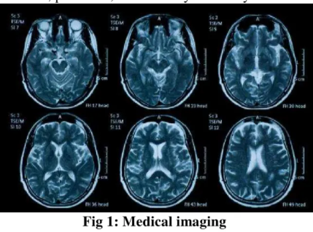
Medical imaging is an art of creating visual representations of the interior of a body for clinical and medical analysis. Medical imaging reveals the internal structures hidden by the skin and bones, as well as to diagnose and treat diseases. Medical imaging also maintains a database of normal anatomy and physiology to make it possible to identify abnormalities.
Early detection and classification of brain disease is very important in clinical practice. Many researchers have projected dissimilar methods for the classification of brain diseases based on different sources of information. Magnetic resonance imaging (MRI) is considered, most of the medical images can be used for the delineation of soft tissue [1]. MRI can help doctors to diagnose the disease as it provides important information about the anatomy, function, perfusion, and viability of the myocardium.
Fig 1: Medical imaging
Segmentation of the cerebral cortical surface is one of the fundamental problems in Medical Imaging. Accurate cortical reconstruction is important for analysis of brain including volume, surface area, thickness and sulcal depth of the cortex [11]. Eventhough accurate and robust segmentation of the cortex remains a challenging task due to the complex anatomy of cortical surface and variability of the folds.
The human cerebral cortex is a convoluted sheet of varying thickness whose two-dimensional macrostructure early anatomists have recognized long before its layered-microstructure its third dimension was discovered [13]. Because of its 2-D macrostructure, the cortical sheet lends itself to representation in a single planar map for each hemisphere. Such maps were first produced manually from postmortem brains by rather coarse techniques. Today a representation of the cortex of a living subject can be unfolded, flattened, and used as a map by computational methods. To this end, the cortical sheet is reconstructed as a polygon mesh from anatomical magnetic resonance (MR) imaging data.
Magnetic resonance imaging (MRI) is a test that uses a magnetic field and pulses of radio wave energy to make pictures of organs and structures inside the body. Magnetic resonance imaging (MRI) is a safe and painless test that uses a magnetic field and radio waves to produce detailed pictures of the body's organs and structures. An MRI differs from a
CAT scan (also called a CT scan or a computed axial tomography scan) because it doesn't use radiation. An MRI scan can be used as an extremely accurate method of disease detection throughout the body. They may be done to provide more information about a problem seen on an X-ray, ultrasound, or CT scan and, in some cases, provide more information than any of these other procedures.
Fig 2: Anatomy of human brain
Because the MRI scan gives very detailed pictures it is the best technique when it comes to finding tumors (benign or malignant abnormal growths) in the brain, including if or how much it may be spread into nearby brain tissue.
MRI can look at the brain for tumors, an aneurysm, bleeding in the brain, nerve injury, and other problems, such as damage caused by a stroke. MRI can also find problems of the eyes and optic nerves and the ears and auditory nerves.
The assessment of human cerebral cortical thickness has massive clinical importance in the determination of pathology and in assessing the processes of “normal” brain maturation and ageing [9]. As the brain develops and matures, from infancy through adulthood to old age, various changes occur in the brains tissues, such that it is difficult to pinpoint when maturation ends and degeneration begins. Sowell et al. [7] have extensively investigated these changes from early childhood using MR. They have found that white matter density and volume continue to increase throughout childhood and adolescence, whereas gray matter density (which they claim to be representative of cortical thickness) increases over the same period, but gray matter volume increases in early childhood and then declines after puberty. Using the methods described in this paper we have been able to provide a systematic method for reconstructing a surface representation of the central cortical layer from MR images of the brain. The LOGISMOS approach starts with an object pre-segmentation step, after which a single graph holding all relationships and surface cost elements is constructed, and in which the segmentation of all
desired surfaces is performed simultaneously in a single optimization process.
II.
RELATED WORKS
Reconstructing the geometry of the human brain cortex from MR images is an important step in both brain mapping and surgical path planning applications. Due to the difficulties with imaging, noise partial volume and image intensity in-homogeneities it becomes more challenging to reconstruct the cortical surfaces. Using fuzzy segmentation algorithm, the problem for reconstructing the entire cortex with correct topology is solved. In this paper, describes a systematic method for obtaining a surface representation of the geometric central layer of the human cerebral cortex such as GM, WM and CSF [1]. The first step in our method is to preprocess the image volume to remove skin, bone, fat, and other noncerebral tissue. Fuzzy segmentations retain more information from the original image than hard segmentations by taking into account the possibility that more than one tissue class may be present in a single voxel and needs to provide some manual intervention.
Reconstruction of the cerebral cortex from magnetic resonance (MR) images is an important step in quantitative analysis of the human brain structure, for example, in sulcal morphometry and in studies of cortical thickness. Existing cortical reconstruction approaches are typically optimized for standard resolution data and are not directly applicable to higher resolution images [2]. A new PDE-based method is presented for the automated cortical reconstruction that is computationally efficient and scales well with grid resolution, and thus is particularly suitable for high-resolution MR images with sub millimeter voxel size.
Thickness Measurement of cerebral cortex can aid diagnosis and provide valuable information about the temporal evolution of diseases such as Alzheimer’s, Huntington’s and Schizophrenia [3]. Methods that measure the thickness of the cerebral cortex rely on an accurate segmentation on MR data. But it still poses a challenge due to the presence of noise, intensity non-uniformity and limited resolution of MRI and high cortical folds. A new segmentation method with anatomical tissue priors is proposed with three post processing refinements.
similarity. In this paper, multispectral gray-level intensity MR brain images are used [4]. T1, T2 and PD-weighted images provide different and complementary information about the tissues. Segmentation is performed in order to classify each pixel of the resulting image according to four possible classes: cerebro-spinal fluid (CSF), white matter (WM), gray matter (GM) and background.
The surface of the human cerebral cortex is a highly folded sheet with the majority of its surface area buried within folds. As such, it is a difficult domain for computational as well as visualization purposes [5]. We have therefore designed a set of procedures for modifying the representation of the cortical surface to (i) inflate it so that activity buried inside sulci may be visualized, (ii) cut and flatten an entire hemisphere, and (iii) transform a hemisphere into a simple parameterizable surface such as a sphere for the purpose of establishing a surface-based coordinate system.
Difficulties with imaging noise, partial volume averaging, image intensity inhomogeneities, convoluted cortical structures, and the requirement to preserve anatomical topology make the development of accurate automated algorithms particularly challenging. In this paper, addresses each of these problems and describe a systematic method for obtaining a surface representation of the geometric central layer of the human cerebral cortex [6]. Using fuzzy segmentation, an isosurface algorithm, and a deformable surface model, the method reconstructs the entire cortex with the correct topology, including deep convoluted sulci and gyri.
This paper describes methods for white matter segmentation in brain images and the generation of cortical surfaces from the segmentations. We have developed a system that allows a user to start with a brain volume, obtained by modalities such as MRI or cryosection, and constructs a complete digital representation of the cortical surface [8]. The methodology consists of three basic components: local parametric modeling and Bayesian segmentation; surface generation and local quadratic coordinate fitting; and surface editing. Segmentations are computed by parametrically fitting to represent the boundary of the gray and white matter we use triangulated meshes generated using isosurface generation algorithms.
Due to noise and partial volume sampling, however, conventional segmentation methods rarely yield a voxel object whose outer boundary represents the folded cortical sheet without topological errors. These errors, called handles, have particularly deleterious effects when the polygon mesh constructed from the segmented voxel representation is inflated or flattened. So far handles had to be removed by cumbersome manual editing,
or the computationally more expensive method of reconstruction by morphing had to be used, incorporating the a priori constraint of simple topology into the polygon-mesh model [13]. In this paper, describe a linear time complexity algorithm that automatically detects and removes handles in pre segmentations of the cortex obtained by conventional methods. The algorithm’s modifications reflect the true structure of the cortical sheet.
III.
EXISTING SYSTEM
Medical imaging provides effective and non-invasive mapping of the anatomy of subjects. Common medical imaging modalities include X-ray, CT, ultrasound, and MRI. Medical imaging analysis is usually applied in one of two capacities: a) to gain scientific knowledge of diseases and their effect on anatomical structure in vivo, and b) as a component for diagnostics and treatment planning. MRI provides detailed images of tissues and is used for both human brain and body studies. Data obtained from MR images is used for detecting tissue deformities such as cancers and injuries. It aims to partition an image into a set of non-overlapping regions whose union is the original image.
Segmentation of brain tissues in MRI (Magnetic Resonance Imaging) images plays a crucial role in three-dimensional volume visualization, quantitative morph metric analysis and structure-function mapping for both scientific and clinical investigations.
LOGISMOS-B clustering algorithm, an unsupervised clustering technique, has been successfully used for image segmentation. Compared with hard C-Means algorithm, LOGISMOS-B is able to preserve more information from the original image. Its advantages include a straightforward implementation, fairly robust behavior, applicability to multichannel data, and the ability to model uncertainty within the data [11]. A major disadvantage of its use in imaging applications, however, is that LOGISMOS-B does not incorporate information about spatial context, causing it to be sensitive to noise and other imaging artifacts. The pixels on an image are highly correlated, i.e. the pixels in the immediate neighborhood possess nearly the same feature data. Therefore, the spatial relationship of neighboring pixels is an important characteristic that can be of great aid in imaging segmentation. The spatial function is the weighted summation of the membership function in the neighborhood of each pixel under consideration.
because of its abnormal feature data. This project introduces an improved LOGISMOS-B algorithm for clustering by incorporating spatial information and altering the membership weighting of each cluster with weighting exponent. The proposed algorithm greatly attenuates the effect of noise and biases the algorithm toward homogeneous clustering.
2.1 Limitations of Existing System
Difficult to provide accurate measurement of cortical surface thickness
Tissue classification can’t be implemented accurately
Accuracy in measurement of regions such as the cingulated cortex is less
Signed error range is large
IV.
PROPOSED SYSTEM
In the proposed system we implemented the improved LOGISMOS-B algorithm for accurate segmentation
3.1 PRE-PROCESSING
The input to our algorithm is a single T1-weighted structural MR image. First, this T1w image is input to the LOGISMOS software for pre-processing [10][11]. LOGISMOS deformably registers the input image to an atlas and performs atlas-based tissue classification. This classification is limited to high level tissue classes sch as white matter (WM), grey matter (GM) and cerebellar white, as well as non brain tissue types such as skull. The bias corrected T1-weighted image is de-noised using gradient anisotropic diffusion filtering for five iterations using the ITK implementation with the default conductance value of 1. For the white matter surface, the gradient magnitude of the T1-weighted image is used as the cost function. For the gray matter surface, a weighted sum of the first and second order derivatives of the T1-weighted image is used, as given by the gradient magnitudes.
3.2 GRAPH CONSTRUCTION
The LOGISMOS approach provides a framework for optimal segmentation of multiple interacting surfaces. This is achieved by modeling the problem as a complex geometric graph. Build columns of this geometric graph, based on the generalized gradient vector flows. The first step of segmentation process is by selecting the pattern of input image automatically. GGVF is dense vector field that token from image by minimizing the energy function. The minimizing energy function process is done by measuring a pair of partial differential equation which spreads gradient vector from edge map of gray-level image that measured from image [2]. The energy in here are two kinds,
there are internal power which comes from curve in deformation process and external power which comes from the image itself.
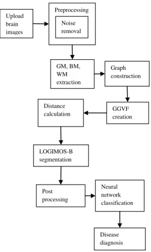
Fig 3: System Architecture
To this end, the preliminary segmentation is converted to a triangular mesh using the marching cubes algorithm. This mesh representation of the preliminary segmentation is referred to as the “base graph” in the LOGISMOS framework [11]. From each node of this base graph (i.e., the vertices of the triangle mesh), a “column” is built, such that the final segmentation will “choose” exactly one node from each column. Consecutive nodes within a column are connected to each other by intra-column arcs. Nodes in neighboring columns are connected to each other by inter-column arcs, which introduce hard constraints on the smoothness of the final surface.
3.3 LOGISMOS Segmentation
Segmentation of brain tissue on magnetic resonance (MR) images normally determines the type of tissue present for each pixel or voxel in a 2D or 3D data set respectively, based on the information gathered from both MR images and prior knowledge
Upload brain images
Preprocessing
Noise removal
GM, BM, WM extraction
Graph construction
GGVF creation Distance
calculation
LOGIMOS-B segmentation
Post processing
Neural network classification
of the brain [14]. It is one of the most vital preprocessing steps in several medical research and clinical applications, such as quantification of tissue volume, visualization and analysis of anatomical structures, multimodality fusion and registration, functional brain mapping, identification of pathology, surgical planning, surgical navigation, and brain substructure segmentation. Segmentation at preliminary stage is important and necessary for the analysis of medical images for computer-aided diagnosis and treatment.
Each node in the graph is assigned a cost related to the segmentation task such that a desirable segmentation has a low cost. Our cost function is based on edge strength, as defined by the first and second derivatives of image intensity [16]. Then, LOGISMOS-B finds the minimum closed set of this graph, which is equivalent to the minimum-cost cut or the optimal surface segmentation [11].
3.4 Post Processing:
Improved LOGISMOS-B segmentation results contains varying amounts of brain stem and cerebellum tissue, which are removed via post-processing on the meshes. This is accomplished by creating a binary mask corresponding to the regions to be removed for each subject. To form the binary mask, cerebellar white and gray matter regions that resulted from LOGISMOS tissue classification during pre-processing are merged and morphologically closed to remove any present holes. The brain stem segmentation is obtained by mapping the atlas segmentation to the subject’s image using image registration also computed by LOGISMOS. The largest connected component in the brain stem segmentation is extracted and morphologically dilated to ensure full coverage of undesired mesh vertices. The brain stem and cerebellum segmentations thus obtained are combined together to create the binary mask to be used for post-processing. And then each corresponding GM vertex are removed.
3.5 Disease Diagnosis
Early detection and classification of brain tumors is very important in clinical practice. Many researchers have proposed different techniques for the classification of brain tumors based on different sources of information. In this module we propose a process for brain tumor classification, focusing on the analysis of Magnetic Resonance (MR) images and Magnetic Resonance Spectroscopy (MRS) data collected for patients with benign and malignant tumors. Our aim is to achieve a high accuracy in discriminating the two types of tumors through a combination of several techniques for image segmentation, feature extraction and classification. The proposed technique has the potential of assisting
clinical diagnosis. So we implement neural network techniques to find the brain diseases with improved accuracy rate.
Fig 2: Disease affected brain image
V.
CONCLUSION:
In this paper presented an improved LOGISMOS method for reconstructing cortical surfaces from MR brain images. This method combines a neural network classification to reconstruct the surface representation of the cortical layer. Compared to the current method, improved LOGISMOS-B offers accuracy in cortical surface reconstruction and efficient in creating binary map for the boundaries of tissues.
REFERENCES
[1] C. Xu, D. L. Pham, M. E. Rettmann, D. N. Yu, and J. L. Prince, “Reconstruction of the human cerebral cortex from magnetic resonance images,” IEEE Trans. Med. Imag., vol. 18, no. 6, pp. 467–480, Jun. 1999. [2] S. Osechinskiy and F. Kruggel, “Cortical
surface reconstruction from high-resolution MR brain images,” Int. J. Biomed. Imag., vol. 2012, p. 870196, Jan. 2012.
[3] M. J. Cardoso, M. J. Clarkson, G. R. Ridgway, M. Modat, N. C. Fox, S. Ourselin, and A. D. N. Initiative, “LoAd: A locally adaptive cortical segmentation algorithm,” NeuroImage, vol. 56, no. 3, pp. 1386–1397, Jun. 2011.
[4] Guillermo N. Abras and Virginia L. Ballarin “A Weighted K-means Algorithm applied to Brain Tissue Classification” JCS&T, vol. 5, no. 3, Oct 2005.
[5] A. M. Dale, B. Fischl, and M. I. Sereno, “Cortical surface based analysis. I. Segmentation and surface reconstruction,” NeuroImage, vol. 9, no. 2, pp. 179–194, Jan. 1999.
[6] C. A. Davatzikos and J. L. Prince, “An active contour model for mapping the cortex,” IEEE Trans. Med. Imag., vol. 14, no. 1, pp. 65–80, Jan. 1995.
Memo No. 2004-007, Medical Image Analysis, Oct. 2008.
[8] M. Joshi, J. Cui, K. Doolittle, S. Joshi, D. V. Essen, L. Wang, and M. I.Miller, “Brain segmentation and the generation of cortical surfaces,” NeuroImage, vol. 9, no. 5, pp. 461–476, May 1999.
[9] C. Vachet, H. C. Hazlett, M. Niethammer, I. Oguz, J. Cates, R. Whitaker, J. Piven, andM. Styner, “Group-wise automatic mesh-based analysis of cortical thickness,” SPIE Med. Imag., 2011.
[10] Y. Yin, X. Zhang, R.Williams, X.Wu, D. D. Anderson, and M. Sonka, “LOGISMOS -layered optimal graph image segmentation of multiple objects and surfaces: Cartilage segmentation in the knee joint,” IEEE Trans. Med. Imag., vol. 29, no. 12, pp. 2023–2037, Dec. 2010.
[11] Ipek Oguz and Milan Sonka, Fellow, IEEE
“LOGISMOS-B: Layered Optimal Graph Image Segmentation of Multiple Objects and Surfaces for the Brain,” IEEE Trans. Med Imag., vol. 33, no. 6, Jun. 2014.
[12] E. Y. Kim and H. J. Johnson, “Robust multi -site MR data processing: Iterative optimization of bias correction, tissue classification, and registration,” Front. Neuroinf., vol. 7, no. 29, pp. 1–18, Nov. 2013.
[13] N. Kriegeskorte and R. Goebel, “An efficient algorithm for topologically correct segmentation of the cortical sheet in anatomical MR volumes,” NeuroImage, vol. 14, no. 2, pp. 329–346, Aug. 2001.
[14] J. S. Kim, V. Singh, J. K. Lee, J. Lerch, Y. Ad-Dab’bagh, D. Mac- Donald, J. M. Lee, S. I. Kim, and A. C. Evans, “Automated 3-D extraction and evaluation of the inner and outer cortical surfaces using aaplacian map and partial volume effect classification,” NeuroImage, vol. 27, no. 1, pp. 210–221, Aug. 2005.
[15] C. Vachet, H. C. Hazlett, M. Niethammer, I. Oguz, J. Cates, R. Whitaker, J. Piven, andM. Styner, “Group-wise automatic mesh-based analysis of cortical thickness,” SPIE Med. Imag., 2011.