Clinical Case Reports
Medeiros et al., J Clin Case Rep 2015, 5:4 http://dx.doi.org/10.4172/2165-7920.1000515
Volume 5 • Issue 4 • 1000515 J Clin Case Rep
ISSN: 2165-7920 JCCR, an open access journal
Open Access Case Report
J
o
urn
al o f Clinical Case R
epo
rts
ISSN: 2165-7920
Bilateral Inguinal Hernia Containing a Rudimentary Uteri, Ovaries and
Tubes in a Woman with Primary Amenorrhea
Francisco C Medeiros1*, Francisco E Vasconcelos2, Bruno HM Dias2 and Renato MLV Leal3
1Department of Maternal and Child Health, School Maternity Assis Chateaubriand, Brazil
2Federal University of Ceara (UFC), Fortaleza-CE, Brazil
3General surgeon and oncologic surgery, School Maternity Assis Chateaubriand, Brazil
*Corresponding author: Francisco das Chagas Medeiros, Department of Maternal and Child Health, School Maternity Assis Chateaubriand, Brazil, Tel: (55-85)3366-8062; Fax: (55 85)3366-8065; E-mail: [email protected]
Received April 04, 2015; Accepted April 26, 2015; Published April 28, 2015
Citation: Medeiros FC, Vasconcelos FE, Dias BHM, Leal RMLV (2015) Bilateral Inguinal Hernia Containing a Rudimentary Uteri, Ovaries and Tubes in a Woman with Primary Amenorrhea. J Clin Case Rep 5: 515. doi:10.4172/2165-7920.1000515
Copyright: © 2015 Medeiros FC, et al. This is an open-access article distributed under the terms of the Creative Commons Attribution License, which permits unrestricted use, distribution, and reproduction in any medium, provided the original author and source are credited.
Keywords: Amenorrhea; Blind vagina; Nuck channel
Introduction
he processesvaginalis of the peritoneum accompanies the testis or round ligament through the inguinal canal into the scrotum or the labium major. In women, the homologous to this structure is called the Nuck channel. It usually undergoes obliteration ater the seventh month of pregnancy, which explains the high incidence of hernias in premature kids [1]. It is possible, though, that the closure only happens ater birth, during the irst year of life. he failure of the closure of this canal can cause a hydrocele or a hernia [1]. here’s an association between a hydrocele of the Nuck channel and a contralateral inguinal hernia [1], which should make us consider the existence of hydrocele in patients with a inguinal cystic mass and a history of inguinal hernia on the other side [2].
Considering the association between hydrocele and hernia of the Nuck channel, it is important to distinguish one from another. While a hernia can present bowel sound over an inguinal swelling and a bulging that is evident while standing, but disappears while lying on supine position [1], a hydrocele is a painless translucent swelling in the inguino-labial region, without any sound over it [1]. he diagnosis can also be established by ultrasonography or MRI [1].
Congenital vaginal agenesis is frequently present in patients with Mayer-Rokitansky-Kuster-Hauser Syndrome and Complete Androgen Insensitivity Syndrome (CAIS) [3,4]. Woman with Rokitansky syndrome have a 46, XX karyotype and normal functioning ovaries, while patients with CAIS present a 46, XY karyotype and testicular gonads [4]. Both conditions present primary amenorrhea [4]. he treatment for these cases usually consists of lengthening the vagina and, in CAIS, preceding a gonadectomy [4]. Since most of these cases present during adolescence, the patient should be included in the decision of treatment and psychological support must be required [4,5].
We present a case of a 21-year-old woman was presented to the Gynecology Department presenting primary amenorrhea and a blind vagina (Mayer-Rokitansky-Küster-Hauser Syndrome). Physical examination revealed Tanner V, thelarche and adrenarche, a 1-2 cm vaginal dimple (Figure 1), and a suspected bilateral inguinal hérnia (During examination, when asked to stand up, it was observed a bulging of both labia majora (Figure 2). he patient’s karyotype was 46, XX. Pelvic ultrasound showed that no ovaries, uterus, or vagina could be seen. Inguinal ultrasound showed a complex mass bilaterally at the inguinal areas (Figure 3). his could suggest the presence of testicles or of a hernia. Considering the amenorrhea, the blind vagina and the bulging, it was thought that the patient presented the Complete
Abstract
In this case report, we show a case of 21-year-old woman who presented amenorrhea, a blind vagina, a bilateral Nuck channel herniation. After initial examination, it was thought that she had the Testicular Feminization Syndrome. After follow-up, it was observed that she presented uterovaginal agenesis and ovary and uterine tubes hernia of the Nuck channel.
Androgen Insensitivity Syndrome, which is usually associated with these inding, although this is not seen in females with 46, XX vaginal agenesis [3]. herefore, a laparotomy surgery was indicated.
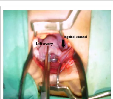
During the surgery, though, it was observed that the bulging was being caused by the presence of a ovary and uterine tubes hernia of the Nuck channel (Figure 4). In a woman presenting the Mayer-Rokitansky-Kuster-Hauser syndrome and herniation of the ovaries, the hernia sac must be opened, in order to release the ovaries into the abdomen and prevent infarction and maintain hormonal function [3]. Ater noticing the hernia, the correction procedure was executed and the lengthening of the vagina was realized.
Discussion
In this case, the patient presented an association of the Mayer-Rokitasky-Küster-Hauser (MRKH) Syndrome and a hernia of the Nuck channel. Considering the pathophysiology of the hernias and hydrocele of the aforementioned channel (failure of closure of the channel) [1] and the malformation represented by the syndrome [5], it is not illogic to question if there is a correlation between their genesis.
Citation: Medeiros FC, Vasconcelos FE, Dias BHM, Leal RMLV (2015) Bilateral Inguinal Hernia Containing a Rudimentary Uteri, Ovaries and Tubes in a Woman with Primary Amenorrhea. J Clin Case Rep 5: 515. doi:10.4172/2165-7920.1000515
Page 2 of 3
Volume 5 • Issue 4 • 1000515 J Clin Case Rep
ISSN: 2165-7920 JCCR, an open access journal
located laterally along the psoas muscle and pelvic sidewall [11]. Primary malformation here can be understood as Mayer-Rokitansky-Kuster-Hauser syndrome or its variants. Herniation of malformed uteri can be attributed to the imperfect closure of congenital openings and failure of fusion of Müllerian ducts, which remained attached in the original position of the inguinal fold. Ten cases have previously been reported until now [12,13].
Oppelt et al., in a study with 284 women showed an important correlation between MRKH syndrome and malformations of the renal system, for example [5]. herefore, our goal, besides reporting a rare case is to interrogate a possible relationship between the two malformations. In order to explore this, more studies are needed.
References
1. Choi YM, Lee GM, Yi JB, Yoon KL, Shim KS, et al. (2012) Two cases of female hydrocele of the canal of nuck. Korean J Pediatr 55: 143-146.
2. Jedrzejewski G, Stankiewicz A, Wieczorek AP (2008) Uterus and ovary hernia of the canal of Nuck. Pediatr Radiol 38: 1257-1258.
3. Bar-Joseph KL, Amies-Oelschlager AM, Avansino JR (2012) A painful protuberance: a young woman had primary amenorrhea and an inguinal hernia. Am J Obstet Gynecol 207: 144.
4. Nakhal RS, Creighton SM (2012) Management of vaginal agenesis. J Pediatr Adolesc Gynecol 25: 352-357.
5. Oppelt PG, Lermann J, Strick R, Dittrich R, Strissel P, et al. (2012) Malformations in a cohort of 284 women with Mayer-Rokitansky-Küster-Hauser syndrome (MRKH). Reprod Biol Endocrinol 10: 57.
6. Deeb A, Hughes IA (2005) Inguinal hernia in female infants: a cue to check the sex chromosomes? BJU Int 96: 401-403.
7. Kriplani A, Banerjee N, Aminni AC, Kucheria K, Takkar D (2000) Hernia uterus inguinale in a 46,XX female. A case report. J Reprod Med 45: 48-50.
8. Al Omari W, Hashimi H, Al Bassam MK (2011) Inguinal uterus, fallopian tube, and ovary associated with adult Mayer-Rokitansky-Küster-Hauser syndrome. Fertil Steril 95: 1119e1-1119e4.
9. Riggall FC, Cantor B (1980) 46,XX hernia uterus inguinale and vaginal agenesis. ObstetGynecol 56: 265-266.
10. Helvacioglu A, Gilmore S, Wells Willson, W, Rizk B (2010) Ovarian malposition -Mullerian anomalies revisited. Middle East Fertility Society Journal 15: 115-118.
in diagnosis, but magnetic resonance image (MRI) has been recognized as the gold standard means. However, as it was the case in our patient, MRI may not be able to identify a rudimentary uterine horn, if it is
Figure 1: Vulvo-vaginal image of physical exam conirming vaginal agenesis.
Figure 2: Bulging of the labia majora and groin bilaterally after the Valsalva´s maneuver.
Figure 3. Inguinal Ultrasound showing both bilateral inguinal complex masses.
Citation: Medeiros FC, Vasconcelos FE, Dias BHM, Leal RMLV (2015) Bilateral Inguinal Hernia Containing a Rudimentary Uteri, Ovaries and Tubes in a Woman with Primary Amenorrhea. J Clin Case Rep 5: 515. doi:10.4172/2165-7920.1000515
Page 3 of 3
Volume 5 • Issue 4 • 1000515 J Clin Case Rep
ISSN: 2165-7920 JCCR, an open access journal
11. Economy KE, Barnewolt C, Laufer MR (2002) A comparison of MRI and laparoscopy in detecting pelvic structures in cases of vaginal agenesis. J Pediatr Adolesc Gynecol 15: 101-104.
12. Kamio M, Nagata T, Yamasaki H, Yoshinaga M, Douchi T (2009) Inguinal hernia
containing functioning, rudimentary uterine horn and endometriosis. Obstet Gynecol 113: 563-566.
13. Bell WB (1909) Rudimentary Uterus Didelphys, with Ectopia of each Uterine Body in an Inguinal Hernial Sac; with some Remarks on the Development of the Female Genital Organs. Proc R Soc Med 2: 311-324.
Citation: Medeiros FC, Vasconcelos FE, Dias BHM, Leal RMLV (2015)
Bilateral Inguinal Hernia Containing a Rudimentary Uteri, Ovaries and Tubes in a Woman with Primary Amenorrhea. J Clin Case Rep 5: 515. doi:10.4172/2165-7920.1000515
Submit your next manuscript and get advantages of OMICS Group submissions
Unique features:
• User friendly/feasible website-translation of your paper to 50 world’s leading languages • Audio Version of published paper
• Digital articles to share and explore Special features:
• 400 Open Access Journals • 30,000 editorial team • 21 days rapid review process
• Quality and quick editorial, review and publication processing
• Indexing at PubMed (partial), Scopus, EBSCO, Index Copernicus and Google Scholar etc • Sharing Option: Social Networking Enabled
• Authors, Reviewers and Editors rewarded with online Scientiic Credits • Better discount for your subsequent articles