Comparison between immunofixation and
electrophoresis for the early detection of relapsed
multiple myeloma
Comparação entre imunofixação e eletroforese na detecção precoce
de recidivas do mieloma múltiplo
Marta Helena C. Aita; Luiz Claudio Arantes; Bruno C. Aita; José Edson P. Silva
Universidade Federal de Santa Maria (UFSM).
First submission on 09/12/14; last submission on 16/08/15; accepted for publication on 31/08/15; published on 20/12/15
ABSTRACT
Introduction: Multiple myeloma (MM) is an incurable progressive hematological neoplasia characterized by heterogeneous evolution and by relapses after therapy. Objective: Compare the effectiveness of serum immunoixation (SIF) and electrophoresis (SPE) techniques in the detection of relapses in MM patients undergoing treatment at the University Hospital of Santa Maria (HUSM). Material and methods:
The study was conducted from January 2012 to July 2014 and included 52 patients from HUSM with conirmed diagnosis of MM. The retrospective monitoring based on laboratory analyses indicated that nine of these patients relapsed, in whom it was possible to compare the effectiveness of SIF and SPE techniques for detecting relapses. Results: For the nine patients, SIF always detected MM relapses earlier than SPE, with a precocity ranging from 2.0 to 18.8 months, for an average of 6.6 months. Discussion and conclusion: The results
indicated that SIF was more effective than SPE for the early detection of relapses, regardless of the class and type of M component (mono/ biclonal). Therefore, the use of SIF allows for better monitoring of MM patients, especially for the detection of relapses, thereby helping in choosing the most appropriate therapy and resulting in increased duration of survival period free of disease.
Key words: multiple myeloma; monoclonal immunoglobulins; immunoixation; electrophoresis; relapses.
INTRODUCTION
The multiple myeloma (MM) is a progressive B-cell hematological malignancy, characterized by the unregulated and clonal proliferation of plasma cells of the bone marrow (BM), which produce and secrete anomalous monoclonal immunoglobulin or fragments of these (free light chain or Bence-Jones protein), called M-protein, myeloma protein or paraprotein, which are secreted
into the blood and/or urine(1-4). In hematological neoplasm, MM
is the disease with worse prognosis and lower survival rates, 5 years in 15%-20% of cases(5-7).
International centers of cancer registry have reported an increase in incidence rates and mortality caused by MM in recent decades, although it is not yet clear whether this is due to the new means of diagnosis or an actual increase in new cases of
the disease(5). Although there is not yet a exact and oficial
knowledge of the incidence MM in Brazil, since the disease is not recorded in the annual estimates of the Bazilian National Cancer Institute(8), some studies, such as Hungria et al. (2008)(9), Paula
and Silva (2009)(10), and Keren (2010)(11), indicate that the average
age at diagnosis is 60.5 years, with most cases diagnosed when the disease is already at an advanced stage.
The diagnosis of MM depends on identiication of monoclonal plasmocytes in the BM, M-protein in the serum or urine and evidence of bone lesions(12). The use of eficient and accurate
techniques for MM diagnosis is essential to differentiate it from other monoclonal gammopathy, which facilitates therapeutic decision, besides providing adequate indicators on the effectiveness of therapy(13-16). Currently, serum protein electrophoresis (SPEP)
remains the standard technique for the diagnosis and treatment of patients with MM(17). Although SPE agarose gel can be considered a
relatively simple laboratory method for the detection of M-protein, the immunoixation serum (SIF) technique is considered the gold standard for conirming the presence of these proteins and to distinguish light and heavy chains in MM(18, 19). The combination
of SPE and SIF techniques increases up to 97% sensitivity in the detection of M-protein in patients with MM(12, 20).
Whereas, following treatment there may occur complete remission of MM, but not its cure(21, 22), it is important monitoring
these patients in order to be able to detect relapse as early as possible(23). In this context, a comparison between SIF and SPE
techniques on its effectiveness in early detection of MM relapses, through the retrospective analysis of serum samples from nine patients, was the main objective of this study.
MATERIAL AND METHODS
Patients
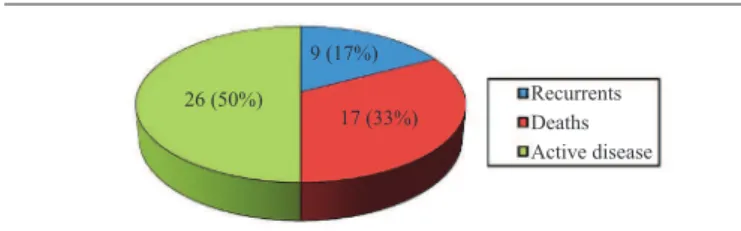
The study population consisted of patients diagnosed with MM monitored at the Outpatient service of Hematology of the University Hospital of Santa Maria (HUSM), RS, Brazil, between January 2012 and July 2014. The evolution of 52 patients followed up during the study is shown at Figure 1. All patients had undergone routine laboratory tests before each visit and received the treatment standardized by HUSM.
For inclusion in the study were considered patients with a conirmed diagnosis of MM. Patients were excluded if: a) lost their clinical follow up and/or has incomplete data from the medical records; b) attended with disease relapsed and advanced state; and c) attended at the place where the research was conducted (HUSM) to chemotherapy and after performed clinical laboratory monitoring in the city of origin.
As the study of the patients was done retrospectively, the results of serum analysis (SPE and laboratory measures) were interpreted
at each clinic visit, spreadsheets were developed for each study patient for better monitoring, especially for those who were in complete remission of the disease and could relapse.
Laboratory analysis
Samples (pre-analysis)
Blood samples were collected at the Hematologic-Oncology Laboratory of HUSM. To collect blood, the venipuncture standard technique was used and the material was transferred to Vacutainer®
tubes, BD Diagnostics, USA. Tubes without anticoagulant were used for serological tests, while tubes with 7.2 mg of anticoagulant dipotassium of ethylenediaminetetraacetic acid (EDTA) served for CBC analysis. After centrifuging the samples at 4,000 evolutions per minute (rpm) for 10 minutes, the patients’ serum was stored in aliquots (Eppendorf tubes) in a freezer at -70ºC.
SPE and SIF
SPE technique was performed in electrophoretic tub/CELM, containing 80 ml of CELM buffer – pH 9.5. Serum was applied to agarose ilm (CELM Gel), according to the technique recommended by the manufacturer. The protein fractions reading were performed by a Software Program for Scanning Densitometry.
SIF analyzes were performed in Sebia® HYDRASYS®
instrument with HydraGel IF Sebia 2/4, according to the manufacturer’s instructions. Such analyzes were performed at the Biochemistry Laboratory of the Hospital de Clínicas de Porto Alegre (RS), Brazil. Serum proteins were separated in an alkaline buffer (pH 9.1) for 9 minutes at 20 W (42 VH). The types of antisera to speciic classes of immunoglobulins (immunoglobulins class G, A and M [IgG], [IgA] and [IgM], respectively) and light chains
(κ and λ) were applied, and the identiication was performed after antigens-antibodies complex staining, which resulted in immunoprecipitation. All reagents are included in the IF/Sebia kit.
Gels reading were performed according to the presence or absence of monoclonal and/or biclonal band(s) of immunoglobulin chains (IgG, IgA and IgM) linked to its light-chain κ and λ or
only free light chain (κ and/or λ). Two patterns are considered, one normal (absence of monoclonal component), and the other abnormal (presence of monoclonal or biclonal component).
Serological measures
Serologic measures were performed in the Biochemistry Department of the Clinical Analysis Laboratory of HUSM, using
FIGURE 1 − Patients with MM (n = 52) included in the study. Progress during the period from January 2012 to July 2014: death, response to treatment with relapse and active disease with no appropriate therapeutic response
MM: multiple myeloma.
Recurrents Deaths Active disease 26 (50%)
Siemens Dimension Pand Plus analyzers. Immunoturbidimetic method was used for immunoglobulin (IgG, IgA e IgM) analysis. For creatinine and albumin dosages, we used the colorimetric method; for lactate dehydrogenase (LDH) analysis, we used the ultraviolet (UV) method.
Blood counts were performed at Hematology Department of HUSM, using Sysmex XE 2100 equipment, while for serum
β2-microglobulin measuring samples were sent to Laboratório de Análises Clínicas Álvaro (PR) for analysis by chemiluminescence
method.
Statistical analysis
Because it is a case study, we only calculated the mean values and standard deviation for some of the variables under study, using the SPSS 15.0 software.
RESULTS AND DISCUSSION
The analysis of medical records of 52 patients with conirmed diagnosis of MM indicated that their average age was 59 years, ranging 28-83 years. The predominant race was white (56%), followed by the brown (23%) and black (21%), and the predominant sex was male (55.8%) (Table 1). The earlier onset
of disease in this study, in relation to the 70-80 years age group, found around 1973(24), can be mainly attributed to advances in
analysis techniques, allowing to diagnose it earlier. Regarding race, our results differ from those reported by Kyle et al. (2002)(25) and
by Klaus et al. (2009)(26), in which there was a higher incidence
of the disease among black people. The MM relationship with population’s race is dificult to clearly establish for Brazilian
conditions, since the ethnicity of the population varies among the different regions of the country(27). The results obtained from
the 52 patients who participated in the present study (Table 1) indicate predominance of IgG immunoglobulin (48%), followed by IgA (29%), free light chain (FLC) (23%) and IgM (2%). The presence of imunoglubulin class D (IgD) and class E (IgE) and nonsecretory MM were not detected. These results conirm those of other studies that the most common myeloma was IgG, with rare cases of IgD, IgE and nonsecretory myeloma(28, 29).
Several studies have reported the existence of risk factors that help the emergence of MM, especially exposure to high doses of ionizing radiation, occupational exposure to agricultural and petrochemical industries in the presence of benzene and other organic, and exposure to insecticides and herbicides(26, 29). Also
lifestyle factors, such as socioeconomic status, smoking and alcohol, may predispose MM occurrence(30). In the population
that constituted the present study, the most prevalent factors, in decreasing order, were: exposure to toxic agents, genetic alterations, smoking and alcohol consumption (Table 1).
In most cases, relapses in patients with MM develop
aggressively(21), therefore the importance of its early detection through
effective methods. SIF method is considered the gold standard(31, 32), with high sensitivity and speciicity to detect the resurgence of
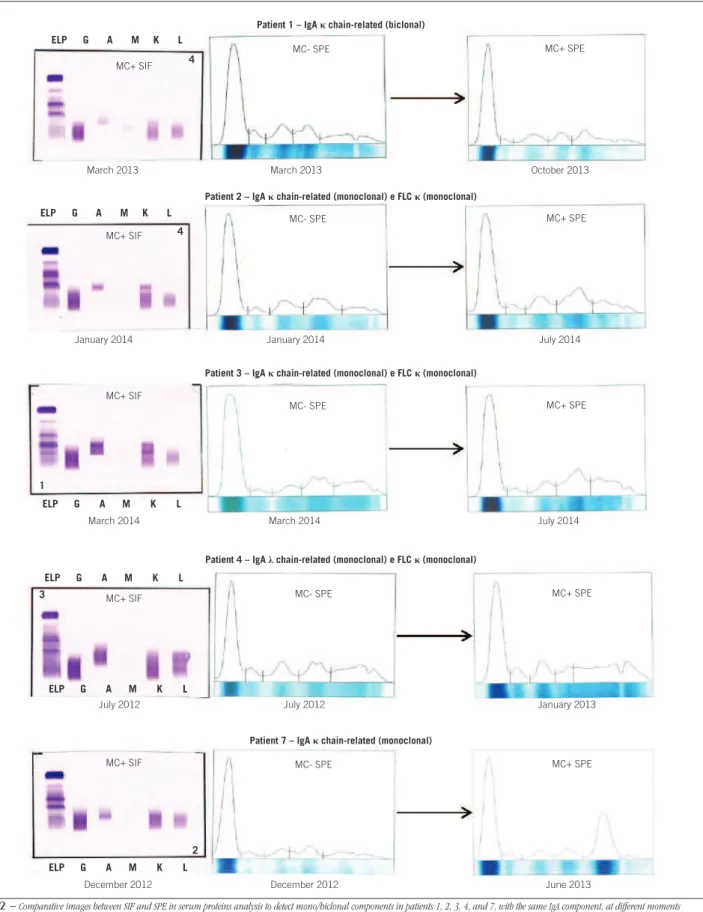
a monoclonal protein and to distinguish the heavy chains from the light chains present in the serum and urine of patients with MM. From the 52 patients who were monitored in this study, nine relapsed during the study period, enabling to observe the evolution of the disease before and after relapse. The comparison between SIF and SPE techniques regarding their effectiveness in detecting relapses of patients in remission is shown in Figures 2, 3 and 4, in which the subjects were grouped according to immunoglobulin classes. One aspect to be highlighted refers to the fact that there was a predominance of IgA standard in the nine patients who relapsed, showing the aggressiveness and high relapse risk in this MM subtype.
Figure 2, shows the results of ive relapsed patients (1, 2, 3, 4 and 7) with the same type of monoclonal/biclonal heavy chain (IgA), it is observed that in the irst serum analysis of all patients, SPE was not sensitive in detecting the monoclonal component, although the patients had clinical symptoms suggestive of relapse, for example, increased intensity of bone pain, asthenia and generally feeling unwell. When SPE results were negative in the same samples of these patients, SIF was applied, and the presence of monoclonal component was detected, conirming the suspicion of MM relapsed.
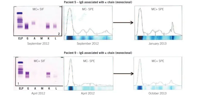
The higher sensitivity of SIF in relation to SPE in detecting MM relapse observed in patients with IgA standards also occurred in the two patients (5 and 9) type IgG protein (Figure 3), and in the two patients (6 and 8) that showed type FLC (Figure 4).
TABLE 1 − Features and general information about 52 patients with MM evaluated in the present study
Features General information
Sex 29 (55.8%) male and 23 (44.2%) female Race 29 (56%) white, 11 (21%) black and 12 (23%) brown
Age Average 59 years ± 13.5 years, 15 (28.8%) 28-48 years, 23 (44.2%) 49-69 years, and 14 (26.9%) above 70 years
Monoclonal component
24 (48%) IgG, 15 (29%) IgA, 12 (23%) FLC, and 1 (2%) IgM
Risk factors
17 (33%) exposure to toxic agentsa, 10 (19%) geneticb, 9 (17%) smoker/alcoholicc, and 16 (31%)
had no risk factor
FIGURE 2 − Comparative images between SIF and SPE in serum proteins analysis to detect mono/biclonal components in patients 1, 2, 3, 4, and 7, with the same IgA component, at different moments
MC+ and MC- indicate the presence and absence of mono/biclonal components, respectively.
SIF: serum immunoixation; SPE: serum electrophoresis; IgA: immunoglobulin A class; MC: monoclonal component; FLC: free light chains.
Patient 1 – IgAκchain-related (biclonal)
MC+ SIF
MC+ SIF
MC+ SIF
MC+ SIF
MC+ SIF
March 2013 March 2013 October 2013
MC+ SPE
MC+ SPE
MC+ SPE
MC+ SPE
MC+ SPE MC- SPE
MC- SPE
MC- SPE
MC- SPE
MC- SPE
January 2014 January 2014 July 2014
Patient 2 – IgAκ chain-related (monoclonal) e FLCκ(monoclonal)
March 2014 March 2014 July 2014
Patient 3 – IgAκchain-related (monoclonal) e FLCκ(monoclonal)
July 2012 July 2012 January 2013
December 2012 December 2012 June 2013
Patient 4 – IgAλchain-related (monoclonal) e FLCκ(monoclonal)
Patient 7 – IgAκchain-related (monoclonal)
ELP G A M K L ELP G A M K L ELP G A M K L ELP G A M K L ELP G A M K L
ELP G A M K L
2 3
1
FIGURE 3 − Comparative images between SIF and SPE in serum proteins analysis to detect MC in patients 5 and 9, with the same IgG component, at different moments
MC+ e MC- indicate the presence and absence of mono/biclonal components, respectively.
SIF: serum immunoixation; SPE: serum electrophoresis; MC: monoclonal component; IgG: immunoglobulin G class.
Pacient 5 – IgG associated withκchain (monoclonal)
MC+ SIF MC- SPE MC+ SPE
September 2012 September 2012 January 2013
Pacient 9 – IgG associated withκchain (monoclonal)
MC+ SIF MC- SPE MC+ SPE
April 2012 April 2012 October 2013
FIGURE 4 − Comparative images between SIF and SPE in serum proteins analysis to detect MC in patients 6 and 8, with the same FLC component, at different moments
MC+ and MC- indicate the presence and absence of mono/biclonal components, respectively.
SIF: serum immunoixation; SPE: serum electrophoresis; MC: monoclonal component; FLC: free light chains.
Pacient 6 – FLCλ(monoclonal)
MC+ SIF MC- SPE MC+ SPE
MC+ SIF MC- SPE MC+ SPE
December 2013 December 2013 June 2014
June 2014 June 2014 August 2014
Paciente 8 – FLCκeλ(monoclonal)
ELP G A M K L ELP G A M K L ELP G A M K L ELP G A M K L
4 3
1
Table 2 was developed from the results presented in Figures 2, 3 and 4. It shows the date of relapse detection by SIF and SPE serum analysis, which allows quantitative comparison of the two techniques as its sensitivity in the early detection of relapses. In the nine relapsed patients, the early average of monoclonal pattern detection by SIF exceeded SPE in 6.6 ± 4.8 months, varying only 2.0 months in the worst case (pacient 8) and 18.6 months in the most favorable situation (pacient 9).
This greater sensitivity of SIF in relation to SPE in detecting monoclonal immunoglobulin, found in this study, conirms the results of several studies conducted mainly in the1980s and 1990s(33-38). Working with a group of 101 patients with monoclonal
gammopathy, Potdevin et al. (1983)(36) found that they were
correctly identiied by SIF in 97 pacients, compared with only 50 cases when SPE was employed. For Vrethem et al. (1993)(38), the low
sensitivity SPE is due to the inability of that technique to detect low concentrations of monoclonal immunoglobulins (˂ 1 g/l-1) when
they are hidden or next to other protein bands. Working particularly with IgM monoclonal immunoglobulin, Keren (1990)(34) identiied
its presence by SIF in a concentration as high as 20 g/l-1, without
that the IgM in question was detected by SPE. For the author, the negativity occurred by SPE is due to the fact that IgM molecules have large volumes and, therefore, they diffuse slowly in the agarose gel used in SPE. One thing to highlight in these works is that they have not been conducted with the speciic objective of comparing the two techniques on the ability for early detection of relapses in patients with MM, as it was done in this study.
By comparing SIF and SPE techniques in a MM patient that underwent chemotherapy, Reichert et al. (1982)(37) found that it
was wrongly considered free from gammopathy by SPE technique, since, when analyzing the same samples retrospectively using SIF, as it was done for the nine patients of this study (Figures 2, 3 and 4), the authors reported they found positive results for IgAλ
TABLE 2 − Comparison between SIF and SPE techniques for sensitivity in the early detection of relapse in nine patients with MM
Patients (n = 9)
Relapse detection by SIF (date)
Relapse detection by SPE (date)
Anticipate the detection of relapse by SIF (months)
1 10/03/2013 22/10/2013 7.4
2 08/01/2014 09/07/2014 6
3 13/03/2014 02/07/2014 3.6
4 12/07/2012 02/01/2013 5.7
5 03/09/2012 10/01/2013 4.2
6 23/12/2013 30/06/2014 6.2
7 13/12/2012 14/06/2013 6
8 01/06/2014 31/07/2014 2
9 10/04/2012 27/10/2013 18.6
SIF: serum immunoixation; SPE: serum electrophoresis; MM: multiple myeloma.
monoclonal gammopathy. Marshall (1980)(35) also compared
SPE and SIF techniques for identiication of IgG, IGA and IgM monoclonal immunoglobulins in three serum samples with high concentrations and in three samples with low concentrations of such immunoglobulin, and found that SIF was able to identify immunoglobulins in all samples, while SPE shown ambiguous results in the three samples with lower concentration and also in one samples with high concentration. This was attributed to the greater capacity of SIF in determining the reaction of antibodies with monoclonal immunoglobulin.
This set of indings from the literature, added to the present study, proves that SPE, when used alone, is not a suitable technique for the early detection of MM relapses, and should always be performed in conjunction with other techniques more sensitive, such as SIF and FLC measurements, among others. Thus, patients can be monitored with greater safety, in both active and remission phase, and the relapses may be detected early and treatment may be quickly introduced.
Besides the determination of monoclonal immunoglobulin and their light chains and FLC -related (Figures 2, 3 and 4), we also analyzed the serum proteins β2-microglobulin and albumin, which are important in monitoring MM relapses. According to Casaretto (2005)(39), measurement of β
2-microglobulin is
a signiicant prognostic factors because it relects the tumor mass and renal function of each patient affected by the disease.
Based on the determination of β2microglobulin and albumin
measurements, Greipp et al. (2005)(40) proposed an international
classiication for MM staging, in which patients are divided into I, II and III, whose median survival corresponds to 62, 44 and 29 months, respectively.
TABLE 3 − ISS of recurrent patients at early positive SIF and SPE
Early positive SIF Early positive SPE Patients
(n = 9)
β2-m
(mg/l)
Serum albumin (g/dl) ISS
β2-m
(mg/l)
Serum
albumin (g/dl) ISS
1 2.4 3.6 I 2.3 4.5 I
2 3.2 3.7 I 4.2 3.7 II
3 1.5 4.8 I 1.5 4 I
4 4.1 3.2 II 6.9 3.3 III
5 1.9 3.7 I 2.1 3.6 I
6 4.3 3.6 II 8.3 3.6 III
7 2 4.1 I 3.3 3.2 II
8 2 4 I 1.7 4 I
9 2.2 3.6 I 2.2 4.1 I
Reference value: β2-m (0.6-2.1 mg/l); serum albumin(3.4-5.0 g/dl); staging according to ISS; stageI (β2-m < 3.5 mg/l; serum albumin≥ 3.5 g/dl); stageII: neither stagesI nor III. There are two categories for this stage: 1) β2-m < 3.5 mg/l, but serum albumin
< 3.5 g/dl or 2) β2-m from 3.5 to < 5.5 mg/l, regardless serum albumin level; stageIII:
β2-m ≥ 5.5 mg/l.
ISS: International Staging System; SIF: serum immunoixation; SPE: serum electrophoresis; β2-m: serum β2-microglobulin.
increase of survival and improvement in quality of life of patients.
One of the analytical limitations of this study is that it does not offer the possibility of comparing SIF with SPE in detecting monoclonal immunoglobulins (heavy chains and/or FLC) in urine samples from the nine patients studied. This was due to the dificulty of collecting 24-hour urine sample in relapsed patients, since they were already weakened and living elsewhere. To overcome this deicity, it would be important to perform serum free and heavy chains measurements in order to obtain the corelation between them. Through these analyzes, it would be possible to increase the sensitivity to detect the remission state of these patients, as well as their relapses.
Some laboratory parameters that assist in monitoring the nine patients in study were evaluated and are shown in Table 4, in which it is observed that there was worsening of anemia in the patients studied. In 66.6 % pacients (1, 2, 4, 6, 7 and 8), this had already occurred even when the relapse was detected early by SIF. This result can be explained by the fact that such patients have MM for several years (average of six years) and, therefore, are already debilitated by the disease itself as well by the frequent use of therapies and/or chemotherapies in previous relapses.
Although the quantiication of complete monoclonal immunoglobulins (IgG, IgA and IgM) assist in monitoring patients with MM, it must be used in conjunction with high
sensitivity methods(32). As seen in Table 4, patients 1, 2, 3, 4 and
7 showed average normal values of IgA 345.8 mg/dl (128.9-775.0 mg/dl) when the relapse was detected early by SIF, and average values 3.6 times increased (787.3-2,240.0 mg/dl) when the relapse was detected late by SPE. In both relapsed patients with monoclonal component IgG (5 and 9), although this immunoglobulin values are at normal levels, we observed that there was an average increase of 989.6 mg/dl in SIF to 1497.5 (51.3 %) in SPE, which reinforces tha advantage of using SIF in relation to SPE in the early detection of this monoclonal component in relapsed MM.
Serum creatinine relects renal function and, therefore, patients 2 and 6 already had higher value (2.0 and 1.4 mg/dl, respectively), even when the relapse was detected by SIF. This occurred because these patients had MM type FLC at diagnosis, which caused damage to renal tubules. As relapse was detected later by SPE, the amount of creatinine level increased from 2.0 to 2.6 mg/dl in patient 2, and 1.4 to 2.1 in patient 6, indicating worsening of renal function.
TABLE 4 − Laboratories parameters at early positivity by serum immunofixation (SIF) and at early positivity by serum electrophoresis (SPE) of relapsed patients
Patients (n = 9)
Laboratories values at early positivity by SIF Laboratories values at early positivity by SPE
CBC IgG (mg/dl) IgA (mg/dl) IgM (mg/dl) LDH (UI/I) CRE (mg/dl) CBC IgG (mg/dl) IgA (mg/dl) IgM (mg/dl) LDH (UI/I) CRE (mg/dl)
1 NC/NC 612.5 128.9 140.5 218 0.6 ANN 691.4 1538.9 58.5 218 0.9
2 NC/NC 785.8 158.2 17 122 2 ANN 637.3 787.3 15.8 151 2.6
3 N 1242 508.6 62 - 1 N 1183.3 804.2 49.8 - 1.1
4 NC/NC 1591 775 70.4 89 1 ANN 1982.4 867.7 54 102 1.6
5 N 1030.6 156.8 55.6 231 0.8 N 1440.2 103.9 63.3 176 0.9
6 NC/NC 1549 119.5 35.8 230 1.4 ANN 1964 160 27.2 254 2.1
7 NC/NC 717.7 158.5 18.1 167 0.7 ANN 582.7 2240 11.1 98 0.7
8 NC/NC 1530 153.3 41.8 160 0.7 ANN 1605.4 190.5 44.8 155 0.9
9 N 948.6 33.8 16.6 254 0.6 ANN 1554.9 76.8 29.7 200 0.7
Reference values: IgG (681-1.648 mg/dl); IgA (87-474 mg/dl); IgM (48-312 mg/dl); LDH (81-234 UI/I); CRE (Male: 0.8-1.3; Female: 0.6-1.0 mg/dl).
TABLE 5 − Behavior of monoclonal immunoglobulins at the initial diagnosis of MM and after detection of relapse by SIF
Patients (n = 9)
Time to progres-sion in MM (years)
Nº of relapses
Type of MC at the initial diagnosis
Type of MC in the last relapse
1 12 3 (monoclonal)IgA/κ IgA/κ (biclonal)
2 10 2 (monoclonal)FLC κ and λ
IgA/κ (monoclonal)
and FLC κ
( monoclonal)
3 5 1 (monoclonal)IgA/κ
IgA/κ (monoclonal)
and FLC κ
(monoclonal)
4 4 1 (monoclonal)IgA/λ
IgA/λ (monoclonal)
and FLC λ
( monoclonal)
5 2 2 (monoclonal)IgG/κ (monoclonal)IgG/κ
6 2 1 (monoclonal)IgG/λ (monoclonal)FLC λ
7 8 2 (monoclonal)IgA/κ (monoclonal)IgA/κ
8 4 1 (monoclonal)IgG/κ (monoclonal)FLC κ e λ
9 3 1 (monoclonal)IgG/κ (monoclonal)IgG/κ
MM: multiple myeloma; SIF: serum immunoixation; MC: monoclonal component; FLC: free light chains; IgA/κ: immunoglobulin A class κ light chain-related; IgA/λ immunoglobulin A class λ light chain-related; IgG/κ: immunoglobulin G class κ light chain-related; IgG/λ: immunoglobulin G class λ light chain-related.
REFERENCES
1. Barber FD. Multiple myeloma: early recognition by primary care nurse practitioners. J Nurse Pract. 2006; 2(10): 665-72.
Besides providing early detection of MM relapses in relation to SPE, SIF also allows us to evaluate in general the evolution of clonality, in both intact immunoglobulins and FLC. In Table 5, it is observed that with the exception of patients 5, 7 and 9, there was a change in the type of monoclonal component during the course of the disease. It is likely that this heterogeneity clonal also
noted by Magrangeas et al. (2013)(41), Ahn et al. (2014)(42) and Brioli et al. (2014)(43), is due to the prolonged use of chemotherapy
drugs and maintenance after the relapses occurred. This change of MM clonality implies intrinsic cellular resistance to subsequent therapies, requiring new therapeutic approaches.
CONCLUSION
The comparison between SIF and SPE techniques performed in this study, via retrospective analysis of MM relapsed patients, showed that SIF was more effective than SPE in the early detection of relapses, regardless of the class of monoclonal immunoglobulins present. The average of the nine relapsed patients, SIF has detected a monoclonal standard 6.6 months earlier than SPE, varying only two months in the worst cases to 18.6 months in a more favorable
situation.
RESUMO
Introdução: O mieloma múltiplo (MM) é uma neoplasia hematológica progressiva e incurável, caracterizada pela evolução
heterogênea e pela ocorrência de recidivas nos pacientes após o tratamento. Objetivo: Comparar as técnicas de imunoixação (IFS) e eletroforese (EFS) séricas quanto à eicácia em detectar precocemente as recidivas em pacientes com MM e em tratamento junto ao Hospital Universitário de Santa Maria (HUSM). Material e métodos: O estudo foi realizado no período de janeiro de 2012 a julho de 2014, sendo incluídos 52 pacientes do HUSM com diagnóstico conirmado de MM. O monitoramento retrospectivo, realizado por meio de análises laboratoriais, indicou que nove desses pacientes recidivaram, nos quais foi possível comparar a eicácia das técnicas de IFS e EFS na detecção de tais recidivas. Resultados: Nos nove pacientes em estudo, a IFS sempre detectou as recidivas do MM antes da EFS, sendo que essa precocidade variou de dois a 18,8 meses, com tempo médio de 6,6 meses. Discussão e conclusão: Os resultados indicaram que a IFS foi mais eicaz do que a EFS em detectar as recidivas, independentemente da classe e do tipo de componente M (mono/biclonal). Portanto, o uso da IFS permite monitorar melhor os pacientes com MM, principalmente na detecção das recidivas, o que pode auxiliar na escolha da terapia mais adequada, além de aumentar o tempo de sobrevida livre da doença.
Unitermos: mieloma múltiplo; imunoglobulinas monoclonais; imunoixação; eletroforese; recidivas.
2. Mangan P. Recognizing multiple myeloma. Nurse Pract. 2005; 30: 14-27.
4. Sarasquete ME, García-Sanz R, González D, et al. Minimal residual disease monitoring in multiple myeloma: a comparison between allelic-speciic oligonucleotide real-time quantitative polymerase chain reaction and low cytometry. Haematologica. 2005; 90(10): 1365-72.
5. Altieri A, Chen B, Bermejo JL, Castro F, Hemminkia K. Familial risks and temporal incidence trends of multiple myeloma. Eur J Cancer. 2006; 42: 1661-70.
6. Parkin DM, Bray F, Ferlay J, Pisani P. Global cancer statistics, 2002. CA Cancer J Clin. 2005; 55: 74-108.
7. Rajkumar SV. Treatment of multiple myeloma. Nat Rev Clin Oncol. 2011; 8: 479-91.
8. Instituto Nacional de Câncer. José Alencar Gomes da Silva. Coordenação de Prevenção e Vigilância. Estimativa 2014: incidência de câncer no Brasil. Rio de Janeiro: INCA; 2014. p. 124.
9. Hungria VTM, Maiolino A, Martinez G, et al. International Myeloma Working Group Latin America. Conirmation of the utility of the International Staging System and identiication of a unique pattern of disease in Brazilian patients with multiple myeloma. Haematologica. 2008; 93(5): 791-2.
10. Paula e Silva RO, Brandão KMA, Pinto PVM, et al. Mieloma múltiplo: características clínicas e laboratoriais ao diagnóstico e estudo prognóstico. Rev Bras Hematol Hemoter. 2009; 31(2): 63-8.
11. Keren DF. Multiple myeloma laboratory testing for plasma cell proliferative processes. Clin Lab News. 2010; 36(8): 8-10.
12. Kyle RA, Rajkumar SV. Criteria for diagnosis, staging, risk stratiication and response assessment of multiple myeloma. Leukemia. 2009; 23: 3-9. 13. Lahuerta JJ, Martinez-Lopez JN, Serna J, et al. Remission status deined by immunoixation vs. electrophoresis after autologous transplantation has a major impact on the outcome of multiple myeloma patients. Br J Haematol. 2000; 109: 438-46.
14. Lapalus E, Chevailler A. Diagnostic biologique d’une immunoglobuline monoclonale. Revue Française des Laboratoires. 2000; 327: 67-74. 15. San Miguel JF, Gutiérrez NC, Mateo G, Orfao A. Conventional diagnostics in multiple myeloma. Eur J Cancer. 2006; 42: 1510-9. 16. Von Sucro L, Moraes JC, Silva L, et al. Mieloma múltiplo: diagnóstico e tratamento. Rev Med Minas Gerais. 2009; 19(1): 58-62.
17. San Miguel JF, Paiva B, Gutiérrez NC. New tools for diagnosis and monitoring of multiple myeloma. Am Soc Clin Oncol Educ Book. 2013; 313-8.
18. Ghrairi N, Bouakkez H, Dahmouni A, et al. Dificultés au cours de l’immunoixation sérique. Immunoanalyse & Biologie Spécialisée. 2009; 24(2): 100-3.
19. The International Myeloma Working Group. Criteria for the classiication of monoclonal gammopathies, multiple myeloma and related disorders: a report of the International Myeloma Working Group. Br J Haematol. 2003; 121: 749-57.
20. Rajkumar SV. Multiple myeloma. Curr Probl Cancer. 2009; 33(1): 7-64. 21. Ludwig H, Sonneveld P. Disease control in patients with relapsed and/ or refractory multiple myeloma: what is the optimal duration of therapy? Leuk Res. 2012; 36(1): 27-34.
22. Stevenson JD, Wall C, Patel A, Lim J. Multiple myeloma: a review. Orthopaedics and Trauma. 2014; 28(3): 187-93.
23. Bianchi G, Ghobrial IM. Does my patient with a serum monoclonal spike have multiple myeloma? Hematol Oncol Clin North Am. 2012; 26: 383-93.
24. Fassas A, Tricot G. Results of high-dose treatment with autologous stem cell support in patients with multiple myeloma. Semin Hematol. 2001; 38: 231-42.
25. Kyle RA, Therneau TM, Rajkumar SV, et al. A long-term study of prognosis in monoclonal gammopathy of undetermined signiicance. N Engl J Med. 2002; 346(8): 564-9.
26. Klaus DG, Carvalho DC, Baldessar MZ. Caso clássico de mieloma múltiplo: uma revisão. Arquivos Catarinenses de Medicina. 2009; 38(4). 27. Hungria VTM, Maiolino A, Martinez G, et al. Multiple myeloma in Brazil: clinical and demographic feature and the utility of ISS in patients, mostly with advanced disease. Haematologica. 2006; 91: 96.
28. Bouatay A, Hizem S, Youssef BY, et al. Myélome multiple: aspect clinique, diagnostic biologique et pronostic. Immunoanalyse & Biologie Spécialisée. 2013; 28(1): 30-5.
29. Faria RMD, Silva ROP. Gamopatias monoclonais – critérios diagnósticos e diagnósticos diferenciais. Rev Bras Hematol Hemoter. 2007; 29(1): 17-22.
30. Morgan GJ, Davies FE, Linet M. Myeloma aetiology and epidemiology. Biomed Pharmacother. 2002; 56: 223-34.
31. Bender LM, Cotten SW, Fedoriw Y, Willis MS, Mccudden CR. Evaluation of digital images for identiication and characterization of monoclonal immunoglobulins by immunoixation. Clin Biochem. 2013; 46: 255-8.
32. Hungria VTM, Crusoe EQ, Quero AA, Sampaio M, Maiolino A, Bernardo WM. Guidelines on the diagnosis and management of multiple myeloma treatment: Associação Brasileira de Hematologia e Hemoterapia e Terapia Celular Project guidelines: Associação Médica Brasileira – 2012. Rev Bras Hematol Hemoter. 2013; 35(3): 201-17.
33. Kahn SN, Bina M. Sensitivity of immunoixalion electrophoresis for detecting lgM paraproteins in serum. Clin Chem. 1988; 34: 1633-59. 34. Keren DF. The use of high-resolution electrophorcsis, kappa and lambda quantiication, and immunoixation to diagnose monoclonal gammopathies in serum. Clin Immunol. 1990; 7(1): 106-10.
35. Marshall MO. Comparison of immunofixation and immunoelectrophoresis methods in the identiication of monoclonal immunoglobulins in serum. Clin Chim Acta. 1980; 104: l-9.
36. Potdevin F, Roncato M, Drupt F, Paris M, Leclerc M. Contribution of immunoixation on agarose gel in the characterization of serum monoclonal immunoglobulins. Ann Institut Pasteur Immunol. 1983; 134(13): 105-23.
37. Reichert CM, Everett DF, Nadler PI, Papadopoulos NM. High-resolution zone electrophoresis, combined with immunoixation in the detection of an occult myeloma paraprotein. Clin Chem. 1982; 28: 2312-3.
39. Casaretto L. Mieloma múltiplo – como o vemos nos dias atuais. Rev Bras Oncol Clin. 2005; 1(4): 13-8.
40. Greipp PR, San Miguel J, Durie BGM, et al. International staging system for multiple myeloma. J Clin Oncol. 2005; 23(15).
41. Magrangeas F, Avet-Loiseau H, Gouraud W, et al. Minor clone provides a reservoir for relapse in multiple myeloma. Leukemia. 2013; 27(2): 473-81.
42. Ahn J, Jung S, Yang D, et al. Patterns of relapse or progression after bortezomib-based salvage therapy in patients with relapsed/ refractory multiple myeloma. Clin Lymphoma Myeloma Leuk. 2014; 14(5): 389-94.
43. Brioli A, Melchor L, Cavo M, Morgan GJ. The impact of intra-clonal heterogeneity on the treatment of multiple myeloma. Br J Haematol. 2014; 165: 441-54.
MAILING ADDRESS
Marta Helena Carlesso Aita