Evaluation of pantoprazol treatment response of patients with asthma
and gastroesophageal reflux: a randomized prospective
double-blind placebo-controlled study*
Leandro Heusi dos Santos1, Iana Oliveira e Silva Ribeiro1, Pablo Gerardo Sánchez1,
Jorge Lima Hetzel2, José Carlos Felicetti3, Paulo Francisco Guerreiro Cardoso4
Abstract
Objectives: To determine the effect that the treatment of GERD has on the clinical management, as well as the respiratory function, of
patients with asthma and to evaluate the clinical characteristics of this group of patients. Methods: Patients with asthma and concomitant GERD, documented using 24-h pH-metry, were evaluated by means of quality of life questionnaires, as well as questionnaires related to respiratory and digestive symptoms. In addition, esophageal manometry, spirometry and the determination of peak expiratory flow were also performed prior to and after the study. Forty-nine individuals who were diagnosed with GERD by means of 24-h esophageal pH-metry were selected and participated in a clinical randomized double-blind placebo-controlled study, involving the administration of 40 mg/day of pantoprazol for 12 consecutive weeks. Results: Forty-four individuals completed the study (n = 22 per group). There was significant improvement in the scores for respiratory symptoms and quality of life only in the group that received pantoprazol (p = 0.01 and p = 0.001, respectively). No respiratory function parameters changed in either group. Conclusions: In this study, the effective treatment of GERD improved patient quality of life, and the symptoms of asthma significantly decreased in the group that received the medication. There were no changes in pulmonary function parameters.
Keywords: Asthma; Gastroesophageal reflux/treatment; Diagnosis; Antacids.
* Study carried out in the Pulmonology Unit, Thoracic Surgery Unit and Esophageal Function Laboratory, Pavilhão Pereira Filho, Santa Casa de Porto Alegre, RS, in cooperation with the Postgraduate Program in Pulmonology at the Federal University of Rio Grande do Sul and Fundação Faculdade Federal de Ciências Médicas de Porto Alegre – FFFCMPA, Federal Foundation School of Medical Sciences of Porto Alegre – Porto Alegre (RS) Brazil.
1. Masters in Pulmonology from the Federal University of Rio Grande do Sul, Porto Alegre (RS) Brazil.
2. Associate Professor of Pulmonology at the Federal Foundation School of Medical Sciences of Porto Alegre, Porto Alegre (RS) Brazil. 3. Assistant Professor of Thoracic Surgery at the Federal Foundation School of Medical Sciences of Porto Alegre, Porto Alegre (RS) Brazil. 4. Associate Professor of Thoracic Surgery at the Federal Foundation School of Medical Sciences of Porto Alegre, Porto Alegre (RS) Brazil.
Correspondence to: Paulo Francisco Guerreiro Cardoso. Santa Casa de Porto Alegre - Pavilhão Pereira Filho, Rua Professor Annes Dias, 285, 1º andar, PPF, CEP 90020-090, Porto Alegre, RS, Brazil.
Introduction
The prevalence of diseases jointly affecting the respiratory and digestive systems is high.(1) It
is estimated that asthma affects 10% of the adult population.(2) However, in epidemiologic studies,
occasional heartburn has been reported by up to 58% of adults, whereas daily heartburn affects up to 7%.(3) In recent decades, the number of studies
asso-ciating gastroesophageal reflux disease (GERD) with respiratory and otorhinolaryngologic manifestations has increased.(4-7) Well-documented
physiopatho-logical mechanisms have been described, helping to explain how these diseases interact. Studies in animals(8,9) and in humans(10,11) have shown that GERD
can aggravate asthma by means of microaspiration, vagal reflex, and increased airway responsiveness. Therefore, drug and surgical anti-reflux therapies should both improve, or eventually resolve, the respiratory symptoms of some patients. However, in two meta-analyses, published in 1998(11) and
in 2000,(12) in which studies on anti-reflux therapy
in patients with asthma were compiled, it was shown that the objective improvement in respiratory symp-toms of patients was followed by only discrete or no detectable improvement in the pulmonary function of those patients. In another systematic review, it was concluded that GERD treatment resulted in no consistent benefit for patients with asthma.(13) The
role of GERD as an aggravating factor for asthma is still controversial, despite the well-known asso-ciation between these diseases. The same is true regarding the treatment of reflux in patients with asthma.
The objective of the present, prospective study was to systematically investigate the clinical and functional response, as well as the quality of life, in patients with asthma and concomitant GERD submitted to anti-reflux therapy using pharmaco-logical acid ablation with a proton pump inhibitor (pantoprazol, 40 mg/day for three consecutive months).
Method
A randomized prospective double-blind placebo-controlled study, with a 90-day follow-up period, was conducted. The Ethics in Research Committee of the Santa Casa de Porto Alegre Hospital approved the study design. The initial inclusion criteria were being older than 18 years of age, having been
clinically/functionally diagnosed with asthma, and having concomitant GERD. In addition, only patients presenting a clinical history consistent with asthma and symptoms stabilized for at least two months were included. Furthermore, spirometry results had to be characteristic: forced expiratory volume in one second/forced vital capacity ratio (FEV1/FVC) < 90% of predicted, indicating airflow obstruction; and obstruction reversibility repre-sented by FEV1 > 200 mL and 7% of predicted. Moreover, patients presenting positivity for bron-chial hyperresponsiveness on the methacholine bronchoprovocation test were included, regard-less of the spirometry findings. The differentiation between a diagnosis of symptomatic GERD and one of asymptomatic GERD was made using stationary esophageal manometry followed by 24-h esopha-geal pH-metry. Exclusion criteria were as follows: recent history of smoking (past eight weeks); abnor-malities in sinus/chest X-rays; history of proton pump inhibitor use within four weeks preceding the study outset; history of H-2 receptor blocker use within two weeks preceding the study outset; systemic arterial hypertension when using angi-otensin-converting enzyme inhibitors, β-adrenergic receptor blockers, or calcium channel blockers that could not be changed; presence of other severe systemic diseases; pregnancy; illiteracy of a complete lack of understanding of the forms that had to be completed.
Pulmonary function tests were carried out using a KOKO® flow spirometry (Pulmonary Data Service Instrumentation, Inc., Louisville, KY, USA). These tests were performed in accordance with the Pulmonary Function Test Guidelines established by the Brazilian Thoracic Society.(14)
Esophageal manometry was carried out with a perforated six-pressure-channel catheter (Synectics, Stockholm, Sweden), with three distal radial and three proximal channels 5 cm apart.
which had been previously located by manometry. The electrode was connected to a portable detector (Mk III; Synectics) and remained connected for 24 h. After the pH-metry catheter was removed, the data were transferred to the analysis software program (Esophogram®; Synectics). The parameters in study and the score adopted were those devised by Johnson & DeMeester,(15) whose description of
normal values is shown in Table 1.
The selected patients underwent pretreatment, in which they performed peak expiratory flow (PEF)
maneuvers using a Mini-Wright® meter (Clement Clarke International Limited, Essex, UK), every day, in the morning and in the evening, for 10 consecu-tive days. The patients also completed a daily diary on asthma and GERD symptoms for 10 consecutive days. This diary was based on the symptoms diary developed by Harding et al.,(16) shown in Table 1.
During this same phase, the technique previously used for inhaled medication, as well as treatment compliance, were reviewed.
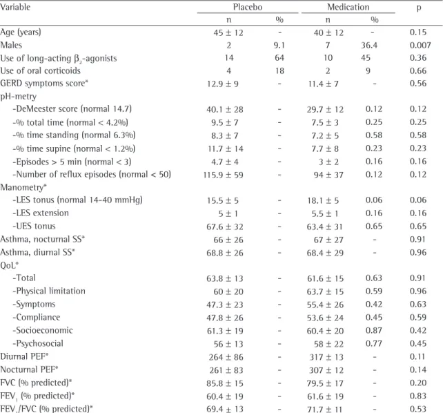
Table 1 - Demographic, clinical and functional characteristics of patients at the study outset.
Variable Placebo Medication p
n % n %
Age (years) 45 ± 12 - 40 ± 12 - 0.15
Males 2 9.1 7 36.4 0.007
Use of long-acting β2-agonists 14 64 10 45 0.36
Use of oral corticoids 4 18 2 9 0.66
GERD symptoms score* 12.9 ± 9 - 11.4 ± 7 - 0.56
pH-metry
-DeMeester score (normal 14.7) 40.1 ± 28 - 29.7 ± 12 0.12 0.12
-% total time (normal < 4.2%) 9.5 ± 7 - 7.5 ± 3 0.25 0.25
-% time standing (normal 6.3%) 8.3 ± 7 - 7.2 ± 5 0.58 0.58
-% time supine (normal < 1.2%) 11.7 ± 14 - 7.7 ± 8 0.23 0.23
-Episodes > 5 min (normal <3) 4.7 ± 4 - 3 ± 2 0.16 0.16
-Number of reflux episodes (normal < 50) 115.9 ± 59 - 94 ± 37 0.12 0.12 Manometry*
-LES tonus (normal 14-40 mmHg) 15.5 ± 5 - 18.1 ± 5 0.06 0.06
-LES extension 5 ± 1 - 5.5 ± 1 0.16 0.16
-UES tonus 67.6 ± 32 - 63.4 ± 31 0.65 0.65
Asthma, nocturnal SS* 66 ± 26 - 67 ± 27 - 0.91
Asthma, diurnal SS* 68.8 ± 26 - 68.4 ± 29 - 0.96
QoL*
-Total 63.8 ± 13 - 61.6 ± 15 0.63 0.91
-Physical limitation 60 ± 20 - 63.7 ± 15 0.59 0.96
-Symptoms 47.3 ± 23 - 55.4 ± 26 0.42 0.63
-Compliance 47.8 ± 26 - 53.6 ± 24 0.45 0.59
-Socioeconomic 61.3 ± 19 - 60.4 ± 20 0.87 0.42
-Psychosocial 56 ± 13 - 58 ± 22 0.77 0.45
Diurnal PEF* 264 ± 86 - 317 ± 13 - 0.11
Nocturnal PEF* 261 ± 83 - 307 ± 12 - 0.14
FVC (% predicted)* 85.8 ± 15 - 79.5 ± 17 - 0.20
FEV1 (% predicted)* 60.4 ± 19 - 61.6 ± 19 - 0.83
FEV1/FVC (% predicted)* 69.4 ± 13 - 71.7 ± 11 - 0.53
At the end of the pretreatment phase, patients completed the asthma quality of life questionnaire developed jointly by the Federal University of São Paulo and the Paulista School of Medicine,(16,17) which
was adapted and validated for use with the Brazilian population based on the domains investigated by Juniper and Guyatt.(18) After the questionnaires
had been completed, the intervention began. The patients were randomly distributed into two treat-ment groups. The patient in one of the groups received pantoprazol (40 mg in a single daily dose), and those in the other group received a placebo. Examiners and patients were both blinded as to the medication being used by any given patient. All subjects were assessed every month by means of a medical visit questionnaire to determine treatment compliance and possible side effects. During the last week of the study, patients were again submitted to a control 24-h pH-metry. In the final medical visit, patients again completed the quality of life ques-tionnaire and performed the PEF maneuvers, as well as being submitted to pulmonary function tests by means of spirometry. The 10-day symptoms diaries were then collected.
Data were compiled in a spreadsheet (Microsoft Office Excel®), and analyses were carried out using the SPSS statistical software program, version 10 (SPSS Inc., Chicago, IL, USA). Demographic, clinical and laboratory variables for both groups were eval-uated for normal distribution and are expressed as means ± standard deviations. In order to evaluate differences between the pretreatment and posttreat-ment phases for each group in terms of pH-metry, symptoms scores and quality of life, the Wilcoxon rank sum test was used. The Mann-Whitney test was used to compare variables between the two groups prior to and after treatment. Comparisons between morning and evening PEF, as well as spirometry values between treatment and placebo groups, were carried out using the unpaired Student’s t-test. For intragroup comparisons prior to and after the study, we used paired Student’s t-test. For all statis-tical tests, the level of significance was set at 5% (p < 0.05).
Results
Of the 73 patients with asthma that met the initial inclusion criteria of the study, 49 were diag-nosed with GERD and were eligible for the study.
Of those, 5 were excluded: 2 due to worsening of symptoms and consequent hospitalization; 1 due to noncompliance with the protocol; 1 due to intol-erance to the medication used in the study; and 1 due to having started smoking. Therefore, the study sample consisted of 44 patients at the study outset. Of those 44, 9 were not submitted to the final pH-metry, and therefore only 35 completed the protocol. However, those 9 patients were also included in the analysis, and each group therefore comprised 22 patients. The groups were designated ‘m’ (patients receiving the active substance) and ‘p’ (patients receiving the placebo). The general charac-teristics of the patients are shown in Table 1. At the study outset, there were no significant differences between the groups in terms of clinical variables (for asthma and for GERD), quality of life scores, or pulmonary function.
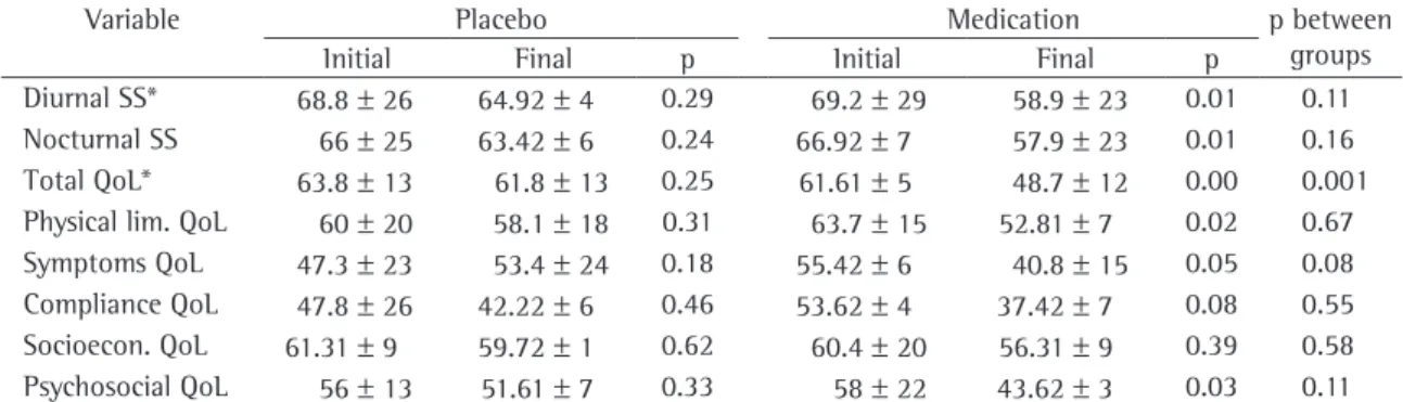
Among the patients with chronic respiratory symptoms and predominance of moderate/severe disease, as determined by the quantity of long-acting β2-agonists and oral corticoids needed to control their symptoms, females predominated. Similarly, female patients with GERD presented high levels of esophageal acid exposure, especially in the evening, and the female patients presenting the greatest alterations were in the placebo group. Of the 44 patients studied, 9 (20%) presented asymp-tomatic GERD. Regarding the control of GERD in both groups, there was pronounced improvement in the ‘m’ group in terms of the symptoms score as well as in terms of the pH-metry readings. Only one patient in this group continued to present an abnormal DeMeester score at the end of the three-month follow-up period. There was an improvement in the symptoms score in both groups. However, this improvement was statistically significant only in the ‘m’ group. Nevertheless, the differences between the groups were not significant when evaluated at the study endpoint. Regarding quality of life, the ‘m’ group presented significant improvement in some of the quality of life score domains. However, when the groups were compared at the study endpoint, statistically significant differences were found only in the overall score. Nevertheless, a tendency toward improvement in all domains was found for the ‘m’ group (Table 2). Pulmonary function test results for both groups are shown in Table 3.
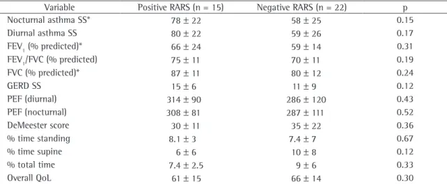
divided into two groups: ‘RARS-positive’ and ‘RARS-negative’. Of the 44 patients evaluated, 7 were excluded from these groups due to the fact that there were insufficient data for this analysis. Table 4 shows the initial characteristics of these two groups; there were no significant differences. Subsequently, in order to test whether the presence of RARS is a predictive factor for the improvement of respiratory parameters, only the RARS-positive group was investigated, by means of comparisons prior to and after the therapeutic intervention, as well as by comparing the RARS-positive members of the ‘m’ group with those of the ‘p’ group. There was no improvement in the respiratory function values in either group, whether studied in isolation or compared at the study endpoint (Table 5).
Discussion
In the present randomized placebo-controlled study, we evaluated the treatment of GERD in patients with asthma over a three-month period.
The correlation between these two diseases has been widely studied in the literature by means of clinical trials in which the role that the treatment of GERD plays in reducing asthma symptoms has been evaluated.(16,19-21) Despite the results, the issue
remains controversial. In recent years, two large reviews were published, both addressing the impact that the treatment of GERD has on asthma control. In the first review,(11) it was concluded that the
treat-ment of GERD reduced asthma symptoms in 69% of the cases, cut the use of asthma medication in 62%, and improved afternoon PEF in 26%. In the second review,(13) it was concluded that treatment of GERD
did not consistently improve asthma symptoms. In addition, it neither reduced the use of medication nor had a significant effect on pulmonary function. However, the authors admitted that the studies included in these reviews presented methodological limitations, highlighting the need for other clinical trials involving this question. One of the contro-versial points reported by these authors was that, among the clinical trials reviewed, only six made
Table 2 - Comparison between the study outset and study endpoint, as well as between the two groups at the study
endpoint, in terms of the clinical control of asthma variables and quality of life.
Variable Placebo Medication p between
groups
Initial Final p Initial Final p
Diurnal SS* 68.8 ± 26 64.92 ± 4 0.29 69.2 ± 29 58.9 ± 23 0.01 0.11 Nocturnal SS 66 ± 25 63.42 ± 6 0.24 66.92 ± 7 57.9 ± 23 0.01 0.16 Total QoL* 63.8 ± 13 61.8 ± 13 0.25 61.61 ± 5 48.7 ± 12 0.00 0.001 Physical lim. QoL 60 ± 20 58.1 ± 18 0.31 63.7 ± 15 52.81 ± 7 0.02 0.67 Symptoms QoL 47.3 ± 23 53.4 ± 24 0.18 55.42 ± 6 40.8 ± 15 0.05 0.08 Compliance QoL 47.8 ± 26 42.22 ± 6 0.46 53.62 ± 4 37.42 ± 7 0.08 0.55 Socioecon. QoL 61.31 ± 9 59.72 ± 1 0.62 60.4 ± 20 56.31 ± 9 0.39 0.58 Psychosocial QoL 56 ± 13 51.61 ± 7 0.33 58 ± 22 43.62 ± 3 0.03 0.11 SS: symptoms score; QoL: quality of life, points; lim.: limitation; Socioecon.: Socioeconomic; and *mean ± standard deviation.
Table 3 - Comparison between the study outset and study endpoint, as well as between the two groups at the study
endpoint, in terms of the respiratory variables.
Variable Placebo Medication p between
groups
initial final p initial final p
use of proton pump inhibitors. In addition, these studies showed that patients with respiratory mani-festations of GERD require higher doses of proton pump inhibitors for the control of symptoms.(22)
Based on these data, we chose to perform another 24-h pH-metry at the study endpoint in order to determine the true efficacy of the acid suppres-sion instituted, with the objective of facilitating the analysis of the results.
In the present study, there was considerable esophageal acid suppression in the ‘m’ group patients, only 1 of which presented an abnormal
DeMeester(15) score at the study endpoint. These
findings might be explained by the use of a proton pump inhibitor, administered in appropriate doses over the course of the study. Other authors made use of this resource with the same objectives and concluded that acid ablation at the study endpoint was an important factor in the context of treatment and verification of its results.(22,23)
The population evaluated in our study included patients with asthma making continuous use of corticosteroids, and most of these patients also needed concomitant use of long-acting β2-agonists
Table 4 - Comparison between the groups with and without reflux-associated respiratory symptoms at the study outset.
Variable Positive RARS (n = 15) Negative RARS (n = 22) p
Nocturnal asthma SS* 78 ± 22 58 ± 25 0.15
Diurnal asthma SS 80 ± 22 59 ± 26 0.17
FEV1 (% predicted)* 66 ± 24 59 ± 14 0.31
FEV1/FVC (% predicted) 75 ± 11 70 ± 11 0.19
FVC (% predicted)* 87 ± 11 80 ± 12 0.24
GERD SS 15 ± 6 11 ± 9 0.12
PEF (diurnal) 314 ± 90 286 ± 120 0.43
PEF (nocturnal) 308 ± 81 287 ± 111 0.52
DeMeester score 30 ± 11 35 ± 22 0.36
% time standing 8.1 ± 3 7.4 ± 7 0.67
% time supine 6 ± 6 10 ± 8 0.12
% total time 7.4 ± 2.5 9 ± 6 0.33
Overall QoL 61 ± 15 66 ± 14 0.30
RARS: reflux-associated respiratory symptoms; SS: symptoms score; PEF: peak expiratory flow; FVC: forced vital capacity; FEV1: forced expiratory volume in one second; QoL: quality of life score (points); and *mean ± standard deviation.
Table 5 - Comparison between the study outset and study endpoint, as well as between the two groups at the study
endpoint, in terms of the variables studied and limited to the RARS-positive patients.
Variables Positive RARS (n = 15)
Placebo (n = 8) Medication (n = 7) p between
groups
Initial Final p Initial Final p
Diurnal asthma SS* 90 ± 16 92 ± 17 0.30 80 ± 21 60 ± 24 0.05 0.02 Nocturnal asthma SS 81 ± 15 80 ± 14 0.39 77 ± 25 59 ± 27 0.04 0.03 Diurnal PEF (L/min)* 284 ± 98 300 ± 92 0.66 323 ± 86 334 ± 82 0.47 0.62 Nocturnal PEF (L/min) 266 ± 94 311 ± 90 0.35 324 ± 67 320 ± 72 0.68 0.84
FVC (%)* 91 ± 16 94 ± 16 0.60 81 ± 11 85 ± 19 0.22 0.49
FEV1 (%)* 64 ± 23 64 ± 27 0.29 68 ± 17 73 ± 20 0.11 0.53
Overall QoL * 60 ± 19 62 ± 16 0.33 62 ± 8 46 ± 8 0.01 0.01
for their treatment. Despite this treatment, many patients presented diurnal and nocturnal symptoms, as well as quality of life limitations, suggesting that this population of patients presented more severe asthma. In very few studies in the litera-ture, comparing the use of proton pump inhibitors with placebo, it has been described or suggested the severity of the respiratory disease. One of the therapeutic trials, in which the clinical severity of the patients studied was mentioned, was an open study.(22) This factor is important because the lack of
information regarding the degree of severity in the beginning of the protocol might be another bias factor in these studies, since patients with more severe diseases are less likely to present pronounced improvement.
In the present study, esophageal manometry showed that, for both groups, the tonus of lower esophageal sphincter was at the lower limit of normality at the study outset. This finding coin-cides with the findings of another study previously conducted in our esophageal function laboratory,(24)
in which the mean lower esophageal sphincter tonus was also at the lower limit of normality (15.3 mmHg). In that same study, the analysis of the esophageal motor profile of 164 patients with asthma submitted to esophageal manometry showed the presence of motor alterations in 32% of the individuals, most of which were represented by hypomotility or inef-fective motility of the distal esophagus. In a similar study,(25) the incidence of manometry alterations
was also reported to be higher in patients with asthma than in control group patients, and the most common disturbance (observed in 53%) was ineffective esophageal motility. In both studies, the authors suggested that the combination of abnormal pH-metry values and esophageal dysmo-tility might indicate microaspiration of gastric acid as a triggering, aggravating, or maintenance factor for respiratory symptoms in this population.
In concordance with other studies described in the literature,(26,27) our analysis revealed no changes
in the respiratory function of the patients treated with acid ablation. This paradox was also found in other studies in which the clinical, or even surgical, treatment of GERD improved asthma symptoms, although there was no improvement in the pulmo-nary function of these patients. The limitations already described in this study and others, such as the small number of patients, or even the extremely
short time of acid ablation, might have contrib-uted to these results. These findings might also be explained by evidence showing an increase in minute ventilation as a triggering factor of dyspnea and thoracic discomfort in patients submitted to esophageal acid perfusion, although there was no airway obstruction.(28)
Field et al.(28) investigated, by means of a GERD/
asthma questionnaire, the presence of RARS. Harding et al.(16) later identified this condition as a predictive
factor for treatment response. It is currently believed that this group of patients, consisting of those diagnosed with asthma and concomitant GERD who report respiratory symptoms directly related to reflux, responds better to anti-reflux measures and drug treatment, consequently presenting better asthma control when being treated for GERD. In our study, we also evaluated the presence of RARS by means of a questionnaire, and most subjects did not present RARS. Some initial characteristics of the patients were also compared by stratifying them on the basis of the presence/absence of RARS in order to better define this group of patients with asthma, and no significant differences were found. Nevertheless, when only the patients with RARS were studied in terms of the study outcome meas-ures, we again found a significant difference in the improvement of the symptoms and the quality of life scores in the ‘m’ group. Therefore, we can infer that the study of this characteristic in patients with asthma and symptomatic GERD might be important for informing decisions regarding the treatment of such patients.
Since this was a randomized prospective double-blind study with a complex methodology, some deficiencies and limitations must be mentioned. The final sample was small, which might have contributed for the lack of more significant findings related to the outcome measures studied. In clinical trials involving larger patient samples, such as that of Kiljander et al.,(19) which involved 57 patients in
a controlled crossover study, there was a 20% func-tional improvement in 35% of the patients treated. In addition, the three-month follow-up period employed in the present study might have been too short for the identification of consistent improve-ment in the respiratory function parameters of the patients evaluated.
a thorough study on the prevalence of GERD in a non-referenced (i.e. bias-free) population of subjects with asthma. Such a study should also include the objective confirmation of abnormal esopha-geal exposure to gastric acid. In addition, there is sufficient evidence to show that there is much to be done regarding therapeutic intervention in this population of patients. It is imperative to determine what characteristics of these patients with asthma could be used as predictors of the response to GERD therapy, as well as what parameters would be the most appropriate for the assessment of GERD. We therefore conclude that, in the present study, GERD treatment significantly improved the quality of life and the symptoms scores of the patients with asthma. However, there were no significant alterations in their respiratory function parameters. Among the patients studied, those who presented RARS and were submitted to pharmacological acid ablation were the ones who also presented objec-tive reductions in their asthma score, as well as an improvement in their quality of life at the end of the evaluation period.
Acknowledgments
The authors would like to thank Altana Pharma, the Postgraduate Program in Pulmonology of the Federal University of Rio Grande do Sul, and the Santa Casa de Porto Alegre Hospital.
References
1. Napierkowski J, Wong RK. Extraesophageal manifestations of GERD. Am J Med Sci. 2003;326(5):285-99.
2. Sociedade Brasileira de Pneumologia e Tisiologia. III Consenso Brasileiro no Manejo da Asma. Jornal de Pneumol. 2002;28(Supl 1):110-20.
3. Johanson JF. Epidemiology of esophageal and supraesophageal reflux injures. Am J Med. 2000;108(Supl 4a):S99-S103. 4. Alexander JA, Hunt LW, Patel AM. Prevalence,
pathophysiology, and treatment of patients with asthma and gastroesophageal reflux disease. Mayo Clinic Proc. 2000;75(10):1055-63.
5. Harding SM, Sontag SJ. Asthma and gastroesophageal reflux. Am J Gastroenterol. 2000;95(Supl 8):S23-S32. 6. Malagelada JR. Review article: supra-oesophageal
manifestations of gastro-oesophageal reflux disease. Aliment Pharmacol Ther. 2004;19 (Supl 1):S43-S8.
7. Mansfield LE, Stein M. Gastroesophageal reflux and asthma: a possible reflex mechanism. Ann Allergy. 1978;41(4):224-6. 8. Tuchman DN, Boyle JT, Pack AI, Scwartz J, Kokonos
M, Spitzer AR, et al. Comparisons of airway responses following tracheal and esophageal acidification in the cat. Gastroenterology 1984;87:872-9.
9. Harding SM, Guzzo MR, Richter JE. 24-h esophageal pH testing in asthmatics: Respiratory Symptom Correlation with Esophageal Acid Events. Chest. 1999;115(3):654-9. 10. Theodoropoulos DS, Pecoraro DL, Efstratiadis SE. The
association of gastroesophageal reflux disease with asthma and chronic cough in the adult. Am J Respir Med. 2002;1(2):133-46.
11. Field SK, Sutherland LR. Does medical antireflux therapy asthma in asthmatics with gastroesophageal reflux?: a critical review of the literature. Chest. 1998;114(1):275-83. 12. Field SK. Gastroesophageal reflux and asthma: can the
paradox be explained? Can Respir J. 2000;7(2):167-76. 13. Gibson P, Henry R, Coughlan J. The effect of treatment for
gastroesophageal reflux on asthma in adults end children. Review [latest version 10 Feb]. In: Cochrane Library. Oxford: Update Software;1999.
14. Sociedade Brasileira de Pneumologia e Tisiologia. Diretrizes para Testes de Função Pulmonar. J Pneumol. 2002;28(3):1-221.
15. Johnson LF, Demeester TR. Twenty-four-hour pH monitoring of the distal esophagus. A quantitative measure of gastroesophageal reflux. Am J Gastroenterol. 1974;62(4):325-32
16. Harding SM, Richter JE, Guzzo MR, Schan CA, Alexander RW, Bradley LA. Asthma and gastroesophageal reflux: acid suppressive therapy improves asthma outcome. Am J Med. 1996;100(4):395-405.
17. Fernandes ALG, Oliveira MA. Avaliação de qualidade de vida nas asma. J Pneumol. 1997;23(3):148-52.
18. Juniper EF, Guyatt GH, Epstein RS, Ferrie JP, Jaeschke R, Hiller TK. Evaluation of impairment of health related quality of life in asthma: development of a questionnaire for use in clinical trials. Thorax. 1992;47(2):76-83.
19. Kiljander TO, Salomaa ER, Hietanen EK, Terho EO. Gastroesophageal reflux in asthmatics: a double-blind, placebo-controlled, crossover study with omeprazole. Chest 1999;116(5):1257-64.
20. Littner MR, Leung FW, Ballard ED 2nd, Huang B, Samra NK; Lansoprazole Asthma Study Group. Effects of 24 weeks of lansoprazole therapy on asthma symptoms, exacerbations, quality of life, and pulmonary function in adult asthmatic patients with acid reflux symptoms. Chest. 2005;128(3):1128-35.
21. Jiang SP, Liang RY, Zeng ZY, Liu QL, Liang YK, Li JG. Effects of antireflux treatment on bronchial hyper-responsiveness and lung function in asthmatic patients with gastroesophageal reflux disease. World J Gastroenterol 2003;9(5):1123-5. 22. Harding SM. Gastroesophageal reflux: a potential
asthma trigger. Immunol Allergy Clin North Am. 2005;25(1):131-48.
23. Maev IV Viuchnova ES, Balashova NN, Shchekina MI. Use of omeprazole and esomeprazole in patients suffering from bronchial asthma with associated gastroesophageal reflux disease. Eksp Klin Gastroenterol 2003; 116(3):26-31. 24. Ribeiro I, Cardoso PFG, Hetzel JL, Camargo JJP, Felicetti
JC. Esofagomanometria e pHmetria esofagiana ambulatorial de 24 horas nos pacientes com asma com e sem sintomas digestivos associados. J Pneumol 2000;26(Supl 3):S16. 25. Fouad YM, Katz PO, Hatlebakk JG, Castell DO. Ineffective
26. Sontag SJ, OConnell S, Khandelwal S, Greenlee H, Schnell T, Nemchausky B, et al. Asthmatics with gastroesophageal reflux: long term results of a randomized trial of medical and surgical antireflux therapies. Am J Gastroenterol. 2003 98(5):987-99.