ISSN 0100-879X
CLINICAL INVESTIGATION
www.bjournal.com.br
www.bjournal.com.br
Volume 44 (1) 1-83 January 2011
Institutional Sponsors
The Brazilian Journal of Medical and Biological Research is partially financed by
Hotsite of proteomics metabolomics developped by:
Braz J Med Biol Res, January 2011, Volume 44(1) 11-15
doi: 10.1590/S0100-879X2010007500114
Wrist ultrasound analysis of patients with early rheumatoid arthritis
Wrist ultrasound analysis of patients with
early rheumatoid arthritis
J.A. Mendonça
1, M.A. Yazbek
1, I.M.M. Laurindo
2and M.B. Bertolo
11Serviço de Reumatologia, Faculdade de Ciências Médicas, Universidade Estadual de Campinas, Campinas, SP, Brasil 2Serviço de Reumatologia, Faculdade de Medicina, Universidade de São Paulo, São Paulo, SP, Brasil
Abstract
In the present study, we evaluated 42 wrists using the semi-quantitative scales power Doppler ultrasound (PDUS) and gray scale ultrasound (GSUS) with scores ranging from 0 to 3 and correlated the results with clinical, laboratory and radiographic data. Twenty-one patients (17 women and 4 men) with rheumatoid arthritis according to criteria of the American College of Rheuma-tology were enrolled in the study from September 2008 to July 2009 at Universidade Estadual de Campinas (UNICAMP). The average disease duration was 14 months. The patients were 66.6% Caucasians and 33.3% non-Caucasians, with a mean age of 42 and 41 years, respectively. A dorsal longitudinal scan was performed by ultrasound on the radiocarpal and midcarpal joints using GE LOGIQ XP-linear ultrasound and a high frequency (8-10 MHz) transducer. All patients were X-rayed, and the Larsen score was determined for the joints, with grades ranging from 0 to V. This study showed significant correlations between clini -cal, sonographic and laboratory data:GSUS and swollen right wrist (r = 0.546), GSUS of right wrist and swelling of left wrist (r = 0.511), PDUS of right wrist and pain in left wrist (r = 0.436), PDUS of right wrist and C-reactive protein(r = 0.466). Ultrasound can be considered a useful tool in the diagnosis of synovitis in early rheumatoid arthritis mainly when the anti-cyclic citrullinated peptide and rheumatoid factor are negative, and can lead to an early change in the therapeutic decision.
Key words: Power Doppler ultrasound; Gray scale ultrasound; Rheumatoid arthritis; Wrist; Synovitis
Introduction
Correspondence: J.A. Mendonça, Rua da Fazenda, 125, Casa 10, Condomínio Dálias, 13175-665 Sumaré, SP, Brasil. E-mail: [email protected]
Received March 10, 2010. Accepted October 7, 2010. Available online October 29, 2010. Published January 17, 2011. Joint ultrasound has proved to be an important tool to
evaluate synovitis, which could be considered the first ab -normality in early rheumatoid arthritis (RA) (1-4). X-ray has been the method most frequently used to assess bone and joints, but an important drawback is that it does not show lesions in soft tissues in the early stage of disease (5-7).
Ultrasound is a sensitive technique for the detection of
synovitis, effusion and bone erosion in RA, often difficult to
determine by clinical assessment and traditional radiography (6,7).Subclinical synovitis can be diagnosed early in inflam -matory diseases by ultrasound and its evolution may even be monitored, thus being a precursor of oligoarthritis and polyar-thritis(8). Ultrasound facilitates the early detection of arthritis and allows an earlier establishment of therapy (9-12).
Material and Methods
Twenty-one patients were enrolled from September 2008 to July 2009 at Universidade Estadual de
Campi-nas (UNICAMP), CampiCampi-nas, Brazil. Seventeen women and 4 men were included: 66.6% Caucasians and 33.3% non-Caucasians, with a mean age of 42 and 41 years, respectively, with RA according to criteria of the American College of Rheumatology. Gray scale (GS) and power Doppler (PD) ultrasound were applied to patients with early RA and correlated with clinical, laboratory and radiological data. The average disease duration was 14 months since
presenting the first sign of arthritis. The medications used
by the patients are listed in Table 1.
Clinical evaluation consisted of Disease Activity Score based on 28 joints (DAS28), calculated using the con-centration of the C-reactive protein (CRP) or erythrocyte sedimentation rate (ESR). The DAS28 can range from 0 to 10, and studies have shown that the lower rate of disease activity is 3.2 (13). This study used DAS28 equal to or greater than 3.2 as inclusion criteria.
12 J.A. Mendonça et al.
gave written informed consent after completing the specific
quality of life instrument - Health Assessment Questionnaire (HAQ), which evaluates disability, discomfort and pain at the time of the exam (14,15).
The ultrasound was performed with the patient seated
and with hands flat on the top of the table. The radiocarpal and midcarpal regions were examined by a dorsal longi-tudinal scan, when evaluating the cross-scan of the wrist joint. The equipment used was a linear GE LOGIQ XP Ultrasound apparatus (USA) with a high-frequency (8-10 MHz) transducer, which accurately shows surface struc-tures such as tendons, synovia, and other strucstruc-tures. All structures were documented to ensure high reproducibility of the results (16,17).
The ultrasound analysis using a GS ultrasound reflects
an increase of the synovium as a hypoechoic image and PD ultrasound demonstrates the microcirculation in the phase of
synovial hypertrophy, identifying inflammatory activity. Using a semi-quantitative scale, a joint capsule was classified as
having a degree of 0 to 3 of synovial proliferation, and a degree of intense or important signal by PD(18-21).
Synovitis was scored by GS ultrasound (GSUS) as 0 = absence, 1 = mild (describes a small hypoechoic/an-echoic line beneath the joint capsule), 2 = moderate (the joint capsule is elevated parallel to the joint area), and 3 = severe or marked (characterizes a strong distension of the joint capsule).
Synovitis was scored by PD ultrasound (PDUS) as: 0 = absence (no intra-articular color signal), 1 = mild (up
to 3 color signals or 2 single and 1 confluent signal in the
intra-articular area, 2 = moderate (greater than grade 1 to
<50% of the intra-articular area filled with color signals), and 3 = marked (≥50% of the intra-articular area filled with
color signals).
The Doppler settings were optimizedto ‘low flow’, with
a medium wall filter (to minimizeflash artifact) and a pulse
repetition frequency of 800 Hz.
Wrist radiography was performed in all patients and the Larsen score was used to evaluate the aspects in the X-ray. Joints received grades of 0 to V, as follows: grade 0 = normal; grade I = mild abnormality (presence of one or more of the following lesions: edema of soft tissues, osteopenia around the joint and a slight decrease in joint
space); grade II = definite abnormality (presence of small
erosions, decreased joint space is not obligatory); grade III
= marked abnormality (presence of erosions and decreased joint space); grade IV = severe abnormality (the original joint surface remains partially preserved), and grade V = muti-lating abnormality (the original joint surface disappeared; huge deformity is present) (22-25).
Results
Clinical, laboratory and imaging results
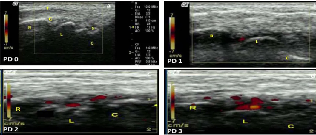
Fourteen (66.6%) patients had pain in the right wrist and 13 (61.9%) had pain in the left wrist; 16 (76%) patients had swelling of the right wrist and 14 (66.6%) had swelling of the left wrist. The mean (± SD) DAS28 for this sample was 4.0 ± 1.1 (data variation: 0 to 10), the mean HAQ was 1.2 ± 0.5 (0 = without disability to 3 = severe disability), with 145.7 IU/dL (normal <22 IU/dL) anti-cyclic citrullinated peptide (anti-CCP) and the average rheumatoid factor was 231.63 IU/dL (normal <20 IU/dL). Mean CRP was 6.97 ± 11.13 mg/dL (normal >6 mg/dL) and mean ESR was 4.7 ± 1.0 mm (normal >10 mm). Synovitis was detected by GSUS in 66.6% (N = 14) and 61.9% (N = 13) of right and left wrists, respectively. In the right wrist, the synovitis determined by GSUS was 28.5% (N = 6) grade 1, 19% (N = 4) grade 2 and 19% (N = 4) grade 3. In the left wrist, synovitis was 33.3% (N = 7) grade 1, 14.2% (N = 3) grade 2, and 14.2% (N = 3) grade 3. PDUS was seen in 23.8% (N = 5) of the right and left wrists. In the right wrist, 9.5% (N = 1) were grade 2 and 19% (N = 4) were grade 3. In the left wrist, 16% (N = 4) were grade 2 and 4.76% (N = 1) were grade 3 (Figures 1 and 2).
The Spearman correlation coefficient at the P < 0.05 level of significance revealed the following significant cor -relations: GSUS and swollen right wrist (r = 0.546), GSUS of right wrist and swelling of left wrist (r = 0.511), PDUS of right wrist and pain in left wrist (r = 0.436), PDUS of right wrist
and CRP (r = 0.466). At the P < 0.001 level of significance,
the following correlations were obtained: GSUS and PDUS of right wrist (r = 0.730), GSUS and PDUS of left wrist (r = 0.758), PDUS of right and left wrist (r = 0.732), GSUS of right and left wrist (r = 0.552), PDUS of left wrist and GSUS of right wrist (r = 0.574), PDUS of right wrist and GSUS of left wrist (r = 0.687), as presented in Table 2.
The detection of synovitis by GSUS was higher than by PDUS. This may have occurred because a unit of frequency for low power Doppler was used. This may have caused the
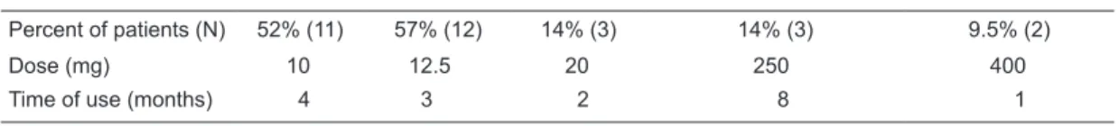
Table 1. Drugs used by the patients at the time of examination.
Prednisone Methotrexate Leflunomide Chloroquine diphosphate Hydroxychloroquine
Percent of patients (N) 52% (11) 57% (12) 14% (3) 14% (3) 9.5% (2)
Dose (mg) 10 12.5 20 250 400
underestimation of the capture of active microcirculation, which is always important in the evaluation of synovitis by GSUS.
There was no significant correlation between wrist GSUS
and PDUS and DAS28, HAQ score or radiography. The
as-sessment of intra-observational (same reader) Larsen score for the X-ray had a mean kappa value of 0.185 and there was no agreement with a semi-quantitative ultrasound scale, since only radiographic osteopenia of the carpal bones and soft tissue edema of both wrists was detected.
Figure 1. Synovitis identified by gray scale (GS) ultrasound in a dorsal longitudinal wrist scan. a =
grade 0; b = grade 1; c = grade 2; d = grade 3. T = tendon; S = synovitis; R = radius; L = lunate; C = capitate bone; c = cartilage.
Figure 2. Synovitis identified by power Doppler (PD) ultrasound in a dorsal longitudinal wrist scan. a
14 J.A. Mendonça et al.
References
1. Filippucci E, Iagnocco A, Meenagh G, Riente L, Delle Sedie A, Bombardieri S, et al. Ultrasound imaging for the rheuma-tologist. Clin Exp Rheumatol 2006; 24: 1-5.
2. Grassi W, Salaffi F, Filippucci E. Ultrasound in rheumatology.
Best Pract Res Clin Rheumatol 2005; 19: 467-485. 3. Kane D, Bruyn G, Arnold E, Grassi W. A rheumatologist’s
perspective on musculoskeletal ultrasound in rheumatology: comment on the editorial by Roemer et al. Arthritis Rheum
2006; 55: 341-342.
4. Kane D, Grassi W, Sturrock R, Balint PV. Musculoskeletal ultrasound - a state of the art review in rheumatology. Part 2: Clinical indications for musculoskeletal ultrasound in rheumatology. Rheumatology 2004; 43: 829-838.
5. Brown AK, O’Connor PJ, Roberts TE, Wakefield RJ, Karim Z, Emery P. Recommendations for musculoskeletal ultrasonog-raphy by rheumatologists: setting global standards for best practice by expert consensus. Arthritis Rheum 2005; 53: 83-92.
6. Grassi W, Filippucci E, Carotti M, Salaffi F. Imaging mo -dalities for identifying the origin of regional musculoskeletal pain. Best Pract Res Clin Rheumatol 2003; 17: 17-32. 7. Bresnihan B, Kane D. Sonography and subclinical synovitis.
Ann Rheum Dis 2004; 63: 333-334.
8. Wakefield RJ, Green MJ, Marzo-Ortega H, Conaghan PG, Gibbon WW, McGonagle D, et al. Should oligoarthritis be reclassified? Ultrasound reveals a high prevalence of sub -clinical disease. Ann Rheum Dis 2004; 63: 382-385. 9. D’Agostino MA, Ayral X, Baron G, Ravaud P, Breban M,
Dougados M. Impact of ultrasound imaging on local
corticos-teroid injections of symptomatic ankle, hind-, and mid-foot in chronic inflammatory diseases. Arthritis Rheum 2005; 53: 284-292.
10. Koski JM, Helle M. Ultrasound guided synovial biopsy using portal and forceps. Ann Rheum Dis 2005; 64: 926-929. 11. Balint PV, Kane D, Wilson H, McInnes IB, Sturrock RD.
Ultrasonography of entheseal insertions in the lower limb in spondyloarthropathy. Ann Rheum Dis 2002; 61: 905-910. 12. Naredo E, Cabero F, Beneyto P, Cruz A, Mondejar B, Uson
J, et al. A randomized comparative study of short term re-sponse to blind injection versus sonographic-guided injec-tion of local corticosteroids in patients with painful shoulder.
J Rheumatol 2004; 31: 308-314.
13. van Gestel AM, Haagsma CJ, van Riel PL. Validation of rheumatoid arthritis improvement criteria that include simpli-fied joint counts. Arthritis Rheum 1998; 41: 1845-1850. 14. Ferraz MB, Oliveira LM, Araujo PM, Atra E, Tugwell P.
Crosscultural reliability of the physical ability dimension of the health assessment questionnaire. J Rheumatol 1990; 17: 813-817.
15. Ciconelli R, Ferraz M, Santos W, Meinão I, Quaresma N. Tradução para a língua portuguesa do questionário de avaliação de qualidade de vida SF-36 (Brasil SF-36). Rev Bras Reumatol 1999; 39: 145-150.
16. Grassi W, Tittarelli E, Blasetti P, Pirani O, Cervini C. Finger tendon involvement in rheumatoid arthritis. Evaluation with high-frequency sonography. Arthritis Rheum 1995; 38: 786-794.
17. Manger B, Kalden JR. Joint and connective tissue
ultra-This study showed an important correlation between the clinical and laboratory data, such as the correlation of the CRP and ultrasound data.
Discussion
In the present study, 42 wrist joints were evalu-ated by ultrasound in a population with very early RA characterized by 14 months of disease. PDUS
showed significant synovitis changes indicated by
the existence of active microcirculation in the joint capsule, as demonstrated by a semi-quantitative scale. PDUS and GSUS are important tools for confirma -tion of synovitis, which can be dissociated from the clinical
findings, as reported in the literature (7). A positive and
significant correlation between GSUS and PDUS of both
wrists may characterize the symmetry of clinical involve-ment of the joints in RA.Thus, these variables detected by ultrasound could enhance the clinical diagnosis of early RA, even when CCP and the rheumatoid factor are negative. When we evaluated the level of intra-observational agree-ment (kappa) between the results of ultrasound and the
X-ray data a discrepancy was identified, e.g., the number
of changes seen by ultrasound in patients with early RA was higher than that seen by the X-ray, and the changes
found in the X-ray of the wrists were edema of soft tissues or osteopenia. The treatment of the RA has greatly pro-gressed and currently intends to induce clinical remission
based on the hypothesis that the inflammatory process
leads to structural damage. Thus, there is a need to change the focus of evaluation, using a method that characterizes structural changes in the joint rather than only edema or osteopenia (26).
The ultrasound is important in the practice of the rheu-matologist to prevent future structural damage, providing more comfort to the patient by fast, unlimited and safer scanning without irradiation. It can be considered a useful toolin the diagnosis of synovitis in early RA and can lead to an early change in the therapeutic decision.
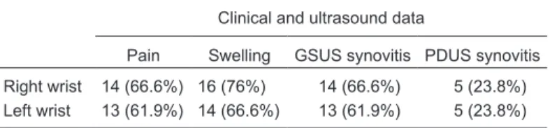
Table 2. Correlation between clinical and ultrasound examination.
Clinical and ultrasound data
Pain Swelling GSUS synovitis PDUS synovitis
Right wrist 14 (66.6%) 16 (76%) 14 (66.6%) 5 (23.8%) Left wrist 13 (61.9%) 14 (66.6%) 13 (61.9%) 5 (23.8%)
sonography - a rheumatologic bedside procedure? A Ger -man experience. Arthritis Rheum 1995; 38: 736-742. 18. Fiocco U, Ferro F, Vezzu M, Cozzi L, Checchetto C, Sfriso
P, et al. Rheumatoid and psoriatic knee synovitis: clinical, grey scale, and power Doppler ultrasound assessment of the response to etanercept. Ann Rheum Dis 2005; 64: 899-905.
19. Taylor PC, Steuer A, Gruber J, Cosgrove DO, Blomley MJ, Marsters PA, et al. Comparison of ultrasonographic assess-ment of synovitis and joint vascularity with radiographic evaluation in a randomized, placebo-controlled study of infliximab therapy in early rheumatoid arthritis. Arthritis Rheum 2004; 50: 1107-1116.
20. Szkudlarek M, Klarlund M, Narvestad E, Court-Payen, Strandberg C, Jensen KE, et al. Ultrasonography of the metacarpophalangeal and proximal interphalangeal joints in rheumatoid arthritis: a comparison with magnetic resonance imaging, conventional radiography and clinical examination.
Arthritis Res Ther 2006; 8: R52.
21. Backhaus M, Ohrndorf S, Kellner H, Strunk J, Backhaus TM, Hartung W, et al. Evaluation of a novel 7-joint ultrasound
score in daily rheumatologic practice: a pilot project. Arthritis Rheum 2009; 61: 1194-1201.
22. Kaye JJ. Radiographic methods of assessment (scoring) of rheumatic disease. Rheum Dis Clin North Am 1991; 17: 457-470.
23. Plant MJ, Saklatvala J, Borg AA, Jones PW, Dawes PT. Mea-surement and prediction of radiological progression in early rheumatoid arthritis. J Rheumatol 1994; 21: 1808-1813. 24. Larsen A, Dale K, Eek M. Radiographic evaluation of
rheu-matoid arthritis and related conditions by standard reference films. Acta Radiol Diagn 1977; 18: 481-491.
25. Larsen A, Thoen J. Hand radiography of 200 patients with rheumatoid arthritis repeated after an interval of one year.
Scand J Rheumatol 1987; 16: 395-401.
26. Hafstrom I, Albertsson K, Boonen A, van der Heijde D, Landewe R, Svensson B. Remission achieved after 2 years treatment with low-dose prednisolone in addition to disease-modifying anti-rheumatic drugs in early rheumatoid arthritis is associated with reduced joint destruction still present after 4 years: an open 2-year continuation study. Ann Rheum Dis