Original Article
CURRENT ROLE OF TRANSRECTAL ULTRASONOGRAPHY IN
THE EARLY DETECTION OF PROSTATIC CANCER*
Viviane Cristine Tavares Santos1, Miguel Angelo Milito2, Edson Marchiori3
* Study developed at the Service of Radiology and Radiodiagnosis, Hospital Universitário Clementino Fraga Filho, Universidade Federal do Rio de Janeiro, Rio de Janeiro, RJ, Brazil.
1. MD, Radiologist, Master graduate student at Department of Radiology, Faculty of Medicine, Universidade Federal do Rio de Janeiro.
2. Assistant MD at Service of Radiology and Radiodiagnosis, Hospital Universitário Clementino Fraga Filho.
3. Titular Professor of Radiology at Universidade Federal Fluminense, Adjunct Coordinator of the Course of Post-Graduation in Radiology at Universidade Federal do Rio de Janeiro.
Mailing address: Dra. Viviane Cristine Tavares Santos. Rua Gonzaga Bastos, 123/302, Vila Isabel. Rio de Janeiro, RJ, Brazil, 20541-000. E-mail: [email protected]; [email protected]
Received March 4, 2005. Accepted after revision August 3, 2005.
Abstract
CONCLUSION: Transrectal ultrasound findings, even in association with color Doppler, are not sufficiently accurate to determine whether a patient should or not be submitted to biopsy.
Keywords: Prostate; Prostatic cancer; Transrectal ultrasound; Color Doppler; Prostate biopsy.
INTRODUCTION
Presently, prostate cancer is a health problem worldwide. In Brazil it has become a true public health problem, since it is the second most usual type of cancer affecting men, and has presented a trend of increase in the last years as a result of the population aging(1).
For this reason, the prostatic carcinoma demands the adoption of tools accurate enough to allow its early detection and adequate treatment, improving the patient survival and reducing morbidity. Currently, the prostate cancer diagnosis is based on digital rectal examination, on blood levels of prostate-specific antigen (PSA) and on transrectal ultrasound, but none of these methods is sensitive or specific enough to be utilized alone for definition of the conduct to be adopted in relation to the patient(2,3).
The PSA is a simple diagnostic tool usually employed for prostate cancer screening although may present a dubious meaning in case of an intermediary index increase. The greatest utility of PSA is its role as an indicator of tumoral volume and of the disease progress after the initial treatment(4,5).
Transrectal ultrasound, although presenting a lower sensitivity and specificity than previously thought, is able to detect more tumors and more early than other methods. Additionally, technical developments have occurred with the introduction of Color Doppler that, in spite of having not fully met initial expectations, is an important adjuvant factor in the prostatic cancer investigation for increasing both positive predictive value and sensitivity(2,6,7).
Along this progress, the utilization of ultrasound is well established as the main method for guiding material collection by means of prostate biopsy in a fast, safe and relatively painless way.(6,7).
The objectives of the present study were: to determine the sensitivity, specificity, transrectal ultrasound positive and negative predictive values, both in gray scale and color Doppler, utilizing biopsy results as a golden standard, as well as to correlate ultrasound and color Doppler findings with histopathological results according to Gleason score.
MATERIALS AND METHODS
Ultrasound studies and biopsies in all patients included in this study were performed by or under supervision of a same physician of the service, assuring the highest possible homogeneity of examination technique and data.
Previously to the biopsies, the patients were submitted to antibioticprophylaxis with 400 mg norfloxacine, two hours before the procedure and were told to utilize a “fleet enema” for rectal cleansing one hour before the examination. Antibioticprophylaxis was continued for two additional days after biopsy.
All the studies were performed in an Aspen (Acuson Corporation; California, EUA) ultrasound equipment with an “end-fire” type 7 MHz EC7 endocavitary transducer.
The patient was positioned in left lateral decubitus and examination was initiated in B mode in gray scale on the whole prostatic gland and seminal vesicles, performing the scanning in sagital and ortogonal planes (coronal oblique). At this moment also the prostate weight was estimated by multiplication of the three dimensions of the gland (greatest axes of length, width and height) by a π/6 factor (approximately 0.52), assuming that 1 ml prostatic tissue is equivalent to approximately 1 g.(6).
Findings considered as suspects for cancer at ultrasound were: hypoechoic node in peripheral zone, diffuse hypoechogenicity, loss of differentiation between peripheral zone and internal gland, focal swelling or peripheral zone asymmetry, irregularities and rupture of prostatic capsule(6,8,9).
After scanning in gray scale, the color Doppler was performed, with the color box adjusted for covering the whole peripheral zone, neurovascular bundles and the greatest part of the internal gland (which eventually was not possible in glands presenting highly increased volume), aiming at allowing comparison of vascularization standard between both lobes. If any abnormal vascularization area was found at color Doppler, the color box was reduced to increase sensitivity, allowing a better evaluation of the focal change.
The color Doppler gain was set just below the threshold for noise. Low velocity and high sensitivity parameters were utilized. The filters were adjusted to optimize the visualization of low flow and small vessels.
Findings at color Doppler were divided into asymmetry in vascularization (associated or not with focal lesion seen in gray scale) and focal or diffuse increase in vascularization in the peripheral zone.
Ultrasound-guided biopsies were performed using transrectal route, with a Pro.Mag 2.2 (Medical Devices Technologies, Florida – USA) biopsy pistol system with 18 gauge needle.
Four to six random fragments were obtained from each peripheral zone, besides directed biopsies (1 to 2 fragments) in case of any focal changes detected at gray scale or color Doppler.
and identified in separate vials. Later, the fragments were referred to the pathology service for analysis.
The pathology service routine for prostatic fragments consists in preparing slides stained with hematoxiline-eosine and analysis afterwards. For the purposes of this study, pathological findings were rated as benign (normal, benign hyperplasia, prostatitis, infarct) and malign (prostatic intraepithelial neoplasm, cancer). The Gleason score was calculated in specimens diagnosed as cancer.
PSA measures were performed previously to the biopsies, using a polyclonal kit by means of the chemiluminescence method, the values < 4.00 ng/ml being considered as normal.
For calculating sensitivity, specificity, positive and negative predictive values, one has defined as true-positive cases those cases presenting suspect findings at ultrasound and histopathologically positive for cancer; true-negative cases, in the absence of suspect findings at ultrasound and benign finding at pathology; false-positive, those with suspect findings at ultrasound and benign findings at pathology; and false-negative, those with no suspect finding at ultrasound, but histopathologically diagnosed as cancer.
The comparison of numerical values like age, PSA values, gland weight and Gleason score, was made by means of the Student t test for non-paired samples, p values < 0.05 being considered as significant.
RESULTS
Of the 88 patients studied, four were excluded for presenting insufficient material for histopathological analysis. Therefore 84 patients were investigated.
In these 84 patients the biopsy results were positive for adenocarcinoma in 31 (36.9%) and negative for adenocarcinoma in 54 (63.1%). There was no case of neoplasm different from prostate adenocarcinoma.
Mean age of patients was 70.4 years (ranging from 46 to 94 years); 68.9 years (ranging from 46 to 86 years) for patients with negative biopsies and 72 years (ranging from 56 to 94 years) for patients with biopsies positive for adenocarcinoma. The difference was not statistically significant (p > 0.05).
The average prostate weight was 43.8 g in patients with no prostatic neoplasm and 39.6 g in patients with adenocarcinoma (non-statistically significant difference, p > 0.05).
patients presented normal prostatic volume and only 16% of patients presented prostatic weight > 50 g.
PSA values ranged from 0.65 ng/ml to 511 ng/ml. Only nine patients presented PSA, 4.0 ng/ml (10.7%). Mean values of PSA values were 7.6 ng/ml in patients with benign results at biopsy and 48.1 ng/m. in patients with positive results for cancer, with a statistically significant difference (p < 0.05). There was no case of adenocarcinoma with PSA lower than 4.0 ng/ml (100% sensitivity), but the majority of patients with negative biopsy for cancer (44 patients, 83%) also presented values higher than 4.0 ng/ml (17% specificity).
Ultrasound results were considered as normal (Figure 1) in 38 (45.2%) patients and suspect in 46 patients (54.8%). Among suspect findings, the most frequent one was peripheral zone hypoechoic node (Figures 2, 3 and 4), observed in 29 patients (58.7%), while peripheral zone diffuse hypoechogenicity with loss of differentiation between peripheral zone and internal gland (Figures 5 and 6) were observed in 19 patients (36.9%) with suspect ultrasound and two patients (4.4%) presenting isoechoic bulging of the peripheral zone. Fifty-seven alterations were found in 46 patients with suspect ultrasound. Four patients presented both area of diffuse hypoechogenicity and hypoechoic node, and one of them had two nodes besides the heterogeneous area. Five patients presented two hypoechoic nodes in the peripheral zone.
Correlating the US gray-scale findings with histopathological results, one has observed that 21 cases were true-positive, 25 false-positive, 28 true-negative and 10 false negative (Table 1).
Therefore, for one suspect finding at ultrasound we had a 67.7% sensitivity, 52.8% specificity and positive and negative predictive values of 45.6% and 73.6%, respectively.
Dividing data by suspect findings, of 29 patients with hypoechoic nodes in peripheral zone, 12 had positive biopsies for adenocarcinoma, i.e., positive predictive value of 41.4%. On the other hand, among patients presenting loss of zonal differentiation, 11 of 19 had positive biopsies (positive predictive value of 57.9%). Two patients presenting isoechoic bulging had negative biopsies for adenocarcinoma (positive predictive value of 0%).
As regards color Doppler findings, 55 cases were negative and 29 positive, i.e. with some focal of diffuse increase of the flow in prostate gland. All the color Doppler findings were related to findings at US in gray-scale (Figures 3B, 4B and 5B).
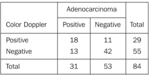
Correlating color Doppler findings with histopathological findings, we obtained 18 true-positive cases, 11 false-positive cases, 42 true-negative cases and 13 false-negative cases (Table 2).
Therefore, taking only color Doppler findings into consideration, sensitivity, specificity, positive and negative predictive values were, respectively, of 58.0%, 79.2%, 62.0% and 76.3%.
45.6% to 62.0%), but, on the other hand, there was a decrease in sensitivity (from 67.7% to 58.0%).
Correlating findings at gray-scale with color Doppler findings, we observed that, most frequently, US studies were normal (38 cases); hypoechoic nodes were found in 29 cases, of which 19 were associated with Doppler flow focal change.
Diffuse hypoechoic change with loss of zonal differentiation was seen in 19 patients, 12 of them presenting Doppler flow abnormality
In three patients, the presence of hypoechoic node and diffuse heterogeneity coexisted, although in different peripheral zones, in two of them both were hypervascular findings. In the other patient, two nodes (one hypervascular) coexisted, besides a hypoechoic heterogeneous area without increased vascularization.
Mean Gleason score was 6.9, and the most frequently value found was 7 (4 + 3), present in 14 of the patients (45.2% of patients with positive biopsy for adenocarcinoma).
There was no statistically significant difference between Gleason values for patients with suspect or unsuspected ultrasound at gray-scale (means of 6.71 and 6.67, respectively) or at color Doppler (means of 7.1 and 6.6, respectively) (p values > 0.05). The difference between Gleason score mean values was significant only when the group with normal ultrasound was compared with the group presenting diffuse heterogeneity with hypervascularization (p value < 0.05). Comparing the group with abnormalities at ultrasound, we observed that although patients presenting diffuse changes with hypervascularization had higher mean Gleason score, this difference is not statistically significant (p value > 0.05).
DISCUSSION
The prevalence of prostate adenocarcinoma histopathologically detected, considered in the present study as gold standard, was of 39.3%. This value is similar to others reported by studies presenting a similar method of patients recruitment in which the prevalence ranged between 40%(2) and 47,5%(10).
The prostate volume was higher, although at non-statistically significant rates, in patients with benign biopsies and this finding may be connected with the higher prevalence of benign hyperplasia whose prevalence increases with ageing(11).
On the other hand, this 4 ng/ml cutting level for normal PSA presented specificity as low as 17%, despite the non-statistically significance of the mean values between groups of patients with cancer and with histopathologically benign findings. This overlapping is observed because both the normal and the hyperplastic prostatic parenchymas affect the PSA levels, so patients with prostatic hyperplasia may present higher than normal PSA levels(5,13).
Gray scale ultrasound demonstrated a sensitivity of 67.7%, with positive predictive value of 45.6%, besides a specificity of 52.8%. These values are within the range reported by several studies where values for sensitivity ranged from 41%(2) to 96%(10), for specificity, from 27%(14) to 81%(2) and for positive predictive value, between 45%(15) and 53%(10).
On the other hand, color Doppler findings demonstrated sensitivity of 58.0%, positive predictive value of 62.0% and specificity of 79.2%. Again, values were similar to those of the literature, where the sensitivity ranged from 43.2%(2) to 86.6%(10), the positive predictive value from 40.8%(2) to 77%(10), and specificity from 38%(15) to 66.4%(2). One may observe that, in the present study, the specificity was higher than in the literature, since the association of methods in our study showed the color Doppler playing a more effective adjuvant role.
Correlating the gray-scale findings with the color Doppler findings, we observed that the finding more frequently associated with prostatic cancer was the peripheral zone node with associated focal hypervascularization in 12 patients. Only one patient with peripheral zone node and adenocarcinoma at biopsy presented a node without hypervascularization.
These findings are compatible with histopathological findings where prostatic tumors usually present as hypoechoic nodular areas due to their cellular arrangement distinct from the normal peripheral zone and that about 76% of nodular cancers are remarkably hypoechoic(16). Similarly, other studies demonstrate that prostatic cancers present lower microvasculature density, with vessels smaller in diameter than those observed in normal tissues (tumoral angiogenesis) and occupying larger area of tissue, appearing as hypervascularization at Doppler(17).
On the other hand, 17 patients with benign biopsy also presented hypoechoic node at ultrasound, eight of them presenting at Doppler as hypervascular nodes. Three of these patients with hypervascular nodes had a diagnosis of acute and/or chronic prostatitis and the other ones had a diagnosis of benign prostatic hyperplasia or normal histology. But, among patients with node without increased vascularization there was a case of infarct, one of nodular hyperplasia, one of chronic prostatitis and the other had normal results or benign prostatic hyperplasia.
Hypervascularization in a benign lesion also may be explained by histopathology, since an increase in the number of vessels with normal or enlarged dimensions is observed (although not similar to the vessels observed in the tumoral angiogenesis), increasing the percentage of lesion area occupied by vessels and appearing at Doppler as hypervascularization(17).
As regards the finding of diffuse heterogeneity, of 11 cases with diagnosis of adenocarcinoma, nine presented a diffuse increase in the vascularization of the region, while two patients presented normal flow in the prostate gland. On the other hand, of 9 patients with benign biopsy with this same finding, only 3 presented hypervascularization, the diagnosis being chronic prostatitis and benign hyperplasia. The other six patients with diffuse heterogeneity without hypervascularization had histological diagnosis of normality/benign hyperplasia.
The diffuse change of loss of zonal differentiation between central and peripheral zones was described by Ezz el Din & de la Rossette(6) as a specific finding for prostate cancer.
In this study, the positive predictive value of diffuse change at ultrasound for the diagnosis of cancer was of 57.9%. When we associate the positiveness at color Doppler with the change at gray-scale, the positive predictive value increased to 75%. On the other hand, the positive predictive value for a focal, nodular, hypoechoic lesion in the peripheral zone was of 41.4% in this study, increasing to 57.9% when such alteration was also positive at color Doppler. The diffuse change was a more specific ultrasound sign of prostatic carcinoma than the hypoechoic node.
In the two cases of isoechoic swelling, the color Doppler was negative and biopsy benign. This probably indicates that the finding was an asymmetry due to benign hyperplasia, or hyperplastic nodes which caused the peripheral zone swelling making it visually asymmetrical.
In ten of 31 patients, the prostate cancer was not evidenced by both methods, being diagnosed only by means of random biopsy. That is, 32.2% of adenocarcinomas in this study presented isoechoic to the adjacent prostatic parenchyma, according to the findings of Shinohara
et al.(9), who found 39% of tumors non-visualized at ultrasound.
In these patients, the diagnosis of cancer resulted from less than half of the fragments and, in all of them the onset was unilateral and there was no case of diffuse prostatic carcinoma. Additionally, in none of these cases perineural invasion was described.
On the other hand, the mean Gleason score for isoechoic cancers was of 6.67, one patient presenting score 8, although with only one fragment positive for adenocarcinoma, i.e., cancers not visible by ultrasound also are clinically significant (Gleason higher than 6), although apparently presenting as less extensive.
found by this method were clinically significant. Also, there is a partial correlation with the study of Shinohara et al.(9), who reported a significant difference between hypoechoic and isoechoic tumors, the first ones being less differentiated. This difference can be partially explained by the size of the tumoral focus, since Shinohara et al.(9) observed that only 18% of tumors with less than 10 mm were identified by ultrasound.
Geason scores of diffuse or nodular lesions (loss of differentiation) and of lesions with or without vascularization increase at color Doppler were not statistically significant, although hypervascular lesions present higher mean values than lesions that do not present any alteration at color Doppler. But, when compared with the group of patients with diagnosis only by biopsy (normal ultrasound), the difference was statistically significant, reflecting the tumors higher aggressiveness in these cases. As a matter of fact, the increase in microangiogenesis observed in cases of tumors(17) and the relation explained by Shinohara et al.(9) of the tumoral aggressiveness may justify this difference, although there is no report in the literature on the positiveness at color Doppler with higher Gleason score.
There was no case of relevant complication due to the biopsy procedure and there was no significant pain complaint that made the procedure impossible. The technique of biopsies performed in the present study consisted of biopsies directed to lesions evidenced by the examination besides, in average, eight to ten random bilateral biopsies fragments.
According to Hodge et al.(19), the utilization of random biopsies may increase in up to 20% the rate of prostatic cancer detection. Slonim et al.(18) have also observed an increase of 28% in the rate of cancer detection with utilization of random biopsy. In this study, 32.2% of malignancy results were diagnosed only through random biopsy.
Any way, the biopsy technique is not a consensus yet. Initially, Hodge et al.(19) established the use of six fragments (three in each lobe) bilaterally apical, median and basal. Later, several authors questioned its detection capacity, as Presti et al.(20) and Ravery et al.(21) saying that a higher number of biopsied fragments was necessary, while other authors like Naugthon et al.(22) have not observed a significant advantage in the increase of the number of fragments for cancer detection. The Sociedade Brasileira de Urologia (Brazilian Society of Urology), in a study in association with the Colégio Brasileiro de Radiologia (Brazilian College of Radiology), also recommend the extended sextant biopsy protocol (with 12 fragments) as the transrectal biopsy standard due its higher diagnostic capacity (23).
than 4 ng/ml associated with ultrasound low sensitivity and specificity, even if it is associated with color Doppler.
Any way, the ultrasound is useful since, besides random biopsies, it allows the performance of biopsies directed to echographic changes with higher positive predictive value for carcinoma.
REFERENCES
1. INCA – Instituto Nacional do Câncer. Câncer de próstata/Epidemiologia. Instituto Nacional do Câncer, 2003. Disponível em: URL:http//:www.inca.gov.br
2. Kuligowska E, Barish MA, Fenlon HM, Blake M. Predictors of prostate carcinoma: accuracy of gray scale and color Doppler US and serum markers. Radiology 2001;220:757–764.
3. Lee F Jr, Bronson JP, Lee F, et al. Nonpalpable cancer of the prostate: assessment with transrectal US. Radiology 1991;178:197–199.
4. Littrup PJ, Kane RA, Williams CR, et al. Determination of prostate volume with transrectal US for cancer screening. Part I: Comparison with prostate-specific antigen assays. Radiology 1991;178:537–542.
5. Stamey TA, Kabalin JH. Prostate specific antigen in the diagnosis and treatment of adenocarcinoma of the prostate. I. Untreated patients. J Urol 1989;141:1070–1075.
6. Ezz el Din K, de la Rosette JJMCH. Transrectal ultrasonography of the prostate. Br J Urol 1996;78:2–9.
7. Rifkin MD, Dähnert W, Kurtz AB. State of the art: Endorectal sonography of the prostate gland. AJR Am J Roentgenol 1990;154:691–700.
8. Rifkin MD, Choi H. Implications of small, peripheral hypoechoic lesions in endorectal US of the prostate. Radiology 1988;166:619–622.
9. Shinohara K, Wheeler TM, Scardino PT. The appearance of prostate cancer on transrectal ultrasonography: correlation of imaging and pathological examinations. J Urol 1989;142:76–82. 10. Kelly IMG, Lees WR, Rickards D. Prostate cancer and the role of color Doppler US. Radiology 1993;189:153–156.
11. Sagalowsky AI, Wilson JD. Hyperplasia and carcinoma of the prostate. In: Fauci AS, Braunwald E, Isselbacher KJ, editors. Harrison’s Principles of internal medicine. 14th ed. New York, NY: McGraw-Hill 1998;596–602.
12. Spencer JA, Alexander AA, Gomella L, Matteucci T, Goldberg BB. Clinical and US findings in prostate cancer: patients with normal prostate-specific antigen levels. Radiology 1993;189:389–393.
14. Rubens DJ, Gottlieb RH, Maldonado Jr CE, Frank IN. Clinical evaluation of prostate biopsy parameters: gland volume and elevated prostate-specific antigen level. Radiology 1996;199:159–163.
15. Lavoipierre AM, Snow RM, Frydenberg M, et al. Prostate cancer: role of color Doppler imaging in transrectal sonography. AJR Am J Roentgenol 1998;171:205–210.
16. Lee F, Torp-Pederssen S, Littrup PJ, et al. Hypoechoic lesions of the prostate: clinical relevance of tumor size, digital rectal examination, and prostate-specific antigen. Radiology 1989;170:29–32.
17. Louvar E, Littrup PJ, Goldstein A, Yu L, Sakr W, Grignon D. Correlation of color Doppler flow in the prostate with tissue microvascularity. Cancer 1998;83:135–140.
18. Slonim SM, Cuttino JT Jr, Johnson CJ, et al. Diagnosis of prostatic carcinoma: value of random transrectal sonographically guided biopsies. AJR Am J Roentgenol 1993;161:1003– 1006.
19. Hogde KK, McNeal JE, Terris MK, Stamey TA. Random systematic versus directed ultrasound guided transrectal core biopsies of the prostate. J Urol 1989;142:71–75.
20. Presti JC Jr, Chang JJ, Bhargava V, Shinohara K. The optimal systematic prostate biopsy scheme should include 8 rather 6 biopsies: results of a prospective clinical trial. J Urol 2000;163:163–167.
21. Ravery V, Goldblatt L, Royer B, Blanc E, Toublanc M, Boccon-Gibod L. Extensive biopsy protocol improves the detection rate of prostate cancer. J Urol 2000;164:393–396.
22. Naughton CK, Miller DC, Mager DE, Ornstein DK, Catalona WJ. A prospective randomized trial comparing 6 versus 12 prostate biopsy cores: impact on cancer detection. J Urol 2000;164: 388–392.
Tabelas e Quadros
Figure 2. Hypoechoic node in the right lobe peripheral zone (A). Increase in focal vascularization is not observed at color Doppler (B). An area of infarct is histopathologically observed.
A B
Figure 1. Normal aspect of the prostatic gland at ultrasound. Observe the homogeneous and more echogenic peripheral zone in relation to the central gland.
Internal gland
Peripheral zone
Chart 2 Correlation of color Doppler findings with histopathological findings. Color Doppler Positive Negative Total Positive 18 13 31 Negative 11 42 53 Total 29 55 84 Adenocarcinoma
Figure 3. Hypoechoic node in the right peripheral zone (A). A hypervascularization focus is observed in its interior at color Doppler (B). Biopsy revealed a Gleason 7 (4 + 3) adenocarcinoma.
A B
Figure 4. Hypoechoic node in the right peripheral zone, presenting permeated cystic area (A). At color Doppler, hypervascularization is observed in the periphery (solid part) of the node (B). Pathological study demonstrated Gleason 7 (4 + 3) adenocarcinoma with perineural infiltration.
Figure 5. Area of diffuse hypoechogenicityde affecting the whole prostate peripheral zone (A). At color Doppler (B) diffuse hypervascularization of the peripheral zone is observed. Biopsy evidenced Gleason 6 bilateral adenocarcinoma.
A B
A