ARTICLE
Vagus nerve stimulation may be a sound
therapeutic option in the treatment of
refractory epilepsy
Estimulação no nervo vago pode ser uma excelente opção no tratamento de epilepsias
refratárias
Murilo S. Meneses1, Samanta F. B. Rocha1, Cristiane Simão1, Heraldo Nei Hardt Laroca dos Santos1, Cleudi Pereira2, Pedro A. Kowacs1
he prevalence of epilepsy is approximately 1%, and be-tween 70 and 80% of these patients have good control of sei-zures with drug therapy1,2. However, the remaining 20 to 30%
who are refractory to antiepileptic drugs have an important burden regarding their labor, social and cognitive aspects as a result of their seizures, its etiology and sometimes the efects of their drug treatment2,3.
People with medically refractory epilepsy may be included in pre-operative protocols for epilepsy surgery, and as a result, to undergo microsurgical resection for curative treatment2-5.
Another group of patients can be treated surgically only with palliative surgery, such as those based on the disconnection of the epileptogenic process. hese surgeries are aimed at reduc-ing the number of attacks and at improvreduc-ing the quality of life3,6,7.
Vagus nerve stimulation (VNS) is a palliative therapy that has been shown to be efective not only in the treatment of epilepsy refractory to medical treatment but also the one re-fractory to surgical treatment8. It is a procedure less invasive
than disconnective surgeries, such as callosotomy and mul-tiple subpial transections3,6,7.
1Unidade de Cirurgia de Epilepsia, Instituto de Neurologia de Curitiba, Curitiba PR, Brazil; 2Politec Saúde, Curitiba PR, Brazil.
Correspondence: Murilo S. Meneses; Instituto de Neurologia de Curitiba; Rua Jeremias Maciel Perretto 300; 81210-310 Curitiba PR - Brasil; E-mail: [email protected]
Conflict of interest: Dr. Pedro A. Kowacs has given lectures on VNS for Cyberonics/Politec Saúde and has received traveling and meeting grants from Cyberonics/Politec Saúde. Cleudi Pereira works for Politec Saúde. Figure 1 was kindly made available by Cyberonics.
Received 17 January 2012; Received in inal form 01 October 2012; Accepted 08 October 2012
ABSTRACT
Introduction: Refractory epilepsy accounts for 20 to 30% of epilepsy cases and remains a challenge for neurologists. Vagus nerve stimulation (VNS) is an option for palliative treatment. Objective: It was to study the eficacy and tolerability of VNS in patients implanted with a stimula-tor at the Curitiba Institute of Neurology (INC). Methods: A case study of six patients with refracstimula-tory epilepsy submitted to a VNS procedure at the INC in the last four years was described and discussed. Results: Mean age at time of implantation was 29 years. Mean follow-up was 26.6 months. Seizure frequency decreased in all patients (40–50% (n=2) and ≥80% (n=4)). Three patients no longer required frequent hospitaliza-tions. Two patients previously restricted to wheelchairs started to walk, probably because of improved mood. Conclusion: In this population, VNS proved to be a sound therapeutic option for treating refractory epilepsy.
Key words: refractory epilepsy, epilepsy surgery, vagus nerve stimulation.
RESUMO
Introdução: Epilepsias refratárias compreendem de 20 a 30% dos casos de epilepsia e constituem desaio clínico. A neuroestimulação do nervo vago (VNS) é uma opção de tratamento paliativo. Objetivos: Foi estudar a eicácia e a tolerabilidade da VNS nos pacientes implantados no Instituto de Neurologia de Curitiba (INC). Métodos: Um estudo de casos de seis pacientes com epilepsia refratária, submetidos à VNS no INC em quatro anos, foi descrito e discutido. Resultados: A média de idade na implantação foi 29 anos. O seguimento médio foi 26,6 meses. A frequência de crises diminuiu em todos os pacientes (40–50% em um paciente e ≥80% em quatro). Três pacientes deixaram de internar fre-quentemente. Dois pacientes restritos a cadeiras de rodas começaram a andar, provavelmente por melhora de seu humor. Conclusão: Nesta população, a VNS provou ser uma excelente opção no tratamento de epilepsia refratária.
In order to study the results of VNS, the patients treated by this approach at the Institute of Neurology, Curitiba have been retrospectively analyzed, and the impact of the proce-dure on the seizures, the implantation proceproce-dure and the neurostimulator calibration procedures have been described in detail.
METHODS
Population
Between October 2007 and June 2012, six patients with generalized epilepsy refractory to medical treatment under-went VNS surgery at the Institute of Neurology, Curitiba. Of these, three were females and three males. he age ranged from 7 to 44 years old. hree had generalized epilepsy in the past and had undergone callosotomy, two had epilepsy with focal and generalized features and one had posterior tempo-ral lobe epilepsy. Patients that underwent callosotomy also showed focal irritative activity/epileptogenic foci. All pa-tients were refractory to drug treatment, three of which had
already undergone previous callosotomy. In ive cases, a vid-eo-electroencephalogram (video-EEG) was performed and showed generalized, focal or multifocal irritative or epilepto-genic activity. Table 1 presents these data.
Surgical procedure
he surgery is performed with the patient under general anesthesia and endotracheal intubation. Two small skin inci-sions of about 4 cm are performed. he irst one is on a hor-izontal fold of skin on the anterolateral surface of the neck, with a section of the platysma muscle, muscle dissection and exposure of the plans until the left vagus nerve, between the common carotid artery and internal jugular vein using an op-erating microscope. he electrode is wrapped around the va-gus nerve (Fig 1). A second incision is made below the clavicle, and the subcutaneous space is opened to place the generator, which is connected subcutaneously to the electrode already placed around the vagus nerve. he system is tested through telemetry to verify the operation of the generator. Finally, the incisions are closed in two layers after assessing hemostasis, and dry dressings are placed over the surgical wounds.
Table 1. General data related to vagus nerve stimulation treated patients.
Gender/ Age (years)
Age at implantation
(years)
Clinical condition Irritative/ epileptogenic zone Neuroimaging findings Time since implantation Efficacy
(patients’ rating)Adverse events
#1 M, 21
21 Multifocal epilepsy mild cognitive impairment Generalized irritative activity, left occipital rolandic epileptogenic zone Bilateral temporal arachnoid cysts, supratentorial asymmetric ventricular dilation Four years 80% decrease in seizure frequency Transient cough, precordialgia, irritability
#2 F, 27
27
Epilepsy since the irst year of life, multifocal, mild cognitive impairment, previous callosotomy Right rolandic irritative zones, indeinable epileptogenic zone Anterior 2/3 callosotomy Three years and nine months 80% decrease in seizure frequency Transient anxiety
#3 M, 50
50
Generalized epilepsy since the irst year of life, tetraparesis, marked cognitive impairment, previous callosotomy Generalized irritative activity, with anterior temporal focal paroxysms in F7
Volumetric reduction of the brain, right frontal encephalomalacia, anterior 2/3 callosotomy Eight months (later explanted) 50% decrease in seizure frequency, mood improvement, started walking again Surgical wound infection
#4 F, 29
29
Seizures onset within the irst year
of life, previous callosotomy, right crural monoparesis, mild cognitive impairment Multifocal neocortical irritative activity and epileptogenic zone Volumetric reduction of the left brain hemisphere, anterior 2/3 callosotomy, left fronto-parieto-temporo-occipital gliosis Three years and one month
80% decrease in seizure frequency, mood improvement, started walking again Transient hoarseness
#5 F, 41
41
Neocortical epilepsies of the left temporal lobe, mild verbal abilities
compromise Left temporal irritative activity, left postero-temporal epileptogenic zone
Frontal horn cystic formation of the left lateral ventricle
One year and four months 40% decrease in seizure frequency Transient cough, chest pain; calibration-dependent dyspnea
#6, M, 7 6
Multifocal epilepsy, severe cognitive impairment Generalized and multifocal irritative activity No signiicant
indings Six months
90% decrease in seizure frequency
Programming the neurostimulator
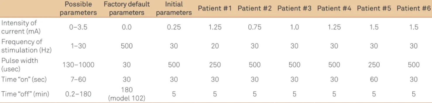
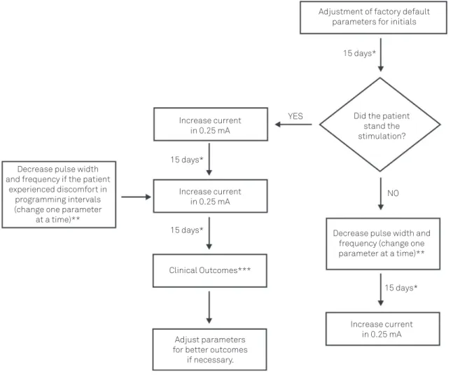
Stimulation has been performed in all cases in the left vagus nerve. he stimulation protocol consisted of activat-ing the neurostimulator just 15 days after the implantation procedure. he initial parameters and current neurostimula-tion parameters are shown in Table 2. Adjustments in neuro-stimulation parameters have been made in monthly visits in the irst six months of follow-up. he default current intensity is 0 mA, set to 0.25-mA increases at every next visit. he de-fault stimulus frequency is 20 to 30 Hz, with a pulse width of 250 to 500 usec current, on-time (time on) of 30 seconds and wait time (time of) of ive minutes. hese parameters can be reduced in case of lack of tolerability to stimulation. he re-lationship between the stimulus frequency, intensity, time-on and time-of is optimized over time. Fig 2 brings a lowchart for setting and adjusting the neurostimulator.
RESULTS
Mean follow-up was 26.6 months. Seizure frequency de-creased in all patients. One patient reported a 40–50% reduc-tion in seizure frequency (patients #3 and #5), and the re-maining four patients estimated an 80% or greater reduction in seizure frequency (n=4). hree patients no longer required
Vagus Nerve Tie-Downs
Anchor Tether
Strain Relief Bend Lead
3 cm (1.18 in.)
1 cm (.39 in.)
Strain Relief Loop Coled Extra Lead
Helical Electrodes
Fig 1. Schematic drawing of a vagus nerve stimulation implant.
Table 2. Data related to the vagus nerve stimulation treated neurostimulator calibration. Possible
parameters
Factory default parameters
Initial
parameters Patient #1 Patient #2 Patient #3 Patient #4 Patient #5 Patient #6
Intensity of
current (mA) 0–3.5 0.0 0.25 1.25 0.75 1.0 1.25 1.5 1.5 Frequency of
stimulation (Hz) 1–30 500 30 20 30 30 30 30 30
Pulse width
(usec) 130–1000 30 500 250 500 500 500 250 500
Time “on” (sec) 7–60 30 30 30 30 30 30 60 30
Time “off” (min) 0.2–180 180
(model 102) 5 5 5 5 5 5 5
frequent hospitalizations. In one of the patients (#5), the re-duction in seizure frequency occurred after her irst year of follow-up. Two patients previously restricted to wheel-chairs started to walk, probably because of improved mood. One system was explanted because of infection (patient #3), but otherwise no other relevant adverse event was seen. Individual follow-up time, eicacy data and adverse events are summed up in Table 1.
DISCUSSION
Effectiveness of VNS
When refractory to medical treatment, symptomatic gen-eralized epilepsy or those epilepsies with secondary bilater-al synchrony are associated with high morbidity. In addition, these patients usually are not candidates for curative resec-tive epilepsy surgery3,6,7. Surgical options include palliative
callosotomy and VNS3,6,7. Callosotomy is a rather aggressive
surgery, but leads to a satisfactory control of tonic-clonic and atonic seizures. Callosotomy leads to a decrease in the fre-quency and severity of epilepsy seizures in a bracket between 40 and 70%. Procedural complications include disconnection syndrome, infection, mutism, hemorrhage and focal neuro-logical deicits6,7. VNS is performed without intracranial
sur-gery, with a minimally invasive technique. According to Nei et al.9, who compared 50 epileptic patients who underwent
cal-losotomy to 21 epileptic patients who underwent VNS, the reduction of tonic and atonic seizures was similar for all sei-zure types, including partial and generalized, but there was a greater reduction in callosotomy. However, there was one death among patients submitted to callosotomy, illustrating the greater morbidity of this procedure as compared to VNS. In another study of 16 adults with medically refractory gener-alized epilepsy, a 43.3% average decrease in seizure frequency has been observed after treatment with VNS10.
he reduction in seizures in three of our patients who had previously undergone callosotomy mirrors that of the avail-able literature. Elliot et al.11 evaluated 110 patients who had
did not afect VNS outcome. Our data add to the previous information and brings thoughts on the possibility of VNS implant prior to surgical procedures, as already suggested by other authors12.
In addition to patients with symptomatic generalized ep-ilepsy, those experiencing idiopathic generalized epilepsies may also respond to the VNS, as reported by Kostov et al.13,
who reported an average 61% decrease in seizure frequency in 12 of these patients undergoing VNS, with concomitant re-duction in the medication they had been on.
Perhaps, the great lesson that we have learned from our cases comes from patient #5, in whom calibration of titration was delayed by the onset of dyspnea, but later on, when pos-sible, led to an improvement in seizure frequency and sever-ity, as would be expected by the current literature, which has reported a greater eicacy in patients with partial epilepsy8.
In addition to drug or surgical refractoriness, there are other indications for VNS. Patwardhan et al.14 described
the case of a 30-year old patient in whom phenytoin,
valproic acid, topiramate and carbamazepine were with-drawn due to Stevens-Johnson syndrome, who went into status epilepticus. After nine days of barbiturate coma, the patient underwent VNS implantation, which allowed total seizure control for 19 days. The association of VNS, phenobarbital and levetiracetam allowed the patient to be discharged seizure-free. Indeed, another case of status epi-lepticus treated with VNS had been previously described in a 13-year old patient15.
An indirect measure of efficacy is the lower utilization of health services by these patients16. We observed this
phenomenon in our population: our patient #1 had a 70% decrease in the frequency of his visits; patients #1 and #2, who were usually admitted to the hospital because of re-current seizures (one to several episodes of status epilep-ticus), no longer require hospital care. After implantation and calibration of the VNS, our patients #2 and #4 stopped attending scheduled visits and attended follow-up visits only when summoned.
* Programming intervals can vary according to tolerability during the rump up.
** Pulse width and frequency can be reset to the previous values depending on tolerability. *** Clinical outcomes are evaluated at each visit, and programming intervals can be increased.
Fig 2. Neurostimulator Programming Flowchart
Adjustment of factory default parameters for initials
15 days*
15 days*
15 days*
15 days* Did the patient
stand the stimulation?
Increase current in 0.25 mA Increase current
in 0.25 mA
Increase current in 0.25 mA Clinical Outcomes***
Decrease pulse width and frequency (change one parameter at a time)**
Adjust parameters for better outcomes
if necessary. Decrease pulse width
and frequency if the patient experienced discomfort in
programming intervals (change one parameter
at a time)**
YES
Other effects of VNS
Another aspect of VNS therapy is the improvement in mood, cognition and quality of life17,18, in which our
sam-ple, though small, echoes the literature. Of our patients, two showed, in addition to the reduction in seizures, a marked antidepressant efect, and, despite being previously bound to wheelchairs, started doing eforts to walk again after VNS. However, two of our patients showed a slight increase in anx-iety-related complaints, which we attribute to increased au-tonomy and greater exposure to daily life situations.
Neurostimulator calibration
Heck et al. described in detail the recommendations for neurostimulation equipment calibration19. he same group
examined 154 patients in diferent calibration parameters, such as pulse duration, frequency, time online, time of and output current, and the relationship of these param-eters with the reduction in the number of seizures20. In their
study, there were no evident changes in the reduction of seizures with diferent parameters, however, in a subgroup with time of ≤1.1 minute, the average reduction changed signiicantly from 21 to 39%.
Our strategy is to change one parameter at a time to monitor through the response which particular change is more appropriate.
he interval between the adjustments is of 15 days until a set of parameters is reached, when the patient begins to show clinical responses. We found that when approaching a current intensity close to 1.5 mA (1.0–1.5 mA), it is desirable to increase the interval between the adjustments to allow a better observation of the clinical response to the new param-eters. Diferent patients may respond better or worse with more or less current; there is not a standard current for all patients. It is important to note that excessively large inter-vals may bring unnecessary delays in seizure control. During the visits, the patient should be observed for their seizures, as well as in issues related to their quality of life, such as behav-ior, way of relating, mood and concentration, among others.
Adverse events
Ben-Menachem21 summed-up side-efects of VNS
stimu-lation in some studies and divided them in early complica-tions of VNS implantation and in side-efects seen in long-term VNS stimulation. Among the early complications of VNS implantation, they described infections (3 to 6%), iatro-genic and reversible vocal cord paralysis, lower facial weak-ness, bradycardia and asystole (0.1%). Side-efects seem in 3 months, 12 months and 5 years, were, respectively: cough (21, 15 and 1.5%); voice alteration (62, 55 and 18.7%); dyspnea (16, 13 and 2.3%); pain (17, 15 and 4.7%); paresthesia (25, 15 and
1.5%); headache (20, 16 and 0%); pharyngitis ( 9, 10 and 0%); depression (3, 5 and 0%); infection (4, 6 and 0%) and death (0, 6 and 6.2%). he two deaths reported in the 12-month fol-low-up study were due to sudden unexpected death (SUDEP) and pneumonia, and the four deaths reported in the ive-year follow-up study were due to SUDEP (n=1) and status epilep-tics (n=3). A larger study on SUDEP in VNS concluded that SUDEP rates dropped to 1.7 per 1,000 after two years, a rate lower than that reported for similar groups not treated with VNS22. Sleep apnea and excessive daytime sleepiness were
described in a 21-year old patient who had obtained a 50% re-duction of seizures after surgery23. Cough, dyspnea and
laryn-geal irritation are known side efects of VNS, however respira-tory changes during sleep were poorly described in literature. Malow et al. studied four epileptic patients with previous ob-structive sleep apnea who have been treated with VNS and concluded that these symptoms may worsen24. To eliminate
this problem, the authors suggest reducing the frequency of the stimulus without changing the other parameters.
Although none of our patients have presented apnea, patients #1, #4 and #5 had transient cough or dysphonia at each calibration, and patient #5 presented dyspnea, which could be circumvented by a slower neurostimulation cali-bration parameters.
Post-operative infections can occur between 3–6%, but most are treated with oral antibiotics, and it is rarely neces-sary to remove the generator or the electrodes25. Patient #4,
despite good reduction in seizure frequency and indepen-dence gain, required the device explantation due to infec-tion. his patient had cognitive impairment, a condition that may have contributed to the contamination of the surgical wound. An irrigation system with vancomycin and Ringer lactate solution was described by Liechty to prevent the re-moval of the generator25.
In conclusion, our data set echoes and adds to previous experience, showing signiicant gains in terms of reduction in seizure frequency, decreased use of health services, au-tonomy and quality of life. Adverse events related to neuro-stimulation were transient and circumvented by the tem-porary reduction in neurostimulation parameters. In one case, the explantation of the neurostimulator was required due to local infection. he set of outcomes shows that VNS treatment had a positive impact on seizure control and in the lives of our patients.
ACKNOWLEDGEMENTS
1. Kwan P, Brodie MJ. Early identiication of refractory epilepsy. N Engl J Med 2000;342:314-319.
2. Kwan P, Sperling MR. Refractory seizures: try additional antiepileptic drugs (after two have failed) or go directly to early surgery evaluation? Epilepsia 2009;50:S57-S62.
3. Benbadis SR, Tatum WO, Vale FL. When drugs don’t work: an algorithmic approach to medically intractable epilepsy. Neurology 2000;55:1780-1784.
4. Meneses MS, Rocha SB, Kowacs PA, et al. Tratamento cirúrgico da epilepsia do lobo temporal: análise de 43 casos consecutivos. Arq Neuropsiquiatr 2005;63:618-624.
5. Meneses MS, Hertz A, Gruetzmacher C, et al. Epilepsia e desordens de malformação do desenvolvimento cortical. J Epilepsy Clin Neurophysiol 2006;12:149-154.
6. McInermey J, Siegel AM, Nordgren RE, et al. Long-term seizure outcome following corpus callosotomy in children. Stereotact Funct Neurosurg 1999;73:79-83.
7. Spencer SS, Spencer DD, Williamson PD, et al. Corpus callosotomy for epilepsy. I: seizure effects. Neurology 1988;38:19-24.
8. Elliott RE, Morsi A, Kalhorn SP, et al. Vagus nerve stimulation in 436 consecutive patients with treatment-resistant epilepsy: long-term outcomes and predictors of response. Epilepsy Behav 2011;20:57-63.
9. Nei M, O’Connor M, Liporace J, Sperling MR. Refractory generalized seizures: response to corpus callosotomy and vagal nerve stimulation. Epilepsia 2006;47:115-122.
10. Holmes MD, Silbergeld DL, Drouhard D, Wilensky AJ, Ojeman LM. Effect of vagus nerve stimulation on adults with pharmacoresistant generalized epilepsy. Seizure 2004;13:340-345.
11. Elliott RE, Morsi A, Geller EB, Carlson CC, Devinsky O, Doyle WK. Impact of failed intracranial epilepsy surgery on the effectiveness of subsequent vagus nerve stimulation. Neurosurgery 2011;69: 1210-1217.
12. Renfroe JB, Wheless JW. Earlier use of adjunctive vagus nerve stimulation therapy for refractory epilepsy. Neurology 2002;59:S26-S30.
13. Kostov H, Larsson PG, Roste GK. Is vagus nerve stimulation a
treatment option for patients with drug-resistant idiopathic generalized epilepsy? Acta Neurol Scand 2007;187:55-58.
14. Patwardhan RV, Dellabadia J Jr, Rashidi M, Grier L, Nanda A. Control of refractory status epilepticus precipitated by anticonvulsant withdrawal using left vagal nerve stimulation: a case report. Surg Neurol 2005;64:170-173.
15. Winston KR, Levisohn P, Miller BR, Freeman J. Vagal nerve stimulation for status epilepticus. Pediatr Neurosurg 2001;34:190-192.
16. Bernstein AL, Hess T. Vagus nerve stimulation therapy for pharmacoresistant epilepsy: effect on health care utilization. Epilepsy Behav 2007;10:134-137.
17. Nemeroff CB, Mayberg HS, Krahl SE, et al. VNS therapy in treatment-resistant depression: clinical evidence and putative neurobiological mechanisms. Neuropsychopharmacology 2006;31:1345-1355.
18. Dodrill CB, Morris GL. Effects of vagal nerve stimulation on cognition and quality of life in epilepsy. Epilepsy Behav 2001;2:46-53.
19. Heck C, Helmers SL, DeGiorgio CM. Vagus nerve stimulation therapy, epilepsy, and device parameters: scientiic basis and recommendations for use. Neurology 2002;59:S31-S37.
20. DeGiorgio CM, Thompson J, Lewis P, et al. Vagus nerve stimulation: analysis of device parameters in 154 patients during the long-term XE5 study. Epilepsia 2001;42:1017-1020.
21. Ben-Menachem E. Vagus nerve stimulation, side effects, and long-term safety. J Clin Neurophysiol 2001;18:415-418.
22. Annegers JF, Coan SP, Hauser WA, Leestma J. Epilepsy, vagal nerve stimulation by the NCP system, all-cause mortality, and sudden, unexpected, unexplained death. Epilepsia 2000;41:549-553.
23. Holmes MD, Chang M, Kapur V. Sleep apnea and excessive daytime somnolence induced by vagal nerve stimulation. Neurology 2003;61:1126-1129.
24. Malow BA, Edwards J, Marzec M, Sagher O, Fromes G. Effects of vagus nerve stimulation on respiration during sleep: a pilot study. Neurology 2000;55:1450-1454.
25. Liechty PG, Tubbs RS, Blount JP. The use of a sump antibiotic irrigation system to save infected hardware in a patient with a vagal nerve stimulator: technical note. Surg Neurol 2006;65:48-50.