ARTICLE
Pleomorphic xanthoastrocytoma: magnetic
resonance imaging findings in a series of
cases with histopathological confirmation
Xantoastrocitoma pleomórfico: achados de ressonância magnética numa série de casos
com confirmação histopatológica
Vinícius Trindade Gonçalves1, Fabiano Reis2, Luciano de Souza Queiroz3, Marcondes França Jr4
Pleomorphic xanthoastrocytoma (PXA), classically described as a superficial supratentorial glioma that af-fects young patients, is associated with extensive menin-geal involvement1. Moreover, it occurs in patients with a
long history of epilepsy. Despite their histologic appear-ance of cellular pleomorphism and the presence of giant cells, literature reviews indicate a good prognosis for this tumor2-7. It is a grade II tumor, according to the World
Health Organization (WHO) classification of tumors of the central nervous system (CNS)8. However, PXA is
as-sociated with high rates of recurrence, anaplastic trans-formation9-11 and death12 when comparing with other
as-trocytic tumors of good prognosis. This study aimed to analyze magnetic resonance imaging (MRI) characteris-tics in a series of patients diagnosed with pleomorphic xanthoastrocytoma in a university hospital.
Study carried out at Departments of Radiology, Pathology and Neurology, Faculty of Medical Sciences, State University of Campinas (UNICAMP), Campinas SP, Brazil. 1Medical Student, School of Medical Sciences, State University of Campinas (UNICAMP), Campinas SP, Brazil;
2MD, PhD, Professor of Department of Radiology, Clinics Hospital, UNICAMP, Campinas SP, Brazil;
3MD, PhD, Professor of Department of Pathology, Clinics Hospital, UNICAMP, Campinas SP, Brazil;
4MD, PhD, Professor of Department of Neurology, Clinics Hospital, UNICAMP, Campinas SP, Brazil.
Correspondence: Fabiano Reis; Departamento de Radiologia, UNICAMP, Cidade Universitária Zeferino Vaz; Rua Tessália Vieira de Camargo 126 / Caixa Postal; 13083-887, Campinas, SP - Brasil; Email: [email protected]
Support: State of São Paulo Research Foundation (FAPESP), Process nº 2010/11378-6. Conflict of interest: There is no conlict of interest to declare.
Received 02 May 2012; Received in inal form 23 June 2012; Accepted 02 August 2012
ABSTRACT
Pleomorphic xanthoastrocytoma (PXA) is a rare glioma. This paper aimed to analyze magnetic resonance imaging (MRI) characteristics in a series of patients diagnosed with PXA. We analyzed MRI indings in 9 patients with histopathologic diagnosis of PXA in our department over the last 12 years. The mean age of patients was 27.3 years. Cortical location was observed in all cases. The lesion imaging was solid-cystic in six cases. In eight cases, the solid component presented hypo or isointense on T1 and iso or hyperintense on T2. Contrast enhancement in the solid component was observed in eight cases. The observed imaging pattern of PXA was supericial location with leptomeningeal involvement, solid-cystic pattern and contrast enhancement in the solid component. We should consider that the association between PXA and other corti-cal tumors may occur, particularly, with gangliogliomas, which tend to be the main differential diagnosis in MRI.
Key words: magnetic resonance imaging, central nervous system, astrocytoma.
RESUMO
Xantoastrocitoma pleomórico (PXA) é um glioma raro. Este estudo teve como objetivo analisar aspectos de imagem por ressonância magnética (RM) de uma série de pacientes com diagnóstico de PXA. Foram analisados exames de RM de 9 pacientes com diagnóstico histopatológico de PXA nos últimos 12 anos. A média de idade dos pacientes foi de 27,3 anos. Localização cortical foi observada em todos os casos. Padrão sólido-cístico foi observado em seis casos. Em oito casos, o componente sólido apresentou-se hipo ou isointenso em T1 e iso ou hiperintenso em T2. Foi observada captação de contraste na porção sólida em oito casos. O padrão de imagem observado do PXA foi de localização supericial com envolvimento lepto-meníngeo, padrão sólido-cístico e captação de contraste pelo componente sólido. Devemos considerar que a associação entre PXA e outros tumores corticais pode ocorrer, particularmente, com ganglioglioma, que tende a ser o principal diagnóstico diferencial em RM.
METHODS
Between January 1999 and December 2010, we obtained MRI indings of all patients with histopathologic diagnosis of PXA in our department. he sample comprised 9 patients (6 males and 3 females) and the age varied between 7 and 63 years (mean age=27.33). MRIs were performed using a 2T scanner (Elscint Prestige®
, Haifa, Israel), with T1 and T2 acquisitions in three or-thogonal planes, including T1-weighted SE gadolinium-enhanced images. MRI acquisition parameters were: sagittal T1 spin echo, 6 mm thick, 180° lip angle; repetition time (TR)=430 millisec-onds, echo time (TE)=12 millisecmillisec-onds, matrix 200×350, ield of view (FOV)=25×25 cm; T2-weighted and proton density “fast spin echo” (FSE), 3 mm thick, 160° lip angle; TR=4.800 milliseconds, TE=108/18 milliseconds, matrix 256×256, FOV=22×22 cm; Axial T1-weighted spin echo (SE): TR=540 milliseconds, TE=28 milli-seconds; axial T2-weighted luid-attenuated inversion recovery (FLAIR) images TR=8.500 milliseconds and 2.000 or 100 millisec-onds, and 2.200 millisecmillisec-onds, TE=72 or 90 millisecmillisec-onds, matrix of 256×296 and FOV of 22×22 cm. T1-weighted SE gadolinium-en-hanced images were obtained in three orthogonal planes.
All patients underwent surgical biopsy or tumor excision, according to clinical indication. he surgical specimens were processed for routine histopathology, and the classiication of tumor type was performed following the World Health Organization (WHO) guidelines.
MRI iles were evaluated by one neuroradiologist (FR). We analyzed the following variables: tumor location, signal on T1 and T2-weighted images, contrast enhancement, ede-ma and association with other tumors in histological exami-nation. he project was submitted to the Ethics Committee of our service, which approved the research protocol (pro-cess nº 0722.0.146.000-10, approved Protocol nº 928/2010). Statistical analysis was performed with assessment of the statistics department of our service.
RESULTS
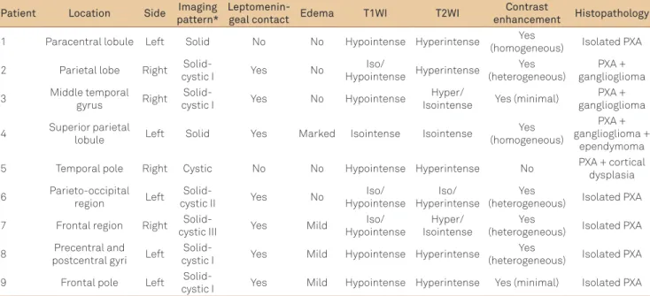
Tables 1 and 2 summarize patient’s data. Patients ages ranged between 7 and 63 years (mean age=27.33 years; me-dian=28.0; standard deviation=19.072). All patients were
Table 1. Clinical data.
Patient Gender Age Symptom and clinical examination Time of
symptom Recurrence (time) – follow-up
1 M 7 Generalized tonic-clonic seizures 3 years
-2 F 28 Seizures 13 years No – 3 yrs
3 F 31 Partial epilepsy, altered alertness and blurred vision 8 years No – 2 yrs
4 F 29 Seizures 2 weeks Yes (10 yrs) – 15 yrs
5 M 15 Seizures 9 years No – 6 yrs
6 M 7 Simple partial epilepsy and left parietal headache - -7 M 63 Confusion, behavior changes, apathy and depression 1 month No – Death (1 mo) 8 M 16 Generalized tonic-clonic seizures 1 year No – 10 yrs
9 M 50 Confusion and syncope 3 months No – 9 yrs
M: masculine; F: feminine; yrs: years; mo: month.
Table 2. Imaging data.
Patient Location Side Imaging pattern*
Leptomenin-geal contact Edema T1WI T2WI
Contrast
enhancement Histopathology
1 Paracentral lobule Left Solid No No Hypointense Hyperintense Yes
(homogeneous) Isolated PXA
2 Parietal lobe Right
Solid-cystic I Yes No
Iso/
Hypointense Hyperintense
Yes (heterogeneous)
PXA + ganglioglioma
3 Middle temporal gyrus Right
Solid-cystic I Yes No Hypointense
Hyper/
Isointense Yes (minimal)
PXA + ganglioglioma
4 Superior parietal
lobule Left Solid Yes Marked Isointense Isointense
Yes (homogeneous)
PXA + ganglioglioma +
ependymoma
5 Temporal pole Right Cystic No No Hypointense Hyperintense No PXA + cortical dysplasia
6 Parieto-occipital region Left
Solid-cystic II Yes No
Iso/ Hypointense
Iso/ Hyperintense
Yes
(heterogeneous) Isolated PXA
7 Frontal region Right
Solid-cystic III Yes Mild
Iso/ Hypointense
Hyper/ Isointense
Yes
(heterogeneous) Isolated PXA
8 Precentral and postcentral gyri Left
Solid-cystic I Yes Mild Hypointense Hyperintense
Yes
(heterogeneous) Isolated PXA
9 Frontal pole Left
symptomatic at diagnosis, and the time of symptom presen-tation was higher than three years in four of them. Seven pa-tients presented history of epilepsy and the other two, mental confusion. Cortical location was observed in all nine cases. More than one lobe was afected in three patients. he pari-etal lobe was the most afected ( ive cases), followed by fron-tal ( four) and temporal lobes (two). he lesion imaging was solid-cystic in six cases. hree imaging patterns were difer-entiated: irst, a cystic mass containing a mural nodule ( four cases); second, a predominantly solid mass that showed cys-tic changes (one); and third, a mixed pattern (one). On T1-weighted images, the solid component presented hypo– or isointense in all nine cases. While on T2-weighted images, the solid component presented isointense in four cases and hyperintense in seven. Leptomeningeal involvement was ob-served in seven cases, and contrast enhancement in the solid component was observed in eight cases. Peritumoral edema was observed in four cases and its magnitude was marked (one) or mild (three). In three cases, PXA was associated with other cortical tumors in histopathology. Ganglioglioma was associated with PXA in two cases and with ependymoma and ganglioglioma in another one. Furthermore, cortical dyspla-sia was associated with PXA in one case. Seven out of nine patients were followed up, one of whom had tumor recur-rence (in the 10th postoperative year) and one died one month
after surgery. he other ive patients are still alive, well and clinically free from recurrence at diferent follow-up periods, ranging from two to ten years.
Statistical analysis showed that the presence of edema was associated with a symptom duration of less than one year prior to diagnosis (p<0.03) by Fisher’s exact test. Older age was associated with atypical clinical presentation (p<0.05) by Mann-Whitney test. No association was found between im-aging patterns and clinical data or histological examination.
DISCUSSION
In 1979, Kepes et al.1 described for the irst time a series
of 12 young patients presenting with a distinctive form of as-trocytoma. In all cases, the tumor was supratentorial and su-pericial, with extensive leptomeningeal involvement and, despite pleomorphism and bizarre giant cells in the micro-scopic picture, the prognosis was relatively favorable. Since this study, over 200 cases of PXA have been reported, most as single cases or small series13.
Typical clinical presentation includes a long history of epilepsy, especially in young patients, most commonly in the second decade of life9. Although 7 patients have
present-ed seizures in our study and 6 of them were younger than 30 years, we had two patients with mental confusion as initial symptom, who were the oldest patients of our series (50 and 63 years). In our sample, the atypical clinical presentation
was signiicantly associated with older age (p<0.05) by Mann-Whitney test. According to our knowledge, there are few re-ports of elderly patients with PXA in the literature; howev-er, Ng et al.14 suggest that elderly patients with PXA may have
a poor prognosis, considering age as an independent risk fac-tor. In our sample, the oldest patient had a bad prognosis and died one month after surgery (Fig 1).
he most common single location is the temporal lobe, followed by parietal and occipital lobes. Moreover, out of the tumors that involve more than one lobe, the contiguous temporal lobe is the most afected9. In our study, temporal,
parietal and frontal lobes were equally afected (two cases each lobe), and, of the three tumors that involve more than one lobe, the parietal lobe was the most afected (three cases), followed by frontal lobe (two cases).
Despite its classic cortical location, PXA is rarely seen in the thalamus, cerebellum, spinal cord or within the eye15-19.
Two cases of intraventricular PXA were recently described20,21.
On MRI, the solid component of PXA is predominantly isointense on T1-weighted images and mildly hyperintense on T2-weighted images, enhancing intensely after intrave-nous contrast administration. Surrounding edema usually is minimal13. In our study, the solid component was hypo —
or hyperintense in eight cases on T1-weighted images and iso — or hyperintense on T2-weighted images in eight cas-es. Contrast enhancement was minimal (two), homoge-neous (three) or heterogehomoge-neous (three). Peritumoral edema
Fig 1. Male, 63 years old. (A) Contrast-enhanced axial T2-weighted magnetic resonance (MR) image shows a frontal expansive solid-cystic lesion, with lobulated delineation. Cystic component shows hyperintensity. Contrast-enhanced axial T1-weighted MR image (B), coronal (C) and sagittal (D) shows enhancement in the solid component and at the periphery of the cystic component.
A
B
was observed in four cases, being predominantly mild. hus, these imaging indings corroborate a typical imaging pattern as described in the literature.
In the four cases in which edema was present in our sam-ple, the duration of symptoms to diagnosis was less than or equal to one year, being signiicantly diferent from the other ive cases, with greater time of evolution (p<0.03) by Fisher’s exact test. his inding may suggest that the presence of edema on PXA tumors may be associated with early tumor growth. Logically, the sample size does not allow generaliza-tions, so further studies are essential.
In a histopathological study, PXA was associated with oth-er tumors in three cases, two with ganglioglioma and anothoth-er with ependymoma and ganglioglioma (Fig 2). Moreover, an-other case of PXA showed association with cortical dysplasia (Fig 3). Furuta et al.22 proposed that PXA and ganglioglioma
grow from a migration failure, resulting in an ectopic posi-tion of neuronal and glial cells. hat would explain the fact that PXA coexists in some cases with ganglioglioma or other cortical tumors. Furthermore, Lach et al.23 presented three
cases of cortical dysplasia associated with PXA that suggest a possible preneoplastic role of cortical dysplasia in the sub-sequent development of PXA.
In conclusion, although this sample comprises only nine patients, most reports of PXA have included only a single case or small series. MRI indings in this series corroborated a typ-ical description of PXA. he observed imaging pattern of PXA was supericial location with leptomeningeal involvement, solid-cystic pattern and contrast enhancement in the solid component. On T1-weighted images, the solid component presented hypo — or isointense and, on T2-weighted images, it was iso — or hyperintense. In our sample, when edema was present, the duration of symptoms to diagnosis was less than
Fig 2. Female, 29 years old. (A) Contrast-enhanced axial T1-weighted magnetic resonance (MR) image shows
expansive cortical lesion in the left parietal lobe, with intense enhancement. (B) Axial T2-weighted MR image shows lesion predominantly isointense and moderate edema. Anatomopathology: Mixed tumor, with PXA, ganglioglioma and ependymoma components.
A
B
Fig 3. Male, 15 years old. (A) Coronal T1-weighted magnetic resonance (MR) image shows a hypointense subcortical lesion and (B) Coronal T2-weighed MR image shows a hyperintense lesion. (C) Area of cortical dysplasia in right temporal lobe with incidental inding of a small pleomorphic xanthoastrocytoma, appearing as a well delimited, basophilic intracortical nodule. HE, X 10. (D) Pleomorphic xanthoastrocytoma is characterized by atypical rounded or fusiform tumor cells, some of which were multinucleated (center). HE, X 100. (E) Pleomorphic tumor cells with cytoplasmic positivity for GFAP, conirming their astrocytic lineage. Immunohistochemistry for GFAP, X 200. (F) Occasional cells displayed cytoplasmic xanthomatous change. Immunohistochemistry for vimentin, X 200. (G) Dysplastic area of cerebral cortex near the xanthoastrocytoma showing a binucleated neuron. HE, X 400. (H) Same area, gliosis of molecular layer of cerebral cortex (top). Immunohistochemistry for GFAP, X 40.
A
B
C
D
E
F
G
H
1. Kepes JJ, Rubinstein U, Eng LF. Pleomorphic xanthoastrocytoma: a distinctive meningocerebral glioma of young subjects with relatively favourable prognosis. A study of 12 cases. Cancer 1979;44:1839-1852.
2. Russel DS, Rubinstein LJ. Pathology of tumours of the nervous system. 5ª ed. Baltimore: Sans Tache; 1989.
3. Kepes JJ. Pleomorphic xanthoastrocytoma: the birth of a diagnosis and a concept. Brain Pathol 1993;3:269-274.
4. Tien RD, Cardenas CA, Rajagopalan S. Pleomorphic xanthoastrocytoma of the brain: MR indings in six patients. AJR 1992;159:1287-1290
5. Fouladi M, Jenkins J, Burger P, et al. Pleomorphic xanthoastrocytoma: favorable outcome after complete surgical resection. Neuro Oncology 2001;3:184-192.
6. Palma L, Maleci A, Lorenzo ND, Lauro GM. Pleomorphic xanthoastrocytoma with 18-year survival. J Neurosurg 1985;63:808-810.
7. Tan TC, Ho LC, Yu CP, Cheung FC. Pleomorphic xanthoastrocytoma: report of two cases and review of the prognostic factors. J Clin Neurosci 2004;11:203-207.
8. Kepes JJ, Louis DN, Giannini C, Paulus W. Pleomorphic xanthoastrocytoma. In: Kleihues P, Cavenee WK (Eds). Pathology and genetics of tumours of the nervous system. Lyon, France: IARC; 2000;52-54.
9. Giannini C, Scheithauer BW, Burger PC, et al. Pleomorphic xanthoastrocytoma: what do we really know about it? Cancer 1999;85:2033-2045.
10. Petropoulou K, Whiteman MLH, Altman NR, Bruce J, Morrison G. CT and MRI of pleomorphic xanthoastrocytoma: unusual biologic behavior. J Comput Assist Tomogr 1995;19:860–865.
11. Tella Jr OI, Herculano MA, Prandini MN, Stavale JN, Aguiar PH. Malignant transformation of pleomorphic xanthoastrocytoma. Arq Neuropsiquiatr 2003;61:104-106.
12. Weldon-Linne CM, Victor TA, Groothuis DR, Vick NA. Pleomorphic xanthoastrocytoma. Ultrastructural and immunohistochemical
study of a case with a rapidly fatal outcome following surgery. Cancer 1983;52:2055-2063.
13. Crespo-Rodríguez AM, Smirniotopoulos JG, Rushing EJ. MR and CT imaging of 24 pleomorphic xanthoastrocytomas (PXA) and a review of the literature. Neuroradiology 2007;49:307-315.
14. Ng WH, Lim T, Yeo TT. Pleomorphic xanthoastrocytoma in elderly patients may portend a poor prognosis. J Clin Neurosci 2008;15:476-478.
15. Koeller KK, Henry JM. From the archives of the AFIP: supericial gliomas: radiologic-pathologic correlation. Armed Forces Institute of Pathology. RadioGraphics 2001;21:1533-1556.
16. Han SB, Choi SJ, Kim L, et al. Cerebellar pleomorphic xanthoastrocytoma: a case report. Korean J Pathol 2006;40:231-234.
17. Naidich MJ, Walker MT, Gottardi-Littell NR, Han G, Chandler JP. Cerebellar pleomorphic xanthoastrocytoma in a patient with neuroibromatosis type 1. Neuroradiology 2004;46:825-829.
18. Hamlat A, Le Strat A, Guegan Y, Ben-Hassel M, Saikali S. Cerebellar pleomorphic xanthoastrocytoma: case report and literature review. Surg Neurol 2007;68:89-95.
19. Kim TJ, Chi JG. Spinal pleomorphic xanthoastrocytoma. Korean J Pathol 1993;27:184-186.
20. Fu YJ, Miyahara H, Uzuka T, et al. Intraventricular pleomorphic xanthoastrocytoma with anaplastic features. Neuropathology 2009;30:443-448.
21. Yang WQ, Huang B, Liang CQ. Pleomorphic xanthoastrocytoma in the lateral ventricle with extensive subarachnoid dissemination: report of a case and review of the literature. Chin Med J 2012;125:396-399.
22. Furuta A, Takahashi H, Ikuta F, Onda K, Takeda N, Tanaka R. Temporal lobe tumor demonstrating ganglioglioma and pleomorphic xanthoastrocytoma components. Case report. J Neurosurg 1992;77:143-147.
23. Lach BL, Duggal N, DaSilva VF, Benoit BG. Association of pleomorphic xanthoastrocytoma with cortical dysplasia and neuronal tumors. A report of three cases. Cancer 1996;78:2551-2563.