DOI: 10.1590/0004-282X20160178
ARTICLE
Modulation of the ultradian human nasal cycle
by sleep stage and body position
Modulação do ciclo nasal humano ultradiano pelo estágio do sono e a posição corporal
Richard E. Frye1,2, Deborah F. Rosin3, Adrian R. Morrison4, Fidias E. Leon-Sarmiento1,5, Richard L. Doty1
he classic nasal cycle, which is present in signiicant num -ber of people, is a 40-minute to four-hour ultradian rhythm of side-to-side nasal mucosal engorgement that results in modu -lation of nasal airlow. he alternating rhythmicity associated with the nasal cycle decreases with age1,2. his cycle has been
demonstrated in both laboratory animals3
and humans4. he
nasal cycle is determined by the autonomic tone of capaci -tance vessels located in the deep portion of the mucosal lin -ing of the nasal turbinates and septum. A hypothalamic and/
or medullary regulator is believed to control the asymmetric lateralization of the autonomic tone5
, although the efects of this regulator can be overridden by other systems associated with asymmetric peripheral nerve stimulation. For exam -ple, localized pressure to a single axilla results in an ipsilat -eral increase and contralat-eral decrease in nasal resistance6
. Lateral recumbency leads to decongestion of the upper-most nasal cavity7
, regardless of the phase of the nasal cycle.
1University of Pennsylvania, Smell and Taste Center, Department of Otorhinolaryngology: Head and Neck Surgery, Philadelphia, PA, USA; 2University of Arkansas for Medical Sciences, Department of Pediatrics, Little Rock, AR, USA;
3 John F. Kennedy Medical Center Section of ENT, Department of Surgery, Edison, NJ, USA;
4University of Pennsylvania, School of Veterinary Medicine, Department of Animal Biology, Philadelphia, PA USA; 5Unicolciencias/Universidad Nacional, Mediciencias Research Group, Bogota, Colombia.
Correspondence: Fidias E. Leon-Sarmiento; University of Pennsylvania, Smell and Taste Center, 5 Ravdin Pavilion, Perelman School of Medicine 3400 Spruce Street, Philadelphia, PA 19104, USA; E-mail: [email protected]
Conflict of interest: There is no conflict of interest to declare.
Support: This research was supported by USAMRAA W81XWH-09-1-0467, and by the Eleanor Dana Center for Sleep Disorders, University of Pennsylvania, Philadelphia, USA.
Received 12 January 2016; Accepted 24 August 2016.
ABSTRACT
Objective: The nasal cycle, which is present in a significant number of people, is an ultradian side-to-side rhythm of nasal engorgement associated with cyclic autonomic activity. We studied the nasal cycle during REM/non-REM sleep stages and examined the potentially confounding influence of body position on lateralized nasal airflow. Methods: Left- and right-side nasal airflow was measured in six subjects during an eight-hour sleep period using nasal thermistors. Polysomnography was performed. Simultaneously, body positions were monitored using a video camera in conjunction with infrared lighting. Results: Significantly greater airflow occurred through the right nasal chamber (relative to the left) during periods of REM sleep than during periods of non-REM sleep (p<0.001). Both body position (p < 0.001) and sleep stage (p < 0.001) influenced nasal airflow lateralization. Conclusions: This study demonstrates that the lateralization of nasal airflow and sleep stage are related. Some types of asymmetrical somatosensory stimulation can alter this relationship.
Keywords: respiratory physiological phenomena; sleep; sleep, REM; activity cicles.
RESUMO
Objetivo: O ciclo nasal é um ritmo ultradiano de lado a lado de ingurgitamento associado com o ciclo da atividade autônoma. O objetivo deste estudo foi abordar a questão assim como a relação presente entre o ciclo nasal e os estágios de sono REM/não-REM. Também analisamos a confusão potencial da influência da posição corporal no fluxo de ar nasal. Métodos: Mensuramos o ciclo nasal em seis sujeitos durante um sono de oito horas usando um termistor nasal. Foi realizada uma polissonografia. Simultaneamente, nós monitoramos a posição corporal usando uma câmera de vídeo juntamente com luzes infravermelhas. Resultados: Um fluxo de ar maior ocorreu através da cavidade nasal direita durante as fases de sono REM do que nos períodos de sono não-REM (p < 0,001). Assim como a posição corporal [F(2.2340) = 86,99, p < 0,001] e o estágio de sono [F(1.2340) = 234.82, p < 0,001] influenciaram a lateralização do fluxo de ar nasal. Conclusões: Este estudo evidencia que a lateralização do fluxo de ar nasal e o estágio do sono estão relacionados. Alguns tipos de estimulação somatosensitiva assimétrica podem alterar esta relação.
Asymmetries in integrated electroencephalographic (EEG) activity have been shown to be correlated with the nasal cycle. Werntz et al.8
found, in 19 subjects, a signiicant negative correlation between the left:right diferences in the EEG and the left:right diferences in nasal airlow (median r = -0.85). Proportionately, more airlow occurred through the right nasal chamber than through the left nasal cham -ber when left hemispheric integrated EEG activity reached its maximum relative to the right. However, the relationship between the nasal cycle and components of the EEG and sleep stage is not clear. Goldstein et al.9 found that the EEG
activity is larger in the left hemisphere during REM sleep periods than during non-REM sleep periods (and vice versa), a inding that would suggest that relative airlow should be greater on the right than on the left during REM episodes. In contrast to this prediction, however, are indings from a num -ber of studies that have used Fourier or period analysis. Such studies found that non-REM sleep periods are associated with a more symmetric EEG across hemispheres, whereas REM sleep periods are associated with a more asymmetric EEG across hemispheres, and that the direction of the asym -metry may not be consistent10,11,12.
In the present study we addressed the issue as to whether a relationship is present between the nasal cycle and REM/non-REM sleep stages. We also examined the poten -tially confounding inluence of body position13,14 on the mea -sure of nasal airlow.
METHODS
Subjects
hree male and three female healthy subjects (mean = 29.9 years, SD = 12.9) participated in a 12-hour recording session. he absence of major airway abnormality was determined by an upper airway examination. No subject reported a history of allergic reactions, sleep disorders, or breathing problems. All had normal left, right, and total nasal resistance values15
, as measured by anterior rhinomanometry. None of the sub -jects were under treatment for any medical disorder, or tak -ing medication at the time of test-ing. All subjects provided informed written consent and the study was approved by the Oice of Regulatory Afairs of the University of Pennsylvania, United States.
Data collection
he participants reported to the sleep laboratory in the late afternoon (usually around 5:30 p.m.). Grass silver disk electrodes, illed with electrolyte gel, were attached to face and scalp areas, which were previously degreased. Each sub -ject was then placed in a hospital bed for four hours of waking recording to facilitate adaptation to the testing environment. Lights were turned out in the test room at 10 p.m., at which time the subjects were allowed to sleep. Polysomnographic
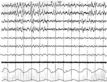
recording was performed with a Grass recorder (Grass Instrument Co., Quincy; Mass). Bilateral EEGs were recorded from the parietal, frontal, and occipital lobes (Figure 1). Two electrodes taped to the chin and two electrodes attached to the outer canthus of each eye provided measurement of elec -tromyography (EMG) and electrooculography (EOG), respec -tively. Nasal thermistors were uniformly placed below the outer edge of each nostril and secured with tape. he body movements of subjects were recorded by a video camera in conjunction with infrared lighting.
At the start of the test session, each subject was asked to cover one nostril and breathe normally. he nasal thermistor ampliier was then adjusted to produce a waveform half the maximum limit of the range of the wave recorded. he same procedure was then repeated for the opposite naris. In this way, the transducers were calibrated to the airlow of the subject’s nares at the start of the testing period. he ampli -ier gain was adjusted to allow proper measurement of wave -forms; a correction factor was later applied to correct for this adjustment. In one subject, a thermistor moved from its posi -tion early in the evening and required reposi-tioning and reca -libration. In this case, the data from the irst portion of the night was not included in the analysis.
Sleep stage was determined using Rechtschafen and Kales’ system16
. he EEGs were scored by a licensed sleep researcher who regularly scored EEGs for the clini -cal sleep laboratory within the Hospital of the University of Pennsylvania. he sleep researcher did not know the hypoth -eses of the study. Epochs containing transition periods or extensive artifacts that precluded the ability to stage the epoch reliably were not included in the analysis. Body posi -tion was classiied as left lateral, right lateral, or supine. he prone body position was never observed for any subject,
probably because the arrangement of electrode leads made this position uncomfortable for the subject.
To determine nasal airlow, the amplitude of one repre -sentative nasal airwave from each nostril was measured from the recording for every minute of study. Correction factors were applied to the airwave amplitude measurement to cor -rect for diferences in ampliier gain when necessary. To cor -rect for the non-linear output of the nasal thermistors, the data were linearized by a natural log function17
. he symme -try of the transformed data (i.e., that it was indeed normal dis -tribution) was conirmed by experiential data analysis meth -odology18
. From these data, two dependent measures were calculated. he irst measure represented relative asymmetry of nasal airlow and was derived by calculating the percent -age of total airlow through the right naris from the trans -formed data [i.e., ([ln(right naris amplitude)] / (ln(right naris amplitude) + ln(left naris amplitude)] * 100]19. he second
measure was the absolute value of the diference between the transformed left and right naris values [i.e., abs[ln(left naris amplitude) - ln(right naris amplitude)]. Both of these mea -sures were then standardized for each subject by converting the subject’s data points to z-scores.
Data analysis
To evaluate the inluences of body position and sleep stage on nasal airlow, a weighted two-way (sleep stage by body position) ANOVA was applied to the dependent measures. his model allowed us to remove bias due to unequal number of observations between subjects. Sleep stages were dichoto -mized as non-REM (which included sleep stages 1, 2, 3 and 4) and REM. Diferences among body positions were compared using one degree-of-freedom post-hoc orthogonal F-Tests. he Bonferroni correction was applied to the resulting p-val -ues to correct for inlated alpha due to multiple comparisons.
RESULTS
Both body position and sleep stage were found to inluence nasal airlow lateralization [body position: F(2,2340) = 86.99, p < 0.001; sleep stage F(1,2340) = 234.82, p < 0.001]. In addi -tion, these two variables signiicantly interacted [body posi -tion by sleep stage interac-tion: F(2,2340) = 26.06, p < 0.001]. he adjusted mean percentage of the airlow through the right nostril during non-REM and REM sleep stages for the three body positions are presented in Table 1. he number of data points within the REM and non-REM periods upon which this analysis was based is shown in Table 2.
Multiple one-degree-of-freedom F-tests were used to com -pare the diferences among body positions. hese post-hoc tests revealed that all positions difered from one another [right vs. left: F(1,2340) = 173.44, p < 0.001; right vs. supine F(1,2340) = 37.56, p < 0.001; left vs. supine: F(1,2340) = 52.01, p < 0.001]. As can be seen in Table 1, airlow was proportionally
greater in the right nasal chamber when a subject was in the left lateral body position. he degree of this efect was less when a subject was in the supine position, and least when in the right lateral position. he sleep-stage-by-body-position interaction relected the fact that the airlow lateralization due to sleep stage was largest in the supine position and smallest in the right lateral position (Figure 2).
In order to establish whether airlow symmetry related to sleep stage similar to its relationship to the EEG10-12,
Table 1. Mean (standard error of the mean) percentage of total airflow through the right nasal chamber as estimated by the analysis of variance model for non-REM and REM sleep stages.
Variable Non-REM
Sleep REM sleep
Overall for body position Right side 50.4 (0.72) 51.7 (0.44) 51.0 (0.42) Supine 52.7 (0.45) 60.2 (0.57) 56.4 (0.27) Left side 54.3 (0.40) 58.8 (0.35) 56.7 (01.37) Overall for
sleep stage 52.5 (0.31) 56.9 (0.28) 54.8 (0.21)
REM: Rapid Eye Movement.
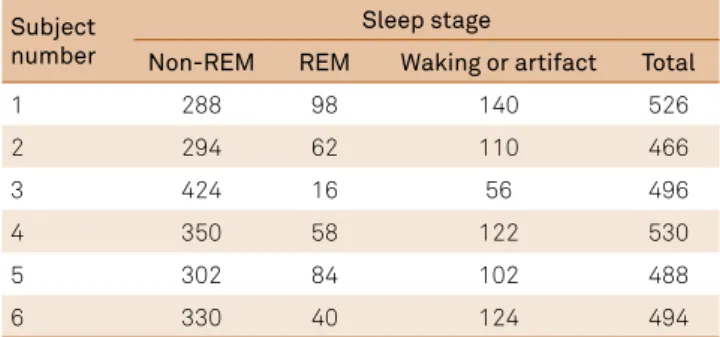
Table 2. Number of data points during REM, non-REM, and waking following the onset of sleep periods for each subject during the test period.
Subject number
Sleep stage
Non-REM REM Waking or artifact Total
1 288 98 140 526
2 294 62 110 466
3 424 16 56 496
4 350 58 122 530
5 302 84 102 488
6 330 40 124 494
REM: Rapid Eye Movement.
Figure 2. Percentage (Y-axis) of total airflow through the right nasal chamber for three body positions during REM (full color bars) and non-REM sleep (light color bars) states. Note the linear relationship between body position and the dependent measure during non-REM sleep. This relationship was not clear during REM sleep, most likely resulting in the significant sleep stage by body position interaction.
40 45 50 55 60 65
we determined if there was a diference in absolute airlow that occurred during REM and non-REM sleep. Although sleep stage was not related to the diference in absolute airlow across nostrils, body position was related to this measure [sleep stage: F(1,2340) = 0.23, p > 0.60; body position F(2,2340) = 46.06, p < 0.001] (Table 3). Multiple one-degree-of-freedom F-tests revealed a signiicant diference between the right lateral posi -tion and the left lateral and supine posi-tions, but did not reveal a signiicant diference between the left lateral and supine posi -tions [right lateral vs. left lateral F(1,2340) = 54.64, p < 0.001; right lateral vs. supine F(1,2340) = 81.87, p < 0.001; left lateral vs. supine F(1,2340) = 2.59, p > 0.10]. However, a signiicant sleep-stage-by-body-position interaction was found [F(1,2340) = 29.53, p < 0.001]. his relects the fact that the magnitude of the abso -lute diference measure was greater during the non-REM than during the REM sleep when subjects were in the right lateral or supine positions, whereas the magnitude of this measure was greater during the REM than the non-REM sleep stage when subjects were in the left lateral position (Table 3).
Since REM sleep is associated with higher frequency EEG than non-REM sleep16
, it is conceivable that a rela -tionship exists between brain wave frequency and nos -tril dominance. We found only a weak relationship, con -fined to the supine position, between sleep stage ranked in terms of brain wave frequency and airflow lateralization (Spearman r = 0.254). Thus, brain wave frequency probably does not account for a significant part of the sleep stage and nasal cycle relationship.
DISCUSSION
his study demonstrates that the lateralization of nasal airlow and sleep stage are related. In fact, greater airlow was found to occur though the right nasal chamber during REM sleep. his study conirms that the side of maximal nasal air -low is inluenced by body position, with the most air-low occurring through the uppermost nasal cavity19. In addition,
the data support the notion that the nasal cycle is not an artifact of asymmetrical body position or other environmen -tal factors20,21,22. Since no asymmetries in body pressure and
posture were present when subjects were in the supine posi -tion, the airlow from this position may be most representa -tive of the endogenous nasal cycle23.
he statistical interaction between sleep stage and body position was likely due to the diferential inluence of body position on the physiological expression of the nasal cycle dur -ing diferent stages of sleep. As can be seen from Table 1, the association between the nasal cycle and sleep stage was great -est when subjects were in the supine position. he smaller magnitude of the sleep stage and nasal cycle relationship when the subjects were in the right or left lateral body position is likely due to asymmetric stimulation of pressure receptors located in supericial ventral, dorsal, and lateral aspects of the pelvic and pectoral girdles and thoracic wall, and deep aspects of the intercostal spaces and parietal pleura24. he relex arc
resulting from such stimulation results in increased vasocon -striction in the inferior nasal cavity and decreased vasocon -striction in the superior nasal cavity. Others have suggested that asymmetric body pressure induces a sustained inhibition of the nasal cycle25. Our data suggest that the irst of the men
-tioned efects had a greater inluence in this study.
A body position by sleep stage interaction was found for the absolute left:right airlow diference measure. his interaction arose from the fact that there was no consistent relationship between the absolute airlow diference and sleep stage across the three body positions. An examination of the sleep stage mean demonstrates that, overall, any efect from sleep stage on the absolute diference in the airlows is opposite to the EEG efect reported by Armitage et al.10. hus, our data suggest it is
unlikely that a relationship exists between the absolute difer -ence in airlow from the two sides of the nose and sleep stage.
he results of the present study clarify and expand obser -vations of previous studies. Alexiev and Roth13
failed to ind an overall relationship between sleep stage and nasal air -low in an eight-hour test session in four normal subjects, although an increase in airlow through the right naris was observed during the third REM sleep cycle. Since body posi -tion data were not collected, their inconsistent results may have arisen from the inluence of asymmetric stimulation of pressure receptors. Hudgel and Robertson14 investigated
which factors, other than body position, inluenced the lat -eralization of nasal airlow during sleep. Unfortunately, the nares were only classiied in terms of their position relative to the lateral body position (i.e., superior or inferior), rather than their anatomic body side (i.e., right or left). hus, it was not possible for them to analyze left-right changes in nasal airlow. Haight and Cole24, while providing evidence for the
nasal cycle during sleep, were unable to demonstrate a clear relationship between sleep stage and nasal cycle phase since measurements were made after the patients were aroused from sleep. Kimura et al.22 found an association between the
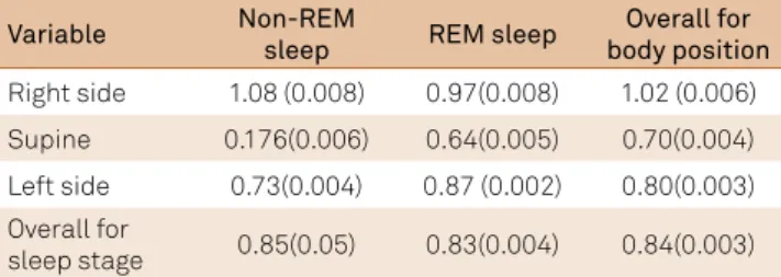
reversal of the nasal cycle and sleep stage in 69% of 16 sub -jects studied. None of the reversals occurred in the slow-wave sleep. Due to the limited statistical information provided by Table 3. Adjusted mean (standard error of the mean) absolute
difference between the logged left and right naris airflow measurements for non-REM and REM sleep stages by each body position.
Variable Non-REM
sleep REM sleep
Overall for body position Right side 1.08 (0.008) 0.97(0.008) 1.02 (0.006) Supine 0.176(0.006) 0.64(0.005) 0.70(0.004) Left side 0.73(0.004) 0.87 (0.002) 0.80(0.003) Overall for
sleep stage 0.85(0.05) 0.83(0.004) 0.84(0.003)
the authors, a more elaborated discussion on their indings cannot be done. Rohrmeier et al.23 studied the nasal cycle in
20 healthy subjects. Long-term rhinolowmetry was mea -sured during wakefulness and sleep. With these indings, these researchers concluded that shifts in body position dur -ing sleep modulate the nasal cycle (p < 0.01). Since polysom -nography was not performed, it is diicult to ascertain which sleep stage related to their indings.
It is unknown whether the REM/non-REM rhythm influences the nasal cycle directly or if a common cen -tral oscillator influences both the nasal cycle and the REM/non-REM rhythm26. A central oscillator hypothesis is
supported by studies suggesting that the REM/non-REM rhythm is derived from a common 90-minute central rhythm known as the basic rest-activity cycle (BRAC)27.
Since the BRAC has been proposed to drive asymmetric autonomic rhythms associated with the relative tonus of blood vessels of both the nasal mucosa and the cerebral hemispheres, it is possible that the mechanism underly -ing the BRAC rhythm is responsible for the synchroni -zation of nasal mucosal engorgement and asymmetric
hemispheric brain activity. Although the REM/non-REM cycle is strongly associated with asymmetric brain activ -ity during sleep, the precise mechanism that relates these rhythms has not been defined.
In conclusion, this study demonstrates a relationship between the rhythms of the nasal cycle and sleep stage; however, the precise association between these rhythms and the cerebral hemisphere EEG rhythm remains enigmatic. Werntz et al.8
assumed that a single oscillator drove cyclic EEG activity; however, other studies suggest that the two hemi -spheres may be driven by separate oscillators, possibly located in the hypothalamus, having slightly diferent periods28. Such
a phenomenon could explain the discrepancies in the cyclic relationship between the two hemispheres reported in the lit -erature. Separate control of turbinate engorgement on either side of the nose probably does not exist since this endoge -nous cycle is robust; moreover, the nasal cycle has been docu -mented under various conditions4 and during various times of
the day5
. he other possibility is that homeostatic regulation by the hypothalamus that characterizes REM29 gets disrupted.
Future research is needed to assess this hypothesis.
References
1. Mirza N, Kroger H, Doty RL. Influence of age on the ‘nasal cycle’. Laryngoscope. 1997;107(1):62-6. doi:10.1097/00005537-199701000-00014
2. Shilenkova VV, Kozlov VS. [A nasal cycle in healthy children]. Vestn Otorinolaringol. 2008;(1):11-6. Russian.
3. Eccles R. The domestic pig as an experimental animal for studies on the nasal cycle. Acta Otolaryngol. 1978;85(5-6):431-6. doi:10.3109/00016487809121472
4. Saroha D, Bottrill I, Saif M, Gardner B. Is the nasal cycle ablated in patients with high spinal cord trauma?. Clin Otolaryngol Allied Sci. 2003;28(2):142-5. doi:10.1046/j.1365-2273.2003.00679.x
5. Eccles R. The central rhythm of the nasal cycle. Acta Otolaryngol. 1978;86(5-6):464-8. doi:10.3109/00016487809107526
6. Davies AM, Eccles R. Reciprocal changes in nasal resistance to airflow caused by pressure applied to the axilla. Acta Otolaryngol. 1985;99(1-2):154-9. doi:10.3109/00016488509119158
7. Haight JJ, Cole P. Reciprocating nasal airflow resistances. Acta Otolaryngol. 1984;97(1-2):93-8. doi:10.3109/00016488409130968
8. Werntz DA, Bickford RG, Bloom FE, Shannahoff-Khalsa DS. Alternating cerebral hemispheric activity and the lateralization of autonomic nervous function. Hum Neurobiol. 1983;2(1):39-43.
9. Goldstein L, Stoltzfus NW, Gardocki JF. Changes in interhemispheric amplitude relationships in the EEG during sleep. Physiol Behav. 1972;8(5):811-5. doi:10.1016/0031-9384(72)90289-2
10. Armitage R, Hoffmann R, Loewy D, Moffitt A. Variations in period-analysed EEG asymmetry in REM and NREM sleep. Psychophysiology.
1989;26(3):329-36. doi:10.1111/j.1469-8986.1989.tb01928.x
11. Moffitt A, Hoffmann R, Wells R, Armitage R, Pigeau R, Shearer J. Individual differences in pre- and post-awakening correlates of dream reports following awakening from different stages of sleep. Psychiatr J Univ Ott. 1982;7:111-25.
12. Pivik RT, Bylsma F, Busby K. Sawyer S. lnterhemispheric EEG changes: relationship to sleep and dreams in gifted adolescents. Psychiatr Univ Ottawa. 1982;7:56-76.
13. Alexiev AD, Roth B. Some peculiar changes in the pattern of respiration connected with REM sleep: a preliminary report. Electroencephalogr Clin Neurophysiol. 1978;44(1):108-11. doi:10.1016/0013-4694(78)90110-4
14. Hudgel DW, Robertson DW. Nasal resistance during wakefulness and sleep in normal man. Acta Otolaryngol. 1984;98(1-2):130-5. doi:10.3109/00016488409107544
15. Pallanch JF, McCaffrey TV, Kern EB. Normal nasal resistance. Otolaryngol Head Neck Surg. 1985;93:778-85.
16. Rechtschaffen A, Kales A. A Manual of standardized terminology, techniques and scoring system for sleep stages of human subjects. Washington, DC: Public Health Service; 1968.
17. Allocca JA, Stuart A. Transducers: theory and application. Reston, VA: Reston; 1984.
18. Behrens JT. Principles and procedures of exploratory data analysis. Psychol Methods. 1997;2(2):131-60. doi:10.1037/1082-989X.2.2.131
19. Rao S, Potdar A. Nasal airflow with body in various positions. J Appl Physiol. 1970;28(2):162-5.
20. Haight JS, Cole P. Is the nasal cycle an artifact? The role of asymmetrical postures. Laryngoscope. 1989;99(5):538-41. doi:10.1288/00005537-198905000-00013
21. Lal D, Gorges ML, Ungkhara G, Reidy PM, Corey JP. Physiological change in nasal patency in response to changes in posture, temperature, and humidity measured by acoustic rhinometry. Am J Rhinol. 2006;20(5):456-62. doi:10.2500/ajr.2006.20.2939
22. Kimura A, Chiba S, Capasso R, Yagi T, Ando Y,
Watanabe S et al. Phase of nasal cycle during sleep tends to be associated with sleep stage. Laryngoscope. 2013;123(8):2050-5. doi:10.1002/lary.23986
23. Rohrmeier C, Schittek S, Ettl T, Herzog M, Kuehnel TS. The nasal cycle during wakefulness and sleep and its relation to body position. Laryngoscope. 2014;124(6):1492-7. doi:10.1002/lary.24546
25. Brown TH, McAfee DA. Long-term synaptic potentiation in the superior cervical ganglion. Science. 1982;215(4538):1411-3. doi:10.1126/science.6278593
26. Atanasov AT, Dimov PD. Nasal and sleep cycle: possible synchronization during night sleep. Med Hypotheses. 2003;61(2):275-7. doi:10.1016/S0306-9877(03)00169-5
27. McPartland RJ, Kupfer DJ. Rapid eye movement sleep cycle, clock time and sleep onset. Electroencephalogr
Clin Neurophysiol. 1978;45(2):178-85. doi:10.1016/0013-4694(78)90002-0
28. Gordon HW, Stoffer DS, Lee PA. Ultradian rhythms in specialized cognitive function. J Clin Exp Neurophysiol. 1990;12:40.
29. Lanfranchi PA, Fradette L, Gagnon JF, Colombo R, Montplaisir J. Cardiac autonomic regulation during sleep in idiopathic REM sleep behavior disorder. Sleep.