Ventricular Dyssynchrony and Increased BNP Levels in Right
Ventricular Apical Pacing
Cláudia Drummond Guimarães Abreu
1, Maria do Carmo Pereira Nunes
1,2, Márcia Melo Barbosa
2,
Manoel Otávio Costa Rocha
1, Antônio Luiz Pinho Ribeiro
1Hospital das Clínicas e Faculdade de Medicina da UFMG1; Departamento de Ecocardiografia do Hospital Socor2, Belo Horizonte, MG - Brazil
Abstract
Background: Long-term right ventricular apical pacing can cause ventricular dyssynchrony and, secondarily, neurohumoral alterations and increase in cardiac morbimortality.
Objective: To analyze ventricular dyssynchrony and its effects on BNP levels in patients with pacemakers and long-term right ventricular (RV) apex pacing.
Methods: Cross-sectional study of 85 patients with single or dual chamber pacemaker, NYHA functional class I or
II and left ventricular ejection fraction (LVEF) ≥ 35%. The dyssynchrony assessment was carried out using several
echocardiographic techniques, including Tissue Synchronization Imaging (TSI), with the analysis of the 12 segments. BNP was measured at the same time when the echocardiogram was performed, but the examiner was blinded to the results.
Results: Forty-six women and 39 men, aged 58 ± 12 years, with Chagas’ disease (56%) and controlled hypertensive individuals (62%), were included in the study. LVEF was 52 ± 8% and the mean QRS duration was 139 ms (120-180 ms). BNP levels were altered in 36.5% of the sample (cutoff = 60 pg/ml). At the multivariate linear regression analysis, BNP was correlated with age (p = 0.024), LVEF (p < 0.0001) and left ventricular (LV) pre-ejection time (p = 0.009), which is an intraventricular dyssynchrony index.
Conclusion: In clinically stable patients receiving conventional cardiac pacing, the intraventricular dyssynchrony was an independent predictor of BNP level increase after adjusted for age and LVEF. (Arq Bras Cardiol 2011; 97(2) : 156-162)
Keywords: Pacemaker, artificial; ventricular dysfunction; natriuretic peptides; echocardiography.
Mailing address: Cláudia Drummond Guimarães Abreu •
Rua Padre Marinho 49/602 - Santa Efigênia - 30140-040 - Belo Horizonte, MG - Brazil
E-mail: [email protected], [email protected]
Manuscript received September 04, 2010; revised manuscript received September 10, 2010; accepted April 06, 2011.
Introduction
Currently, cardiac pacemakers (PM) represent an effective treatment for symptomatic bradycardias cause by sinus node disease (SND) and atrioventricular block (AVB). For many years, the apical region of the right ventricle (RV) was one of the most frequently used sites in conventional implants due to its accessibility and lead cable stability1. However, in spite
of the evident improvement in quality of life for most patients with an artificial implant, the left bundle-branch block (LBBB) induced by right ventricular apical pacing (RVAP) can cause hemodynamic, structural and functional alterations in the heart, with deleterious consequences on the clinical evolution of some patients2,3.
The cardiac pacing at any point of the ventricle alters the natural heart activation and contraction pattern, as the stimulus conduction velocity is slower across the ventricular myocardium, when compared to that resulting from the specialized His-Purkinje system4.
Studies have demonstrated that, in response to an artificial pacing, the myocardial fibers contract erratically, resulting in heterogeneous stretching of myocardial segments that can interfere with the metabolism of the cardiac cell and cause global deterioration of the organ function5,6. Hence, it
could be presupposed that an abnormal electrical ventricular activation sequence associated to the inappropriate stretching of ventricular walls could result in dyssynchrony between the electrical ventricular activity and its contractility, determining, in the long term, ventricular dysfunction, neurohumoral alterations and increased cardiac morbidity and mortality.
Based on this hypothesis, the main objective of the present study was to evaluate the presence of ventricular dyssynchrony (VD) after long-term RVAP and its effects on brain natriuretic peptide (BNP) levels in clinically stable patients with no significant LV dysfunctions.
Methods
From June 2007 to March 2008, 85 Chagasic and non-Chagasic patients were consecutively selected during routine telemetric assessments carried out at the aforementioned laboratory, who had been submitted to single or dual-chamber pacemaker implantation with RVAP, and depended on artificial pacing (percentage of RV pacing ≥ 80%), with a LBBB pattern of ventricular activation at the surface electrocardiogram (ECG).
Patients of both sexes aged 18 to 75 years, with NYHA functional class I or II and left ventricular ejection fraction (LVEF) ≥ 35% were included. Preexisting diseases and medications currently being used were identified.
Systemic arterial hypertension (SAH) was defined as a clinical history of hypertension and systolic blood pressure (SBP) levels ≥ 140 mmHg or diastolic blood pressure (DBP) ≥ 90 mmHg. Only controlled hypertensive and normotensive individuals were included in the study.
Diabetes mellitus was defined as a clinical history of diabetes and regular use of oral hypoglycemiants and/or insulin and only diabetic patients with controlled glycemic levels were included in the study.
Chagas’ disease was identified by the presence of at least two distinct positive serological tests (ELISA, indirect hemagglutination or indirect immunofluorescence test) and relevant epidemiological history.
Kidney function was assessed through creatinine level measurement requested by the assistant physicians during the routine clinical/cardiologic follow-up.
The exclusion criteria considered coronary artery disease, characterized by the presence of chest angina and/or evidence of LV segmental contractility impairment at the echocardiogram, atrial fibrillation (AF), chronic obstructive pulmonary disease (COPD), pregnancy, recent heart surgery (less than 4 weeks) or refusal to participate in the study.
Analysis of the QRS complex and chest X-ray
The duration of the QRS complex consisted in the measurement of the time interval between the emission of the pacemaker spike and the end of the QRS complex (ms), assessed at the surface ECG D2 derivation.
The position of the ventricular lead-electrode was verified through a chest X-ray in the posteroanterior and profile views.
Echocardiographic study
The echocardiographic study protocol consisted in obtaining one and two-dimensional images with pulsed and continuous Doppler guided by color flow mapping, in addition to tissue Doppler, including Tissue Synchronization Imaging (TSI), with analysis of 12 segments.
The evaluations were carried out by en experienced professional in a Vivid 7 equipment (GE Vingmed Ultrasound AS, Horten, Norway), equipped with an electronic transducer of variable frequency (4 to 12 MHz) and high resolution.
Ventricular dyssynchrony was analyzed through different echocardiographic techniques, according to the recommendations of the current Consensus of the American
Society of Echocardiography7. The following measurements
were obtained:
1. DifLV-RVPET - Difference between pre-ejection time of left and right ventricles (interventricular delay) obtained by pulsed Doppler (cutoff ≥ 40 ms); 2. IVSPW delay - Contraction delay between the
interventricular septum and the posterior wall in M mode (cutoff ≥ 130 ms);
3. LVPET -LV pre-ejection time measurement (aortic) at pulsed Doppler (cutoff ≥ 140 ms);
4. MaxDif12seg -Maximum difference of time between two distinct segments obtained with TSI technique (cutoff >100 ms);
5. SD12seg -Analysis of the standard deviation of the aforementioned 12 segments (cutoff > 32.6 ms); 6. IVSLW delay - Contraction delay between the
interventricular septum and the lateral wall also obtained through TSI (cutoff ≥ 65 ms);
BNP measurement
BNP measurement was carried out in a venous blood sample, preferably obtained from the antecubital vein with the patient in the supine position for 30 minutes, before the echocardiogram was performed, with the examiner blinded for the results. Blood pressure (BP) and heart rate (HR) were measured before the collection. The whole blood samples were collected into plastic tubes containing EDTA (1 mg/ ml of blood) and immediately processed using the Triage™ BNP Test kits (Biosite™ Inc., San Diego, USA). In the present study, the cutoff was defined as 60 pg/ml for BNP levels, which was based on previous studies of Chagasic patients with preserved LV function8,9.
Statistical analysis
The categorical variables were described by proportions and the continuous by means and standard deviations, medians and interquartile intervals, according to the distribution pattern. Uni- and multivariate linear regression analyses were used, considering BNP levels as the dependent variable. The level of statistical significance was defined for p values < 0.05.
Results
Only clinically compensated patients with NYHA functional class I or II were included in the study. There was a predominance of dual-chamber pacing (74%) and the median implant time was 63 months (1 - 137 months), and 94% of the sample had been undergoing artificial pacing for six months or more. Total Atrioventricular Block (TAVB) was the most common indication for the implant (85%) and in 48 patients (56%) Chagas’ disease was the cause of the underlying heart disease. The mean QRS duration was 139 ± 14.2 ms and none of the patients had significant LV dysfunction (mean LVEF = 51.8 ± 8%).
subgroup of patients with LVEF ≤ 40%. Its mean ejection fraction was 56.4 ± 11.2%.
BNP level median was 38.9 pg/ml with a minimum measured level of 5 pg/ml and a maximum level of 581 pg/ ml. In 34 patients (40%), these levels were considered to be high (cutoff = 60 pg/ml). The main clinical and demographic characteristics and echocardiographic parameters are shown in Table 1.
Interventricular dyssynchrony was observed in 49 patients (59.8%), considering the median of the interventricular delay. Significant intraventricular dyssynchrony was observed in 77 patients (90.6%), when the analyzed parameter was the median of the LV pre-ejection time, as well as when the standard deviation and the maximum difference of systolic peaks measured in the 12 myocardial segments were analyzed. Similarly, the median of the contraction delay between the interventricular septum and the lateral wall was significant in 31 patients (36.5%) (Table 2).
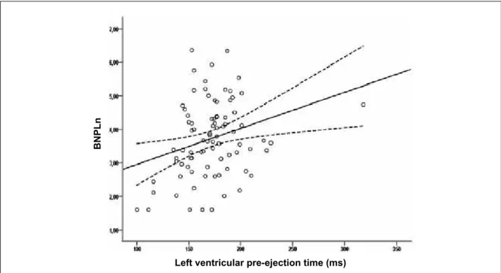
At the univariate analysis, the QRS duration, time of implant, Chagas disease, arterial hypertension and most of the echocardiographic variables did not have a significant correlation with BNP levels. However, age (r = 0.33, p = 0.002) and LVEF (r = -0.28, p = 0.010) were significantly correlated with this parameter. A significant correlation was also observed between the measurement of the LV pre-ejection time (r = 0.27, p = 0.013) and BNP levels (Figure 1). All other measurements performed to assess VD did not show a significant correlation with BNP levels, regardless of the echocardiographic technique used (Table 3). The positive association between LVPET and BNP levels remained even when the first was analyzed as a categorical variable, using the cutoff recommended for the diagnosis of VD according to the current literature (≥ 140 ms) (Figure 2).
At the multiple linear regression analysis, the LVPET remained as the only significant and independent predictor of BNP levels, even after adjustment for covariables, such as age and LVEF. Therefore, the linear regression model obtained disclosed that the older the patient, the more severe the LV dysfunction and the longer the LV pre-ejection time (starting at 140 ms), on average, the higher the BNP levels.
Although it was not the main objective of the present study, an analysis between the subgroups of Chagasic and Non-Chagasic patients was performed, as well as between the different pacing modes, DDD and VVI. However, no significant differences were observed in this series between the groups when the clinical, electrocardiographic and echocardiographic variables were evaluated, except for the mean LVEF, which was lower in Chagasic patients (p = 0,035).
Discussion
The present study showed a high frequency of VD in patients with pacemakers submitted to long-term right ventricular apical pacing who were clinically stable, in addition to a significant correlation between the LV pre-ejection time, which is a measure of intraventricular dyssynchrony and BNP levels.
The study group was characterized by age of 58.5 ± 12.6 years, predominance of Chagasic patients and absence
Table 1 - Clinical and demographic characteristics and main echocardiographic parameters of the 85 patients with long-term RV apical pacing
Variables n (%) or mean ± SD or median (IQL)
Age (years) 58.5 ± 12.6
Women (%) 46 (54)
HR (bpm) 64 ± 3
SBP (mmHg) 125 ± 10
DBP (mmHg) 80 ± 6
Chagasic patients (%) 48 (57) Functional class (NYHA) (%)
I 67 (79)
II 18 (21)
Medications being used
Loop diuretics and thiazides 37 (43.5) ACE Inhibitors 35 (41.2) Calcium-channel blockers 11 (12.9) Digitalis 10 (11.8) Amiodarone 8 (9.4) Beta-blockers 8 (9.4)
ARBs 8 (9.4)
Pacing mode (%)
DDD(R) 63 (74)
VVI(R) 17 (20)
VDD(R) 5 (6)
Ventricular capture (PV%) 96 ± 4
Duration of (ms) QRS: 139 ± 14.2
120-150 (%) 72 (84.7) > 150 (%) 18 (15.3) BNP (pg.ml-1) 38.9 (18.7;81.6)
Echocardiographic parameters
Diastolic diameter of LV (mm) 52.6 ± 6.9 Systolic diameter of LV (mm) 37.1 ± 7.8 LV ejection fraction * (%) 51.8 ± 8.5 RV ejection fraction* (%) 56.4 ± 11.2 E/E’ ratio 10.5 ± 3.6 PASP (mmHg) 34.6 ± 7.1 LA volume index (ml/m2) 36.3 ± 11.2
Data expressed as means (± standard-deviation) or proportion or median (interquartile interval); HR - heart rate; SBP - systolic blood pressure; DBP - diastolic blood pressure; NYHA - New York Heart Association; ACE - angiotensin-converting enzyme; ARBs - angiotensin-receptor blockers; DDD - dual-chamber pacing; VVI - single-chamber pacing; PV% - % of right ventricular paced beats; BNP - natriuretic peptide type B; LV - left ventricle; RV - right ventricle; PASP - pulmonary artery systolic pressure; LA - left atrium. (*)Simpson’s method.
beta-Table 2 - Echocardiographic measurements of inter and intraventricular dyssynchrony after long-term right ventricular apical pacing
VD
measurements (ms)
Median (IQL)
Altered recordings
n (%)
Echocardiographic method
Interventricular
DifLV-RVPET 44 (26;55.3) 49 (59.8) Pulse Doppler Intraventricular
LVPET 172 (153;186) 77 (90.6) Pulse Doppler MaxDif12seg 112 (81;138) 49 (59.0) TDI/TSI SD12seg 38.5 (28.6;50.2) 52 (62.7) TDI/TSI IVSLW delay 59 (30.5; 82) 31 (36.5) TDI/TSI IVSPW delay 70 (50;90) 04 (4.8) M Mode
IQL- interquartile interval (25th; 75th percentiles); DifLV-RVPET - Difference
between pre-ejection time of left (aortic) and right (pulmonary) ventricles. LVPET - left ventricular pre-ejection time ; MaxDif12seg - maximum difference among the 12 segments; SD12seg - standard deviation among the 12 segments; IVSLW - delay between the interventricular septum and lateral wall; IVSPW - delay between the interventricular septum and the posterior wall; TDI/TSI - Tissue Doppler Imaging/Tissue Synchronization Imaging.
Figure 1 - Dispersion diagram showing a positive correlation between BNP and LVPT.
B
N
PL
n
Left ventricular pre-ejection time (ms)
blockers and diuretics by more than 80% of the assessed patients indicates that all were submitted to optimized clinical treatment and were NYHA functional class I or II.
Around 40% of the patients had high levels of BNP, considering 60 pg/ml the cutoff used for the measured levels. This information was based on a previous study that analyzed Chagasic patients with preserved ventricular function and
observed similar mean BNP levels between the control group and patients with LVEF > 40%. Significantly higher levels were found in patients with LVEF ≤ 40%8.
In accordance with other studies9,10, it was observed that
an increase in BNP levels was correlated with an increase in age (r = 0.33, p = 0.002) and LVEF worsening (r = -0.28, p = 0.010). However, considering that most of the sample was NYHA functional class I, more than the increase in LV filling pressures, the VD observed in the study also contributed to the increase in BNP levels. This fact was demonstrated by the significant correlation between the LV pre-ejection time and BNP (r = 0.38, p < 0.0001), regardless of LVEF and age.
Previous studies have demonstrated that dyssynchronous ventricular activation can contribute to asymmetric hypertrophy, myofibrillar disarray, increase in catecholamine concentrations, neurohumoral activation and regional perfusion disorders5. The
studies MOST11, DAVID12 and MADIT II13, in turn, indicated
that VD can create an anatomofunctional substrate capable of impairing heart function in the long term, by observing an increase in the risk of AF, mitral regurgitation and hospital admissions due to heart failure in patients with a high percentage of right ventricular paced beats, particularly those with ventricular dysfunction prior to the implant14. However,
whether VD is an acute phenomenon that could deteriorate LV function in the long term and, consequently, result in HF in the absence of cardiomyopathy, remains to be clarified15,16.
Our results are in accordance, however, with those of other studies that verified that VD secondary to long-term RVAP can impair the LV systolic and diastolic functions and contribute to the increase in BNP levels. According to Chiladakis et al17,
Figure 2 - Association between BNP and LVPT (≥ 140 ms) in patients with long-term RV apical pacing.
B
N
PL
n
Left ventricular pre-ejection time
Table 3 - Correlation between BNP levels and age,
electrocardiogram and echocardiographic parameters of ventricular dyssynchrony
Variables r p
Age (yrs) 0.33 0.002
QRS (ms) 0.10 0.371
Time of implant (months) 0.02 0.850 Ventricular capture (PV%) 0.17 0.112 Chagas’ disease 0.15 0.174 LVEF (%)* -0.28 0.010
LVPET 0.27 0.013
IVSPP delay 0.04 0.722 Dif LV-RDPET 0.08 0.500
SD12seg 0.01 0.918
MaxDif12seg 0.06 0.617 IVSLW delay 0.06 0.569
PV% - % of right ventricular paced beats; LVEF - left ventricular ejection fraction; LVPET - left ventricular pre-ejection time; IVSPP - interventricular septum and posterior wall; DifLV-RVPET - Difference between pre-ejection time of left and right ventricles; SD12seg - standard deviation among the 12 segments; MaxDif12seg - maximum difference among the 12 segments; IVSLW - interventricular septum and lateral wall. (*) Simpson’s Method.
severity detected in patients with RVAP, as well as initial signs of HF in patients with TAVB and normal LVEF, after RVAP for 6.5 ± 5.7 years18,19.
Currently, the VD caused by RVAP can be quantified by several echocardiographic techniques. Although many types of measurements can be used, the most simple and routine ones have shown a good performance in multicentric assessments20. The LV pre-ejection time, for instance, assessed
in the present study, is useful to quantify intraventricular dyssynchrony with good reproducibility according to previous studies, such as the CARE-HF study21. Recently, Sá
et al22 observed the prolonging of this measurement in a small
group of patients, who were also Chagasic with normal LVEF, throughout an eight-month period. The authors observed that although the LVPET measurements did not reach the cutoff required for the diagnosis of VD (≥ 140 ms), this was the only assessed VD measure that showed increases throughout follow-up. It is noteworthy the fact that, in the aforementioned study, the septal pacing predominated over the apical one, which seems to be more deleterious.
In addition to LVPET, which was altered in more than 90% of the sample, the other echocardiographic measures assessed in the study also identified inter- and intraventricular dyssynchrony alterations, disclosing the high frequency of this disorder in patients submitted to long-term RVAP. Indices such as the interventricular delay, maximum difference and standard deviation of the 12 segments were altered in more than 50% of the sample. However, in spite of the usefulness of the several echocardiographic techniques employed, including Tissue Synchronization Imaging, to quantify VD, no significant isovolumetric relaxation time caused by RVAP in patients with
association between these measures and BNP serum levels were observed in this group of patients, in opposition to LVPET. Our results corroborate current discussions, especially in the field of CRT, which aim at answering whether the echocardiographic measures used to date in the analysis of VD are the ones that can better define its presence. Two important prospective and multicentric studies, PROSPECT23
and ReThinQ24, did not find any correlation between VD
measures and the benefits of CRT when selecting candidates to the procedure according to echocardiographic criteria. The importance of technical factors that contribute for these findings has been highlighted, especially by aspects related to the feasibility and reproducibility of the methods used. It is a consensus, however, that the TSI technique must not be considered unproductive for the VD analysis, but technological advances are necessary for less operator-dependent approaches20.
Limitations
As this is a cross-sectional study, patients with PM were assessed at a certain point during their clinical follow-up, which brings limitations to the results. Although our findings indicate an association between the increase in BNP levels and VD secondary to long-term RVAP, the study design does not allow us to establish a causal association between these variables.
It is important to consider that BNP was assessed together with the VD measures, of which specific studies did not define the best indices or ideal cutoffs to perform its analysis and this is one of the reasons of the non-inclusion of the dyssynchrony measures proposed, until then, by the current Cardiac Resynchronization Guidelines25. In spite of that, it is
a consensus in the current literature that the VD measures be used together with clinical criteria in the selection of candidates considered to be borderline for CRT26.
Limitations that are inherent to the Tissue Synchronization Imaging must be mentioned7. Among them are the systolic
curves, not always contained within the ejection interval, multiple peaks resulting from a same myocardial segment and, in some instances, instead of a well-defined peak, the presence of a plateau, which made it difficult to determine the ideal point for the proposed measure.
Conclusions
Patients with PM and long-term RVAP have a high frequency of VD and in the present study, the intraventricular dyssynchrony was identified through the measurement of the LVPET in around 90% of the patients. Moreover, this measure showed to be an independent predictor of increased BNP levels, indicating that VD was capable of producing early neurohumoral alterations in patients with no significant LV dysfunction.
Further studies are necessary to define whether the VD identified at the echocardiogram or the increase in BNP levels in patients with PM submitted to long-term RVAP are capable of predicting an unfavorable evolution.
Potential Conflict of Interest
No potential conflict of interest relevant to this article was reported.
Sources of Funding
There were no external funding sources for this study.
Study Association
This article is part of the thesis of master submitted by Claudia Drummond Guimarães Abreu, from Universidade Federal de Minas Gerais.
References
1. Melo CS. Temas de marca-passo. 2ª ed. São Paulo: Lemos Editorial; 2004. p.478.
2. Baldasseroni S, Opasich C, Gorini G, Lucci D, Marchionni N, Marini M, et al. Left bundle branch block is associated with increased 1-year sudden and total mortality rate in 5517 outpatients with congestive heart failure: a report from the Italian network on congestive heart failure. Am Heart J. 2002;143(3):398-405.
3. Kashani A, Barold SS. Significance of QRS complex duration in patients with heart failure. J Am Coll Cardiol. 2005;46(12):2183-92.
4. Prinzen FW, Augustijn CH, Arts T, Allessie MA, Reneman RS. Redistribution of myocardial fiber strain and blood flow by asynchronous activation. Am J Physiol Heart Circ Physiol. 1990;259(2 Pt 2):H300-8.
5. Vernooy K, Verbeek XA, Peschar M, Prinzen FW. Relation between abnormal ventricular impulse conduction and heart failure. J Interv Cardiol. 2003;16(6):557-62.
6. Dilaveris P, Pantazis A, Giannopoulos G, Synetos A, Gialafos J, Stefanadis C. Upgrade to biventricular pacing in patients with pacing-induced heart failure: can resynchronization do the trick? Europace. 2006;8(5):352-7.
7. Gorcsan J 3rd, Abraham T, Agler DA, Bax JJ, Derumeaux G, Grimm RA, et al. Echocardiography for cardiac resynchronization therapy: recommendations
for performance and reporting--a report from the American Society of Echocardiography Dyssynchrony Writing Group endorsed by the Heart Rhythm Society. J Am Soc Echocardiogr. 2008;21(3):191-213.
8. Ribeiro AL, dos Reis AM, Barros MV, de Sousa MR, Rocha AL, Perez AA, et al. Brain natriuretic peptide and left ventricular dysfunction in Chagas’ disease. Lancet. 2002;360(9331):461-2.
9. Redfield MM, Rodeheffer RJ, Jacobsen SJ, Mahoney DW, Bailey KR, Burnett JC Jr. Plasma brain natriuretic peptide concentration: impact of age and gender. J Am Coll Cardiol. 2002;40(5):976-82.
10. Daniels LB, Maisel AS. Natriuretic peptides. J Am Coll Cardiol. 2007;50(25):2357-68.
11. Lamas GA, Lee KL, Sweeney MO, Silverman R, Leon A, Yee R, et al. Ventricular pacing or dual-chamber pacing for sinus-node dysfunction. N Engl J Med. 2002;346(24):1854-62.
12. Wilkoff BL, Cook JR, Epstein AE, Greene HL, Hallstrom AP, Hsia H, et al. Dual-chamber pacing or ventricular backup pacing in patients with an implantable defibrillator: the Dual Chamber and VVI Implantable Defibrillator (DAVID) Trial. JAMA. 2002;288(24):3115-23.
in the multicenter automatic defibrillator trial II. J Cardiovasc Electrophysiol. 2005;16(4):359-65.
14. Sweeney MO, Hellkamp AS, Ellenbogen KA, Greenspon AJ, Freedman RA, Lee KL, et al. Adverse effect of ventricular pacing on heart failure and atrial fibrillation among patients with normal baseline QRS duration in a clinical trial of pacemaker therapy for sinus node dysfunction. Circulation. 2003;107(23):2932-7.
15. Tops LF, Schalij MJ, Bax JJ. The effects of right ventricular apical pacing on ventricular function and dyssynchrony. J Am Coll Cardiol. 2009;54(9):764-76.
16. Silva RT, Martinelli Fº M, Oliveira JC, Lima CEB, Martins DGMC, Guirão CI, et al. Remodelamento ventricular na estimulação cardíaca apical do ventrículo direito. Arq Bras Cardiol. 2007;88(2):152-8.
17. Chiladakis JA, Koutsogiannis N, Kalogeropoulos A, Zagli F, Arvantis P, Alexopoulos D. Unfavourable effects of continuous, atrial-synchronised ventricular pacing on ventricular systolic and diastolic function in patients with normal left ventricular ejection fraction: usefulness of tissue and colour Doppler echocardiography. Hellenic J Cardiol. 2007;48(6):335-40.
18. Kawanish Y, Ito T, Suwa M, Terasaki F, Futal R, Kitaura Y. Effect of left ventricular dyssynchrony on plasma B-type natriuretic peptide levels in patients with long-term right ventricular apical pacing. Int Heart J. 2008;49(2):165-73.
19. Xue-Hua Z, Chen H, Chung-Wah S, Kai-Hang Y, Wing-Sze C, Kathy LL. New-onset heart failure after permanent right ventricular apical pacing in patients with acquired high-grade AV block and normal left ventriculr function. J Cardiovasc Electrophysiol. 2008;19(2):136-41.
20. Abraham J, Abraham TP. Is echocardiographic assessment of dyssynchrony useful to select candidates for cardiac resynchronization therapy? Echocardiography is useful before cardiac resynchronization therapy if QRS duration is available. Circ Cardiovasc Imaging. 2008;1(1):79-84.
21. Cleland JG, Daubert JC, Erdmann E, Freemantle N, Gras D, Kappenberger L, et al. The effect of cardiac resynchronization on morbidity and mortality in heart failure. N Engl J Med. 2005;352(15):1539-49.
22. Sá LAB, Rassi S, Batista MAL. Conventional ventricular stimulation effects on patients with normal ventricular function. Arq Bras Cardiol. 2009;93(2):167-73.
23. Chung ES, Leon AR, Tavazzi L, Sun JP, Nihoyannopoulos P, Merlino J, et al. Results of the Predictors of Response to CRT (PROSPECT) trial. Circulation. 2008;117(20):2608-16.
24. Beshai JF, Grimm RA, Nagueh SF, Baker JH 2nd, Beau SL, Greenberg SM, et al; RethinQ Study Investigators. Cardiac resynchronization therapy in heart failure with narrow QRS complexes. N Engl J Med. 2007;357(24):2461-71.
25. Epstein AE, Dimarco JP, Ellenbogen KA, Estes NA 3rd, Freedman RA, Gettes LS, et al. American College of Cardiology/American Heart Association Task Force on Practice; American Association for Thoracic Surgery; Society of Thoracic Surgeons. ACC/AHA/HRS 2008 guidelines for Device-Based Therapy of Cardiac Rhythm Abnormalities: executive summary. Heart Rhythm. 2008;5(6):934-55.