www.jped.com.br
ORIGINAL ARTICLE
Biventricular diastolic function assessed by Doppler
echocardiogram in children vertically infected with human
immunodeficiency virus
夽
Mauricio L. Silva
a,b,∗, Silvia M. Nassar
b,c, André P. Silva
b, Leandro L. Ponce
a,
Maria M. de S. Pires
a,baHospital Infantil Joana de Gusmão, Secretaria de Estado da Saúde de Santa Catarina, Florianópolis, SC, Brazil bHospital Universitário, Universidade Federal de Santa Catarina, Florianópolis, SC, Brazil
cDepartment of Informatics and Statistics, Universidade Federal de Santa Catarina, Florianópolis, SC, Brazil
Received 30 September 2013; accepted 28 November 2013 Available online 11 March 2014
KEYWORDS
Ventricular function; Infectious diseases; Children
Abstract
Objective: to determine, by Doppler-echocardiography, the frequency of cardiac diastolic dys-function in asymptomatic and clinically stable pediatric patients with vertical infection by the human immunodeficiency virus (HIV), from the cardiovascular viewpoint.
Methods: this was an observational, prospective, and cross-sectional study, performed at a regional referral clinic for patients with HIV, in a convenience sample of 94 individuals, assessing biventricular diastolic function by Doppler-echocardiography, and weight, blood hemoglobin, and percentage of lymphocytes T-CD4+.
Results: fifty patients had diastolic dysfunction. Left ventricular dysfunction occurred in 38.7%, and the predominant type of dysfunction was decreased myocardial compliance. Right ventri-cular dysfunction was observed in 29.4% of the sample, and abnormal relaxation was the most prevalent type. Simultaneous biventricular dysfunction occurred in 14.1% of the individuals. There was no association between dysfunction and the immune status.
Conclusions: diastolic dysfunction occurred, individually or simultaneously, with no association with immune status; decreased myocardial compliance was predominant in the left ventricle, and abnormal relaxation in the right ventricle.
© 2014 Sociedade Brasileira de Pediatria. Published by Elsevier Editora Ltda. All rights reserved.
夽 Please cite this article as: Silva ML, Nassar SM, Silva AP, Ponce LL, Pires MM. Biventricular diastolic function assessed by Doppler
echocardiogram in children vertically infected with human immunodeficiency virus. J Pediatr (Rio J). 2014;90:403---7.
∗Corresponding author.
E-mail:[email protected], [email protected] (M.L. Silva).
PALAVRAS-CHAVE
Func¸ão ventricular; Doenc¸as infecciosas; Crianc¸as
A func¸ão diastólica biventricular por meio da análise com ecocardiograma com Doppler em crianc¸as infectadas verticalmente pelo vírus da imunodeficiência humana
Resumo
Objetivo: verificar, por meio do ecocardiograma com Doppler, a frequência de disfunc¸ão cardíaca diastólica em pacientes com infecc¸ão vertical pelo vírus da imunodeficiência humana na faixa pediátrica, assintomáticos e clinicamente estáveis do ponto de vista cardiovascular. Métodos: estudo observacional, prospectivo e transversal, realizado em um Ambulatório de Referência Regional para pacientes portadores do vírus da imunodeficiência humana, com uma amostra de conveniência de 94 pacientes, avaliados pelo sexo, idade, peso, func¸ão diastólica biventricular ao Doppler, hemoglobina sanguínea e percentual de linfócitos T-CD4+.
Resultados: apresentaram disfunc¸ão diastólica 50 pacientes. Disfunc¸ão ventricular esquerda ocorreu em 38,7% deles, e o tipo de disfunc¸ão predominante foi diminuic¸ão da complacência miocárdica. A disfunc¸ão ventricular direita foi evidenciada em 29,4% da amostra, sendo o tipo relaxamento anormal o mais prevalente. Disfunc¸ão biventricular simultânea ocorreu em 14,1% dos indivíduos. Não houve associac¸ão da disfunc¸ão com o estado imunológico.
Conclusões: foi verificada disfunc¸ão diastólica, isolada ou simultânea, sem associac¸ão com o estado imunológico, sendo a diminuic¸ão da complacência miocárdica mais comum no ventrículo esquerdo e relaxamento anormal no ventrículo direito.
© 2014 Sociedade Brasileira de Pediatria. Publicado por Elsevier Editora Ltda. Todos os direitos reservados.
Introduction
Cardiovascular manifestations often occur in children with vertical infection by the human immunodeficiency virus (HIV), and the most likely cause is multifactorial. In a prospective study, the cumulative five-year incidence of car-diac dysfunction in children ranged from 18% to 39%, and was the HIV-related cause of death in 11.8%.1---4
Subclinical cardiac abnormalities may develop in early HIV infection, even among individuals with asymptomatic disease or without cardiac dysfunction.1,4---9
The resolution of dilated cardiomyopathy in vertically infected children has been reported in those treated with a combination of drugs.6,10---12 It is possible that a change in diastolic function will precede systolic dysfunction, as observed in other clinical conditions, both in adults and in children and adolescents.3,13---23
The aim of this study was to determine the frequency of diastolic dysfunction in children vertically infected with HIV, both symptomatic and asymptomatic, and clinically sta-ble from the cardiovascular perspective. The association between diastolic dysfunction and immunological status, malnutrition, and anemia was also investigated.
Methods
This was an observational, cross-sectional study performed in a regional pediatric outpatient clinic for follow-up of patients with the acquired immunodeficiency syndrome (AIDS), consisting of a convenience, non-probabilistic sam-ple.
The protocol and the informed consent were approved by the institutional ethics committee, and all participants consented to the study through their legal guardians. From June to November of 1999, 139 children vertically infected
with HIV were evaluated, of whom 94 were selected accord-ing to the inclusion criteria. Age ranged from 20.3 to 170.6 months (mean 69.7 months) and 52 (55.0%) were males.
The definitive diagnosis was made according to the parameters of the Centers for Disease Control and Pre-vention (CDC, Atlanta, United States) of 1994: positive enzyme-linked immunoassay test (ELISA) and confirmatory test (Western blot).24
Forty-five patients were excluded from the analysis due to at least one of the following conditions: congenital heart disease; congestive heart failure; arrhythmia; aneuploidy; HIV-related infections; use of medications, including digi-talis, beta-blockers, vasodilators, and antiarrhythmic drugs; use or previous use of cardiotoxic chemotherapeutic agents; percentage of T CD4 + lymphocytes obtained at intervals greater than four months before or after the date of inclu-sion; and legal guardian’s refusal of patient’s participation in any phase of the study.
After obtaining the informed consent and determining patient eligibility for the study, blood samples were col-lected and the Doppler study was completed.
Variables
The following variables were observed and recorded: gen-der, age (months), weight/age Z-scores25using the software Epi InfoTM 6.04 (GA, USA), blood hemoglobin level (g/dL), percentage of T CD4 + lymphocytes, peak velocity of the E and A waves in the mitral and tricuspid valves (cm/s), and their ratios. The normal reference values used for the anal-ysis of diastolic function in both ventricles were obtained from the available literature.26,27
Table 1 Categories of mitral valve E/A ratio according to age groups.
Age groups
Infant Preschooler School-aged Adolescent Total
MV E/A n % n % n % n % n %
Normal 1 1.1 36 38.7 14 15.1 6 6.4 57 61.3
Lower 0 0.0 8 8.6 2 2.1 0 0.0 10 10.7
Upper 1 1.1 13 14.0 7 7.5 5 5.4 26 28.0
Total 2 2.2 57 61.3 23 24.7 11 11.8 93 100.0
MV-E/A, ratio between the peak velocities of E wave/A wave in the mitral valve; Infant,≥18 and < 24 months; Preschooler,≥24 and < 60 months; School-aged,≥60 and < 120 months; Adolescent,≥120 and < 180 months.
5.0 MHz (SI450 Sonoline; Siemens- Germany), guided by two-dimensional echocardiography and performed by one of the authors (MLS).
All measurements were the mean result of three consec-utive cardiac cycles, and were performed on the equipment display screen and printed on X-ray films.
Statistical analysis was performed with Statistica 5.0 software (StatSoft, Brasil). Descriptive statistics and esti-mation of parameters were calculated (with 95% confidence intervals of [95% CI]), using factorial analysis (through the analysis of the main components) and multiple correlation analysis.
The ratios of E/A velocities of the atrioventricular valves were considered the main variables. Severe anemia and moderate or severe protein-calorie malnutrition were con-sidered confounders.
Results
Of the 94 children studied, 52 (55%) were males. There was a predominance of preschoolers, followed by school-aged children, but no differences were found between genders. Age ranged from 20.3 to 170.6 months (mean 69.7±31.7 months, and median 65.3 months).
Immunological status evaluation showed that the major-ity of children (57.4%) were non-immunocompromised (CD4 +≥ 25%). Moderate immunological impairment (CD4 + of 15% to 25%) was observed in 29 (30.9%), and severe immunological impairment (CD4 + < 15%) in 11 (11.7%).
Blood levels of hemoglobin (g/dL) ranged from 5.4 to 14.2, with a mean of 11.7 + 1.3, median of 11.7, and no significant differences between genders.
It was observed that ten (10.9%) children, five of each gender, had moderate or severe protein-calorie malnutrition (z-score≤-2.0).
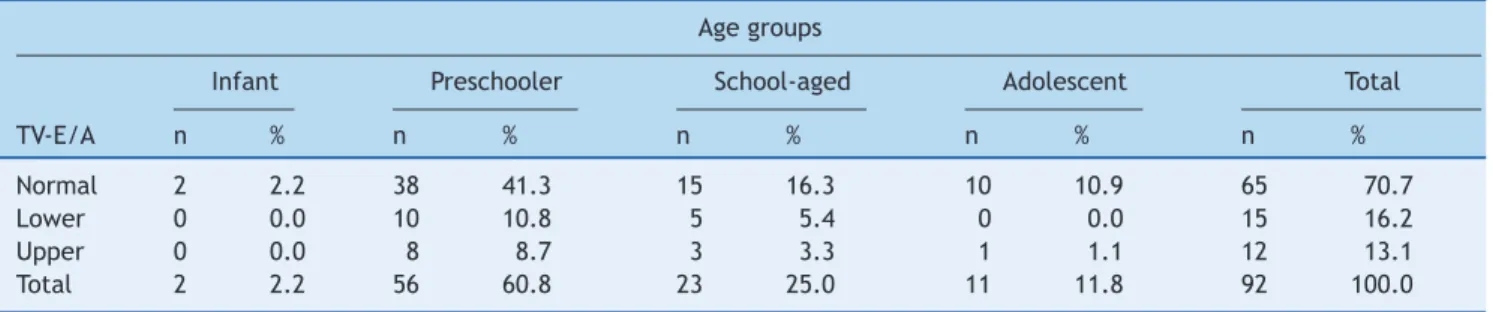
As for the mitral E/A ratio, ten children had values below the minimum normal value (1.1), 26 were above the maxi-mum normal value (3.9), and the remaining 57 were normal. When analyzing the tricuspid E/A ratio, normal values were found in 65 children. In contrast, in 15 the ratio was lower than the minimum normal value (1.58), and 12 had values above the maximum normal value (3.1).
As shown in Tables 1 and 2, the upper category of the mitral E/A ratio included 26 children (28.0%), and when assessing the association between the age groups, a slight predominance of preschoolers was observed (14.0%). The same occurred in the lower category, with eight of ten chil-dren. The upper tricuspid E/A ratio occurred in eight of 12 preschoolers and the lower in ten of the 15 individuals in this age group.
When compassing the E/A ratio of both valves with the immunological status, nutritional status, and blood hemoglobin levels, no trend of association was observed.
The comparison between the mitral and tricuspid E/A ratios showed no association. The left heart abnormalities were not associated with abnormalities in the right side. Fifty of the 92 children had right and/or left ventricular dysfunction during the study.
Factorial analysis identified two distinct groups of chil-dren who were slightly separated, indicating that they would
Table 2 Categories of tricuspid valve E/A ratio according to age groups.
Age groups
Infant Preschooler School-aged Adolescent Total
TV-E/A n % n % n % n % n %
Normal 2 2.2 38 41.3 15 16.3 10 10.9 65 70.7
Lower 0 0.0 10 10.8 5 5.4 0 0.0 15 16.2
Upper 0 0.0 8 8.7 3 3.3 1 1.1 12 13.1
Total 2 2.2 56 60.8 23 25.0 11 11.8 92 100.0
1.0
0.5
0.0
–0.5
–1.0
–1.5
–2.0
–1.5 –1.0 –0.5 0.0 0.5 1.0 1.5 2.0 C1 D1 B2
Dimension 1 : 20.44% of inertia
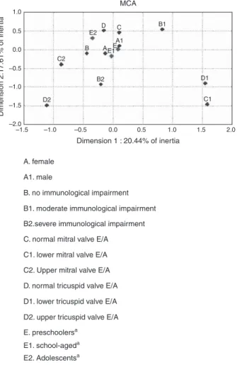
A. female
A1. male
B. no immunological impairment
B1. moderate immunological impairment
B2.severe immunological impairment
C. normal mitral valve E/A
C1. lower mitral valve E/A
C2. Upper mitral valve E/A
D. normal tricuspid valve E/A
D1. lower tricuspid valve E/A
D2. upper tricuspid valve E/A
E. preschoolersa
E1. school-ageda
E2. Adolescentsa
C2
D2
B E2
D C B1
A1
MCA
Dimension 2:17.61% of iner
tia
E1 A E
Figure 1 Multiple correlation analysis (MCA) between cate-gorical variables.
aSupplementary variables.
need further clinical evaluation, as they probably consti-tuted two classes with distinct characteristics.
The results of the multiple correlation analysis for all seven possible dimensions demonstrated that the first dimension had the highest percentage of inertia, or 20.44% of the total inertia. After including the second dimension analysis, the percentage of inertia increased to 38.05%.
The numbers generated from the simple and inertia val-ues for all dimensions of the 11 independent variables, in two dimensions, contributed to the categorical behavior analysis. However, data generated by multiple correlation analysis showed an independent behavior, with no associa-tion between categorical variables (Fig. 1).
Discussion
HIV-infected children are predisposed to cardiac dysfunction as an independent predictor of all-cause mortality, and the exact pathogenesis of cardiac events remains uncertain, but it is most likely multifactorial.9,28
To prevent complications, the assessment of subclinical cardiac abnormalities is suggested. In adults, an indepen-dent association between diastolic dysfunction and HIV infection was reported in a series of studies, even among
asymptomatic HIV-infected patients in the early stages of the disease.3,16---20
It is possible that the diastolic alterations may be an early finding, but their impact on the clinical course of the disease and the possible reversibility of severe complications still need to be elucidated.3,13---17,29
Diastolic dysfunction was found in 50 (54.3%) children, regardless of the side. When the left and right sides were evaluated separately, it was observed that 38.7% (95% CI: 28.8%-48.6%) had left diastolic dysfunction and 29.4% (95% CI: 20.1%-38.7%) had right diastolic dysfunction. On the left side, there was a predominance of decreased myocardial compliance (28.0%; 95% CI: 8.9%-37.1%) compared to 10.7% of abnormal relaxation (95% CI: 4.4%-17.0%). The situation was the opposite on the right side of the heart, with a slight predominance of abnormal relaxation (16.3%; 95% CI: 8.7%-23.7%) compared to the decrease in compliance (13.1%; 95% CI: 6.2%-20.0%).
Plein et al.23reported abnormalities in the left ventricu-lar filling pattern, such as reduction in the maximum velocity of the E wave and increased deceleration time, consistent with diastolic dysfunction in HIV-infected children, classi-fied as stage C according to the classification reviewed by the CDC.
Reports on left ventricular diastolic dysfunction in chil-dren are limited, and the existing are almost always associated with systolic dysfunction, as they are usually symptomatic.22,30
The fact that the immunological status and physical examination did not show a direct association with car-diac involvement demonstrates that HIV-infected children and adolescents should be submitted to Doppler echocar-diographic study as part of the evaluation, even when asymptomatic from a cardiovascular perspective.
Limitations of this study are related to the design, since a non-probabilistic, convenience sample was used, limiting the external validity of these results. The selection bias, inherent to investigations in referral centers, was consid-ered in the sample composition, when the authors planned to study all potentially eligible children.
This sample was characterized as a more selective group. Potential confounders, including severe anemia and mod-erate to severe protein-calorie malnutrition, showed low frequencies, and it was not possible to assess their influence as associated causes of cardiac dysfunction. However, this fact reflects the studied group’s stable condition, as anemia and malnutrition are still prevalent conditions in Brazil.
Cardiac diastolic dysfunction occurred in patients with the selected characteristics, and there was no association with immunological status. Decreased myocardial compli-ance was more frequent in the left ventricle and abnormal relaxation in the right ventricle.
Funding
Infectious Disease Department of HIJG authorized the use of its facilities for the primary care of patients and for the explanation necessary to the informed consent.
Conflicts of interest
The authors declare no conflicts of interest.
References
1. Starc TJ, Lipshultz SE, Easley KA, Kaplan S, Bricker JT, Colan SD, et al. Incidence of cardiac abnormalities in children with human immunodeficiency virus infection: the prospective P2C2 HIV study. J Pediatr. 2002;141:327---34.
2. Langston C, Cooper ER, Goldfarb J, Easley KA, Husak S, Sun-kle S, et al. Human immunodeficiency virus-related mortality in infants and children: data from the pediatric pulmonary and cardiovascular complications of vertically transmitted HIV (P(2)C(2)) Study. Pediatrics. 2001;107:328---38.
3. Hsue PY, Hunt PW, Ho JE, Farah HH, Schnell A, Hoh R, et al. Impact of HIV infection on diastolic function and left ventricular mass. Circ Heart Fail. 2010;3:132---9.
4. Barbarinia G, Barbaro G. Incidence of the involvement of the cardiovascular system in HIV infection. AIDS. 2003;17: S46---50.
5. Lipshultz SE, Easley KA, Orav EJ, Kaplan S, Starc TJ, Bricker JT, et al. Cardiac dysfunction and mortality in HIV-infected children: the Prospective P2C2HIV Multicenter Study. Pediatric Pulmonary and Cardiac Complications of Vertically Trans-mitted HIV Infection (P2C2 HIV) Study Group. Circulation. 2000;102:1542---8.
6. Plebani A, Esposito S, Pinzani R, Fesslova V, Bojanin J, Salice P, et al. Effect of highly active antiretroviral therapy on cardio-vascular involvement in children with human immunodeficiency virus infection. Pediatr Infect Dis J. 2004;23:559---63.
7. Shah I, Prabhu SS, Sumitra V, Shashikiran HS. Cardiac dysfunc-tion in HIV infected children: a pilot study. Indian Pediatr. 2005;42:146---9.
8. Lubega S, Zirembuzi GW, Lwabi P. Heart disease among children with HIV/AIDS attending the paediatric infectious disease clinic at Mulago Hospital. Afr Health Sci. 2005;5:219---26.
9. Brown SC, Schoeman CJ, Bester CJ. Cardiac findings in children admitted to a hospital general ward in South Africa: a compari-son of HIV-infected and uninfected children. Cardiovasc J S Afr. 2005;16:206---10.
10. Cunha Mdo C, Siqueira Filho AG, Santos SR, Abreu TF, Oliveira RH, Baptista DM, et al. AIDS in childhood: cardiac involvement with and without triple combination antiretroviral therapy. Arq Bras Cardiol. 2008;90:11---7.
11. Herdy GV, Pinto CA, Lopes VG, Ribeiro RP, Gomes IM, Tchou HY, et al. Study of the cardiac alterations in HIV-infected children consequent to the antiretroviral therapy. Prospective study of 47 cases. Arq Bras Cardiol. 2003;80:311---20.
12. Diógenes MS, Succi RC, Machado DM, Moisés VA, Novo NF, Car-valho AC. Cardiac longitudinal study of children perinatally exposed to human immunodeficiency virus type 1. Arq Bras Car-diol. 2005;85:233---40.
13. Coudray N, de Zuttere D, Force G, Champetier de Ribes D, Pourny JC, Antony I, et al. Left ventricular diastolic function in asymptomatic and symptomatic human immunode-ficiency virus carriers: an echocardiographic study. Eur Heart J. 1995;16:61---7.
14. Cardoso JS, Moura B, Martins L, Mota-Miranda A, Rocha Gonc¸alves F, Lecour H. Left ventricular dysfunction in human
immunodeficiency virus (HIV)-infected patients. Int J Cardiol. 1998;63:37---45.
15. Longo-Mbenza B, Seghers LV, Vita EK, Tonduangu K, Bayekula M. Assessment of ventricular diastolic function in AIDS patients from Congo: a Doppler echocardiographic study. Heart. 1998;80:184---9.
16. Martínez-García T, Sobrino JM, Pujol E, Galvez J, Benítez E, Girón-González JA. Ventricular mass and diastolic function in patients infected by the human immunodeficiency virus. Heart. 2000;84:620---4.
17. Nayak G, Ferguson M, Tribble DR, Porter CK, Rapena R, Marchi-celli M, et al. Cardiac diastolic dysfunction is prevalent in HIV-infected patients. AIDS Patient Care STDS. 2009;23:231---8. 18. Schuster I, Thöni GJ, Edérhy S, Walther G, Nottin S, Vinet A, et al. Subclinical cardiac abnormalities in human immunodefi-ciency virus-infected men receiving antiretroviral therapy. Am J Cardiol. 2008;101:1213---7.
19. Aggarwal P, Sharma A, Bhardwaj R, Raina R. Myocardial dysfunc-tion in human immunodeficiency virus infecdysfunc-tion: an echocardi-ographic study. J Assoc Physicians India. 2009;57:745---6. 20. Reinsch N, Neuhaus K, Esser S, Potthoff A, Hower M, Brockmeyer
NH, et al. Prevalence of cardiac diastolic dysfunction in HIV-infected patients: results of the HIV-HEART study. HIV Clin Trials. 2010;11:156---62.
21. Meng Q, Lima JA, Lai H, Vlahov D, Celentano DD, Strathdee S, et al. Use of HIV protease inhibitors is associated with left ven-tricular morphologic changes and diastolic dysfunction. J Acquir Immune Defic Syndr. 2002;30:306---10.
22. Lipshultz SE, Chanock S, Sanders SP, Colan SD, Perez-Atayde A, McIntosh K. Cardiovascular manifestations of human immuno-deficiency virus infection in infants and children. Am J Cardiol. 1989;63:1489---97.
23. Plein D, Van Camp G, Cosyns B, Alimenti A, Levy J, Vanden-bossche JL. Cardiac and autonomic evaluation in a pediatric population with human immunodeficiency virus. Clin Cardiol. 1999;22:33---6.
24. Centers for Disease Control and Prevention. 1994 Revised clas-sification system for human immunodeficiency virus infection in children less than 13 years of age; official authorized addenda: human immunodeficiency virus infection codes and official guidelines for coding and reporting ICD-9-CM. MMWR. 1994;43:1---20.
25. Mora JO. A new method for estimating a standardized preva-lence of child malnutrition from anthropometric indicators. Bull World Health Organ. 1989;67:133---42.
26. Grenadier E, Oliveira Lima C, Allen HD, Sahn DJ, Vargas Barron J, Valdes-Cruz LM, et al. Normal intracardiac and great ves-sel Doppler flow velocities in infants and children. J Am Coll Cardiol. 1984;4:343---50.
27. Bu’Lock FA, Mott MG, Martin RP. Left ventricular diastolic func-tion in children measured by Doppler echocardiography: normal values and relation with growth. Br Heart J. 1995;73:334---9. 28. Bowles NE, Kearney DL, Ni J, Perez-Atayde AR, Kline MW, Bricker
JT, et al. The detection of viral genomes by polymerase chain reaction in the myocardium of pediatric patients with advanced HIV disease. J Am Coll Cardiol. 1999;34:857---65.
29. Blanchard DG, Hagenhoff C, Chow LC, McCann HA, Dit-trich HC. Reversibility of cardiac abnormalities in human immunodeficiency virus (HIV)-infected individuals: a serial echocardiographic study. J Am Coll Cardiol. 1991;17:1270---6. 30. Lipshultz SE, Easley KA, Orav EJ, Kaplan S, Starc TJ, Bricker