NeuroimagiNg iN ParkiNsoNism
A study with magnetic resonance and spectroscopy
as tools in the differential diagnosis
Luiz Felipe Rocha Vasconcellos
1, Sergio A. Pereira Novis
2,
Denise Madeira Moreira
3,4, Ana Lucia Z. Rosso
2, Ana Claudia C.B. Leite
5Abstract – The differential diagnosis of Parkinsonism based on clinical features, sometimes may be difficult. Diagnostic tests in these cases might be useful, especially magnetic resonance imaging, a noninvasive exam, not as expensive as positron emission tomography, and provides a good basis for anatomical analysis. The magnetic resonance spectroscopy analyzes cerebral metabolism, yielding inconsistent results in parkinsonian disorders. We selected 40 individuals for magnetic resonance imaging and spectroscopy analysis, 12 with Parkinson’s disease, 11 with progressive supranuclear palsy, 7 with multiple system atrophy (parkinsonian type), and 10 individuals without any psychiatric or neurological disorders (controls). Clinical scales included Hoenh and Yahr, unified Parkinson’s disease rating scale and mini mental status examination. The results showed that patients with Parkinson’s disease and controls presented the same aspects on neuroimaging, with few or absence of abnormalities, and supranuclear progressive palsy and multiple system atrophy showed abnormalities, some of which statistically significant. Thus, magnetic resonance imaging and spectroscopy could be useful as a tool in differential diagnosis of Parkinsonism.
KeY WorDs: Parkinson’s disease, progressive supranuclear palsy, multiple system atrophy, magnetic resonance, spectroscopy.
Neuroimagem no parkinsonismo: estudo com ressonância magnética e espectroscopia por ressonância como ferramentas no diagnóstico diferencial
Resumo – o diagnóstico diferencial do parkinsonismo baseado em parâmetros clínicos pode ser difícil. Alguns exames complementares podem ser úteis, especialmente a ressonância magnética, um método não invasivo, de menor custo quando comparado a tomografia por emissão de pósitrons, proporcionando uma análise anatômica satisfatória. A ressonância por espectroscopia analisa o metabolismo cerebral, com resultados variáveis na literatura no estudo das síndromes parkinsonianas. selecionamos 40 indivíduos para realização de ressonância magnética e espectroscopia, sendo 12 com doença de Parkinson, 11 com paralisia supranuclear progressiva, 7 com atrofia de múltiplos sistemas tipo parkinsoniana e 10 indivíduos sem manifestações neurológicas ou psiquiátricas (grupo controle). As escalas clínicas analisadas foram a de Hoenh e Yahr, unified Parkinson’s disease rating scale e o mini-exame do estado mental. os resultados encontrados revelaram que pacientes com doença de Parkinson e controle apresentavam em geral o mesmo aspecto por imagem enquanto os grupos paralisia supranuclear progressiva e atrofia de múltiplos sistemas com anormalidades, havendo significância estatística em algumas variáveis. A ressonância magnética e a espectroscopia podem ser úteis no diagnóstico diferencial do parkinsonismo.
PAlAvrAs-CHAve: Parkinsonismo, doença de Parkinson, paralisia supranuclear progressiva, atrofia de múltiplos sistemas, ressonância magnética, espectroscopia.
Universidade Federal do rio de Janeiro, Hospital Universitário Clementino Fraga Filho and Instituto de Neurologia Deolindo Couto and Hospital Pró-Cardíaco: 1Hospital dos servidores do estado, rio de Janeiro rJ, Brazil; 2Hospital Universitário Clementino Fraga Filho and 3Instituto de Neurologia Deolindo Couto, Universidade Federal do rio de Janeiro, rio de Janeiro rJ, Brazil; 4Hospital Pró-Cardíaco, serviço de radiologia, rio de Janeiro rJ, Brazil; 5Fundação oswaldo Cruz, rio de Janeiro rJ, Brazil.
received 14 July 2008, received in inal form 16 october 2008. Accepted 25 November 2008.
The Parkinsonian syndrome or parkinsonism (PK) cor-respond to clinical signs of rigidity, bradykinesia, tremor, and postural instability, and the presence of two of them is required to deine probable PK, and one of these two signs must be tremor or rigidity. The PK is classiied as pri-mary, secondary, atypical or plus, and hereditary1,2. The
ac-curate diagnosis may be dificult based upon clinical signs, especially at early stages, and in some cases only after the performance of neuropathological studies it could be possible to deine the diagnosis3-7. It is important to
deter-mine this due to the different prognosis, pharmacothera-py, and epidemiological analysis8,9.
Magnetic resonance imaging (MrI) and spectroscopy by MrI (Mrs) are noninvasive tools helping the physician to establish a more accurate diagnosis. MrI offers an ad-equate analysis of abnormalities in the basal nuclei, mid-brain, pons, medulla, and cerebellum, which are impaired in atypical PK10-23.
We selected patients with diagnosis of PK and ana-lyzed the usefulness of neuroimaging (MrI and Mrs) in the differential diagnosis of this condition.
method
We designed a prospective, case-control, double-blind, 24 months study. The MrI was performed in a Ge machine, 1.5 Te-sla sigma Horizon model, the sequences analyzed were T 1, T 2,
lair, diffusion, axial-oblique in T 2 in Fast spin-echo (Fse) and Proton Density (PD) and T 2 in spin-echo (se). In addition to 5 mm slices, we included 3 mm slices in the lentiform nucleus. The Mrs was single voxel (8 cc), Press technique (Tr/Te=1500/50) bilaterally in lentiform nucleus, midbrain, white matter of fron-tal lobe and hippocampus.
Informed consent was obtained from all patients or their immediate relatives, and the study was approved by the ethics Committee of the institutions involved.
Forty individuals were included in this study (age range: 50 to 85 years), 30 with Parkinsonian syndrome and 10 without any neurological or psychiatric disorders. Four patients were
exclud-ed, two due to cerebrovascular disease showed in MrI, and two related to technical problems during MrI.
All individuals were examined by the same neurologist, and 26 patients met the criteria for probable Parkinson’s disease (PD) [n=10], (Gelb et al.24 ), progressive supranuclear palsy (PsP) [n=10],
(Tolosa et al.25), and multiple system atrophy-parkinsonian type
(MsA-P) [n=6], (Gilman et al.26). For clinical assessment, the scales
adopted were Hoehn-Yahr stage27, uniied Parkinson’s disease
rat-ing scale (UPDrs) Part III28 and mini-mental status examination
(MMse)29. The patients performed the Tilt Table test for
evalu-ation of dysautonomia.
The indication for MrI was the same for all individuals: “parkin-sonism”, so that the radiologist did not know the actual diagnosis.
The variables in MrI were: anteroposterior diameter of the
medulla, pons, midbrain and fourth ventricle, transverse diam-eter of lateral and third ventricles, presence of cerebral and/or cerebellar atrophy, and signal abnormalities in white matter, len-tiform, midbrain, pons and medulla, linear posterolateral hyper-signal in lentiform nucleus, and transverse hyper-signal in the pons.
Mrs was performed bilaterally on white matter of frontal lobe, lentiform nucleus, midbrain and hippocampus. We used the N-acetyl aspartate/creatine (NAA/Cr) and N-acetyl aspar-tate/choline (NAA/Cho) relation. The value adopted was the mean of both white matter from the frontal lobe and the hip-pocampus, and the contralateral relation of the most affected side on the lentiform and midbrain, and when there was sym-metry, the mean was obtained.
Statistical analysis
For quantitative variables the statistical analysis adopted was student’s t-test or the Mann-Whitney test, and for quali-tative the X2, Fisher and Mantel Haenszel. There was statistical signiicance when p value was <0.05.
results
The clinical variables that did not show differences statistically signiicant among the three groups were: age, disease duration, and sex (Table 1).
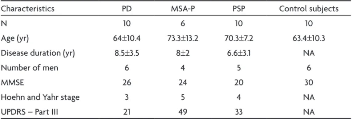
Table 1. Demographic and clinical characteristics by patient group.
Characteristics PD MsA-P PsP Control subjects
N 10 6 10 10
Age (yr) 64±10.4 73.3±13.2 70.3±7.2 63.4±10.3
Disease duration (yr) 8.5±3.5 8±2 6.6±3.1 NA
Number of men 6 4 5 6
MMse 26 24 20 30
Hoehn and Yahr stage 3 5 4 NA
UPDrs – Part III 21 49 33 NA
hn Yahr p=0.021 and UPDrs p=0.029), and a trend to sta-tistical signiicance in PD and PsP (Hoehn and Yahr, and UPDrs p=0.08).
Patients with PsP presented lower scores in MMse, followed by MsA-P and PD, and there was statistical sig-niicance in the three groups comparing to controls (PD p=0.046; MsA-P p=0.002, and PsP p=0.0004) (Table 1).
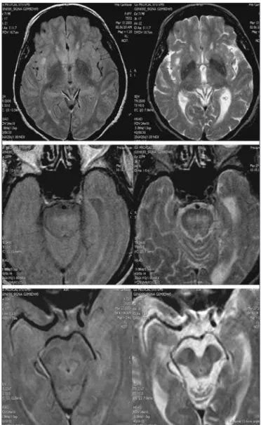
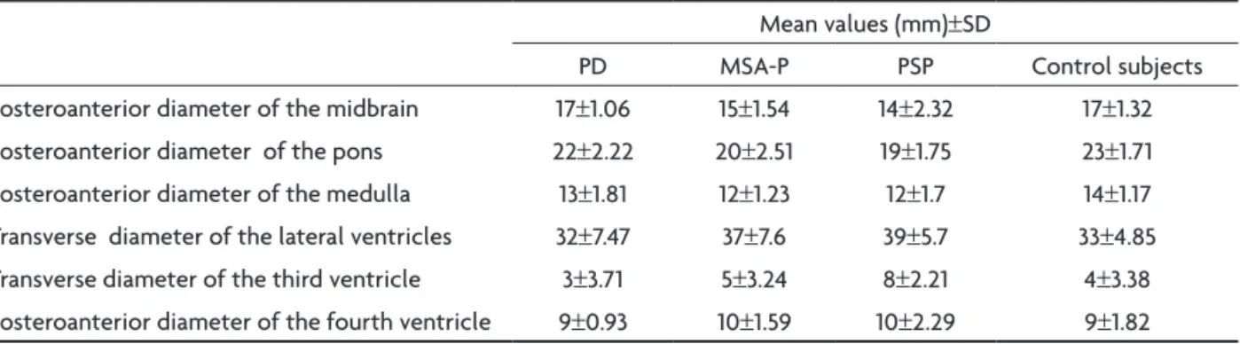
Image variables demonstrated cerebral atrophy in all cases of PsP and MsA-P, having statistical significance in PD versus PsP (p=0.001), PD versus MsA-P (p=0.006), controls versus PsP (p=0.011), and controls versus MsA-P (0.043). Cerebellar atrophy was more common in MsA-P and PsP, with statistical signiicance in PD versus MsA-P (p=0.043), controls versus PsP (p=0.034) and controls ver-sus MsA-P (p=0.010). We observed a higher prevalence of white matter alterations in atypical PK with no statistical signiicance. signal change in the lentiform nucleus was observed more commonly in MsA-P and PsP, but no sta-tistical signiicance was documented (Figs 1–3).
The posterolinear increased signal in the lentiform nucleus was demonstrated only in the MsA-P and PsP groups, presenting statistical signiicance when
compar-Fig 1. Hyposignal in the lentiform nucleus (found in 67% of MSA-P group), and hypersignal in the pons (found in 33% of MSA-P group) and the midbrain on T2, lair or DP sequences (found in 70% of PSP group).
Dysautonomia was documented in 20% of PD and 100% of MsA-P.
In the motor scales (UPDrs and Hoehn and Yahr), the results showed higher scores in PsP and MsA-P than in PD. There was statistical signiicance in PD versus MsA-P
(Hoe-Fig 2. Posterolateral linear hypersignal in the lentiform nucleus, with asymmetric symptoms, T2 sequence (found in 50% in MSA-P group).
ing PD versus MsA-P (p=0.03), and controls versus MsA-P (p=0.03).
signal changes in the midbrain were more commonly observed in PsP, and in MsA-P in the pons, with statistical signiicance in midbrain (p=0.0015).
The quantitative variables detailed in Table 2 demon-strated that some measurement of brainstem and ven-tricular system had statistical signiicance to differenti-ate atypical PK and PD/control group. The measurements that revealed statistic signiicance according to region and groups were:
Midbrain – PD versus PsP (p=0.002), PD versus MsA-P (p=0.012), controls versus PsP (p=0.002) and controls ver-sus MsA-P (0.010).
Pons – DP versus PsP (p=0.012), PsP versus controls (p=0.007) and MsA-P versus controls (p=0.01).
Medulla – DP versus MsA-P (p=0.041), PsP versus con-trols (p=0.008) and MsA-P versus concon-trols (p=0.001).
Lateral ventricles – PD versus PsP (p=0.041) and controls versus PsP (p=0.045).
Third ventricle – DP versus PsP (p=0.015) and PsP versus controls (p=0.009).
Fourth ventricle – DP versus PsP (p=0.037) and DP versus MsA-P (p=0.024).
The values of Mrs are related in Table 3 and some re-duction showed statistical signiicance:
NAA/Cr in the lentiform nucleus – PD versus PsP (p=0.049) and PsP versus controls (p=0.036).
NAA/Cr in the hippocampus – PsP versus control (p=0.0007). NAA/Cho in the midbrain – PsP versus MsA (p=0.028) and PsP versus control (p=0.046).
discussioN
The increase of life expectancy results in a raise of degenerative disorders. PD is one of the most common neurodegenerative disease (followed by Alzheimer dis-ease), as epidemiological studies show in the literature30.
Parkinsonian signs may be seen in different medical condi-tions, having variable course, treatment and prognosis so it is important to determine an accurate diagnosis as soon as possible8,9. Based only in clinical data, especially in the
early stages of the disease, physicians may not establish a correct diagnosis3-7.
The accuracy of clinical diagnosis of PK is variable, in PD ranging from 76% to 90%, and in others PK the accuracy is even lower3-7. one study conducted in a movement
dis-orders specialized center, showed that the positive
predic-Table 2. Magnetic resonance variables (quantitative).
Mean values (mm)±sD
PD MsA-P PsP Control subjects
Posteroanterior diameter of the midbrain 17±1.06 15±1.54 14±2.32 17±1.32
Posteroanterior diameter of the pons 22±2.22 20±2.51 19±1.75 23±1.71
Posteroanterior diameter of the medulla 13±1.81 12±1.23 12±1.7 14±1.17
Transverse diameter of the lateral ventricles 32±7.47 37±7.6 39±5.7 33±4.85
Transverse diameter of the third ventricle 3±3.71 5±3.24 8±2.21 4±3.38
Posteroanterior diameter of the fourth ventricle 9±0.93 10±1.59 10±2.29 9±1.82
PD, Parkinson disease; MsA-P, multiple system atrophy; PsP, progressive supranuclear palsy.
Table 3. Spectroscopy variables.
variables PD
(N=10)
PsP (N=10)
MsA (N=6)
Control subjects (N=10)
NAA/Cr lentiform nucleus Mean 1.46 1.31 1.4 1.45
NAA/Cr midbrain Mean 1.77 1.5 1.74 1.65
NAA/Cr frontal lobe Mean 1.51 1.47 1.53 1.56
NAA/Cr Hippocampus Mean 1.36 1.2 1.33 1.42
NAA/Chol lentiform nucleus Mean 1.64 1.43 1.55 1.56
NAA/Chol midbrains Mean 1.53 1.34 1.69 1.56
NAA/Chol frontal lobe Mean 1.71 1.57 1.55 1.62
NAA/Chol Hippocampus Mean 1.4 1.41 1.42 1.5
tive value of PD was 98.6%, and to atypical parkinsonism 71.4%, conirming that the diagnosis of atypical PK, even in specialized centers, is sometimes dificult to establish7.
some diagnostic tests could be useful for the differ-ential diagnosis of PK, and MrI is one of the most impor-tant10-23. our objective was to determine the usefulness of
MrI and Mrs in a PK group, based on well known imaging aspects according to the subtype of PK, assessing which variables had statistic signiicance in these groups.
We included the three PK that most frequently lead to misdiagnosis: PD, MsA-P, and PsP, all compared to control group. The criteria used to clinical diagnosis was the most speciic, as showed in the literature24-26.
We used three clinical scales: motor part of UPDrs, Hoehn and Yahr and MMse27-29. These scales showed
in-creased motor impairment (higher scores in UPDrs and Hoehn-Yahr) in the MsA-P, followed by PsP, and increased cognitive impairment (lower scores of MMse) in PsP, fol-lowed by MsA-P. We did not observe a correlation be-tween the duration of the symptoms with Mrs abnor-malities, but with the clinical diagnosis of patient.
MrI variables demonstrated that some are helpful to differentiated PK syndromes, as the presence of cerebral and cerebellar atrophy and signal enhancement of some encephalic structures (lentiform nucleus, midbrain and pons), more common in atypical PK.
The decreased signal enhancement in the lentiform nucleus may be observed in normal aging, so in our study we only considered it as “abnormal” if the hypointensity was moderate to severe15,31. our data showed that
moder-ate to severe decrease hypointensity in lentiform nucleus was observed more frequently in MsA and PsP, with no difference between PD and control groups and when it was associated with posterolateral linear hypersignal in putamen, suggested the diagnosis of atypical PK (more frequently in MsA group).
The most useful measurement of encephalic diameter in our study was the midbrain, as it had been shown by Warmutth et al.18. values below 15 mm in the midbrain
sug-gested PsP or MsA-P, with lower values seen in PsP. some values of Mrs had statistical signiicance, the most useful were from the lentiform nucleus, hippocam-pus, and midbrain, depending on the diagnosis, indicating a severe neuronal impairment (neuronal death). There are few studies in which the brainstem is evaluated by Mrs, due to technical dificulties (bone proximity). In our study we demonstrated that it is feasible, but we had to repeat the exam, in some cases several times, to achieve a con-sistent chart. The study done by Watanabe et al.23
dem-onstrated the usefulness of Mrs of the pons in MsA
pa-tients. As the midbrain is the most affected area in PsP, we analyzed it by Mrs. We have found NAA/Cho decrease in midbrain of PsP group with statistical signiicance, indicat-ing neuronal loss.
Based on our data we concluded that: (1) Patients with PsP and MsA-P presented increased motor and cognitive impairment in the scales used, correlating with decrease in NAA/Cr in lentiform nucleus and NAA/Cho in mid-brain in the PsP group; (2) Cerebral and cerebellar atrophy were more prevalent and severe in PsP and MsA-P groups; (3) linear hypersignal in the lateral portion of the puta-men, hypersignal in midbrain and in pons, all suggest the diagnosis of PsP or MsA-P; (4) Midbrain or pons atrophy suggests atypical parkinsonism, the former PsP, and the latter MsA-P; (5) Comparing the two methods, MrI and Mrs, the former had better applicability.
our study showed that anatomical analysis through MrI and Mrs of some areas could be useful in the differ-ential diagnosis of PD and atypical PK, helping physicians to establish a more accurate diagnosis of PK.
refereNces
1. Shannon KM. Movement disorders. In: Bradley WG, Daroff RB, Fen-ichel GM, Jankovic J (Eds). Neurology in clinical practice. 4th ed. Phila-delphia: Butterworth and Heinemann 2004:2125-2168.
2. Fahn S, Przedborski S. Distúrbios do movimento. In: Rowland LP (Ed). Tratado de neurologia – Merritt´s. 10ª ed. Tradução: Araújo CLC, Mun-dim FD. Revisão técnica: Cavalcanti JLS. Rio de Janeiro: Guanabara Koogan 2000:589-604.
3. Rajput AH, Rozdilsky B, Rajput A. Accuracy of clinical diagnosis in par-kinsonism: a prospective study. Can J Neurol Sci 1991;18:275-278. 4. Hughes AJ, Daniel SE, Kilford L, Lees AJ. Accuracy of clinical
diagno-sis of idiopathic Parkinson’s disease: a clinico-pathological study of 100 cases. J Neurol Neurosurg Psychiatry 1992;55:181-184.
5. Jankovic J, Rajput AH, McDermott MP, Perl DP. The evolution of diag-nosis in early Parkinson disease. Parkinson Study Group. Arch Neurol 2000;57:369-372.
6. Carrilho PE, Barbosa ER. Progressive supranuclear palsy in a sample of Brazilian population: clinical features of 16 patients. Arq Neuropsiquiatr 2002;60:917-922.
7. Hughes AJ, Daniel SE, Ben-Shlomo Y, Lees AJ. The accuracy of diagno-sis of parkinsonian syndromes in a specialist movement disorder ser-vice. Brain 2002;125:861-870.
8. Tison F, Yekhlef F, Chrysostome V, et al. Parkinsonism in multiple sys-tem atrophy: natural history, severity (UPDRS-III), and disability assess-ment compared with Parkinson’s disease. Mov Disord 2002;17:701-709. 9. Watanabe H, Saito Y, Terao S, et al. Progression and prognosis in multi-ple system atrophy: an analysis of 230 Japanese patients. Brain 2002;125: 1070-1083.
10. Bhattacharya K, Saadia D, Eisenkraft B, et al. Brain magnetic resonance imaging in multiple-system atrophy and Parkinson disease: a diagnos-tic algorithm. Arch Neurol 2002;59:835-842.
11. Warmuth-Metz M, Naumann M, Csoti I, Solymosi L. Measurement of the midbrain diameter on routine magnetic resonance imaging: a sim-ple and accurate method of differentiating between Parkinson disease and progressive supranuclear palsy. Arch Neurol 2001;58:1076-1079. 12. Righini A, Antonini A, Ferrarini M, et al. Thin section MR study of the
basal ganglia in the differential diagnosis between striatonigral degen-eration and Parkinson disease. J Comput Assist Tomogr 2002;26:266-271. 13. Schocke MF, Seppi K, Esterhammer R, et al. Diffusion-weighted MRI
differentiates the Parkinson variant of multiple system atrophy from PD. Neurology 2002;58:575-580.
15. Kraft E, Trenkwalder C, Auer DP. T2*-weighted MRI differentiates mul-tiple system atrophy from Parkinson’s disease. Neurology 2002;59: 1265-1267.
16. Hutchinson M, Raff U. Structural changes of the substantia nigra in Par-kinson’s disease as revealed by MR imaging. Am J Neuroradiol 2000;21: 697-701.
17. Schrag A, Good CD, Miszkiel K, et al. Differentiation of atypical par-kinsonian syndromes with routine MRI. Neurology 2000;54:697-702. 18. Asato R, Akiguchi I, Masunaga S, Hashimoto N. Magnetic resonance
imaging distinguishes progressive supranuclear palsy from multiple system atrophy. J Neural Transm 2000;107:1427-1436.
19. Vymazal J, Righini A, Brooks RA, et al. T1 and T2 in the brain of healthy subjects, patients with Parkinson disease, and patients with multiple system atrophy: relation to iron content. Radiology 1999;211:489-495. 20. Tedeschi G, Litvan I, Bonavita S, et al. Proton magnetic resonance
spec-troscopic imaging in progressive supranuclear palsy, Parkinson’s dis-ease and corticobasal degeneration. Brain 1997;120:1541-1552. 21. Abe K, Terakawa H, Takanashi M, et al. Proton magnetic resonance
spec-troscopy of patients with parkinsonism. Brain Res Bull 2000;52: 589-595. 22. Federico F, Simone IL, Lucivero V, et al. Usefulness of proton magnet-ic resonance spectroscopy in differentiating parkinsonian syndromes. Ital J Neurol Sci 1999;20:223-239.
23. Watanabe H, Fukatsu H, Katsuno M, et al. Multiple regional 1H-MR spectroscopy in multiple system atrophy: NAA/Cr reduction in pontine base as a valuable diagnostic marker. J Neurol Neurosurg Psychiatry 2004;75:103-109.
24. Gelb DJ, Oliver E, Gilman S. Diagnostic criteria for Parkinson disease. Arch Neurol 1999;56:33-39.
25. Tolosa E, Valldeoriola F, Marti MJ. Clinical diagnosis and diagnostic cri-teria of progressive supranuclear palsy (Steele-Richardson-Olszewski syndrome). J Neural Transm 1994;42(Suppl):S15-S31.
26. Gilman S, Low PA, Quinn N, et al. Consensus statement on the diag-nosis of multiple system atrophy. J Neurol Sci 1999;163:94-98. 27. Hoehn MM, Yahr MD. Parkinsonism: onset, progression and
mortali-ty. Neurology 1967;17:427-442.
28. We Move. Disponível em <http://www.wemove.org. Acesso em: 17, maio de 2002.
29. Brucki SMD, Nitrini R, Caramelli P, Bertolucci PHF, Okamoto IH. Sug-estões para o uso do mini-exame do estado mental no Brasil. Arq Neu-ropsiquiatr 2003;61:777-782.
30. Prusiner SB. Shattuck lecture-neurodegenerative diseases and prions. N Engl J Med 2001;344:1516-1526.
31. Drayer BP. Imaging of the aging brain: Part I. Normal indings. Radiology