VIEWS AND REVIEWS
Physiopathology of symptoms and signs in
multiple sclerosis
Fisiopatologia dos sintomas e sinais na esclerose múltipla
Maria José Sá
Maybe in accordance with its name, multiple sclerosis (MS), as well as the ield of primary demyelinating diseases of the central nervous system (CNS) as a whole, generates daily an impressive number of publications, dedicated to a variety of multiple aspects, turning impossible one to keep up with the immense body of new data. As expected, most studies ad-dress issues that are still unknown or need to be better under-stood, as the aetiology and pathogenesis of MS. In the last two decades, the literature has been looded with reports of new therapies that allegedly modify the natural history of MS, as such denominated disease-modifying drugs (DMD). It is un-deniable that the discovery of DMD represents a remarkable
step forward in the control of the disease, is a non-stop re-search ield and brings hope to the possibility of cure. Even so, the positive efects of irst-line DMD are considered to be rather modest, since the drugs act in the inlammatory, but not in the neurodegenerative mechanisms of the disease, hence the impact in the long term disability is small, where-as the better eicacy of second-line drugs must be weighed against a poorer safety proile1.
herefore, in the clinical setting, the people alicted with MS must be subjected to a comprehensive and individualized therapeutically intervention, able to control the symptoms and signs of the disease, thus ameliorating their well-being.
MD, PhD Senior Neurologist and Head of MS Clinic, Department of Neurology, Centro Hospitalar São João. Associate and Aggregate Professor, Faculty of Health Sciences, Universidade Fernando Pessoa, Porto, Portugal.
Correspondence: Maria José Sá; Department of Neurology, Centro Hospitalar São João; Alameda Professor Hernâni Monteiro 4200/319; Porto - Portugal; E-mail: [email protected]
Conflict of interest: There is no conlict of interest to declare.
Received 23 March 2012; Received in inal form 16 April 2012; Accepted 23 April 2012
ABSTRACT
The physiopathology of symptoms and signs in multiple sclerosis (MS) is a less divulged topic albeit its importance in the patients’ manage-ment. Objective: It was to summarize the main biophysical and biochemical mechanisms which produce the clinical manifestations in MS.
Results: The mechanisms underpinning neurological deicits are described in the relapsing and in the progressive phases, stressing in-lammatory and neurodegenerative components, especially demyelination, axonal damage and conduction impairment. Transient worsening based in Uhthoff’s phenomenon, mechanisms producing positive symptoms, as paraesthesias and Lhermitte sign due to axonal hiperexcit-ability and ephaptic interactions, and development of cortical symptoms will also be addressed. The variety of processes leading to neural repair and functional recovery in the remitting phase is focused, as remyelination and adaptive changes due to neural plasticity. Conclusion:
The awareness of mechanisms producing symptoms in MS emphasises the role of symptomatic and rehabilitation therapies in the improve-ment of patients’ well-being.
Key words: multiple sclerosis, /physiopathology, demyelination, axonal damage.
RESUMO
A isiopatologia dos sintomas e sinais na esclerose múltipla (EM) é um tópico pouco divulgado apesar da sua importância na abordagem dos doentes. Objetivo: Foi apresentar os principais mecanismos biofísicos e bioquímicos que produzem manifestações clínicas da EM. Resul-tados: Descrevem-se os mecanismos subjacentes aos déices neurológicos nas fases de surto e progressivas, realçando as componentes inlamatória e neurodegenerativa, especialmente desmielinização, lesão axonal e alterações da condução. Serão igualmente referidos os sintomas transitórios explicados pelo fenômeno de Uhthoff, a produção de sintomas positivos, como as parestesias e o sinal de Lhermitte por hiperexcitabilidade axonal e interações efáticas, e o desenvolvimento de sintomas corticais. Apresentam-se os diversos processos de reparação neural e de recuperação funcional nas fases de remissão, como a remielinização e as alterações adaptativas por neuroplastici-dade. Conclusão: O conhecimento dos mecanismos que produzem os sintomas da EM realça o papel das terapêuticas sintomáticas e de reabilitação na melhoria do bem-estar dos doentes.
It must be remembered that MS is a chronic and not curable neurological disease that usually begins in young adults, be-ing the leadbe-ing cause of non-traumatic neurological disabil-ity in early ages, and is unpredictable regarding the clinical manifestations, course and disability. For these reasons, MS represents a major burden to the patients, families, society and health economy2.
In efect, the experience of following many patients in our MS Clinic along more than 25 years emphasises the im-portance of the symptomatic therapy and the rehabilitation approaches, among the global therapeutic armamentarium employed in the disease. As a result and to better understand the role of symptomatic therapies, either pharmacological or non-pharmacological, the physiopathological changes tak-ing place in the demyelinattak-ing lesions which compromise the functioning of the nerve ibres, along the disease evo-lution, must be identiied. his topic is not very frequently addressed in the literature targeting neurological clinicians, which may be explained by its complexity and the assump-tion that those mechanisms belong to biophysical and bio-chemical disciplines. Despite this supposition, it is certain-ly pertinent to remember what is going on the CNS when a MS patient becomes paretic, as example of a negative sign, or feels paraesthesias, traducing a positive symptom. Briely, it has been known since long that the neurological impair-ment in the early stages is due to the conduction block in de-myelinated ibres3 whereas irreversible disability is ascribed to axonal transection and neuronal loss4. On the other hand, the natural reactive mechanisms that seek to counteract the pathological process thorough myelin repair, in order to nor-malize the neural functions, are seen in early phases5,6, but are progressively lost later7,8.
he aim of this work was to revisit the basic mechanisms that underpin the clinical expression of symptoms and signs in multiple sclerosis, which hypothetically may be targeted by symptomatic therapies, in a practical summarized way that can be easily accessed by health professionals dedicated to MS, who wish to understand their patient’s symptoms day by day, to better manage the disease. he mechanisms that produce the clinical manifestations in MS will be presented in the next sections according to their type — negative/positive — and the phase of the disease — relapse, remission, progressive — bear-ing in mind that all processes lead ultimately to axonal injury.
MYELIN AND AXONAL CONDUCTION
he CNS myelin sheath is a lipid-rich multilayered structure composed of oligodendrocytic plasma membranes wrapping around the CNS axons, which provides insulation for electrical impulses (reviewed in the study of Aggarwal et al.9). Myelin is a highly stable membrane periodically interrupted by the nodes of Ranvier that are rich in voltage-gated sodium channels.
In myelinated ibres, the action potential jumps rapidly in a saltatory way, from one node to another, promoting the fast processing of information. Briely, the biophysical prop-erties underlying these phenomena are as follows: the cur-rent is generated by the opening of sodiumchannels in the nodes of Ranvier with a resultant sodium inlux and depo-larization of the axonal membrane; then, the current moves towards next node and is prevented from lowing out by the myelin sheath in the internodal segments; the electrical low ends due to the opening of the potassium channels and sub-sequent repolarization10.
In normal conditions, myelinated axons have a safety fac-tor for transmission, deined as the current available to depo-larize the axolemma divided by the current necessary to do so11, which is several times higher than threshold. his means that the potential action generated in a node is several times superior to the current necessary to ire the next node.
When myelin is damaged, the safety factor is reduced to around the unit and the conduction of demyelinated axons is successful or unsuccessful depending on the milieu condi-tions12-15. As a result, the axon sufers consequent physiologi-cal changes, such as loss of saltatory properties of electriphysiologi-cal conduction, reduction in conduction velocity and a predispo-sition to conduction block.
Mechanisms of symptoms production in the relapsing phase
During relapses, MS patients lose partial or totally some type of physiological functions, traduced by a variety of neg-ative symptoms, as, for instance, paresis, hyposthesia, visu-al impairment, diplopia or ataxia. As daily testiied in MS Clinics, the variability in the clinical manifestations is high among patients, as well as in diferent phases of the disease in each patient. he symptomatic expression depends not only on the location of MS lesions but also on the variety of conduction properties displayed by afected axons.
he main cause of relapse is the failure of axonal conduc-tion at the site of a lesion, which is due to axonal changes, relat-ed both to acute inlammation and demyelination, which pos-sibly act in sequential phases and ultimately proceed together in a complex way. As well, there is evidence that axons may be directly injured and that glial dysfunction may also play a role.
he impairment in the axonal transmission may be main-ly functional due to inlammation, oedema and toxic action of products released in the MS plaques, which happens in the earlier acute phases of a relapse, or entirely structural, in later phases, when axon is exposed by myelin damage in internod-al segments and internod-also by direct axoninternod-al injury in the nodinternod-al and perinodal regions (Table 1).
Inlammation
he recognition that inlammatory components are able to disclose clinical symptoms derives from the knowledge that the very irst event in the new lesions located in clin-ically eloquent areas, related to relapses, is the appearance of inlammatory markers after the focal breakdown of the blood-brain-barrier (BBB)16. Some substances released upon the formation of a new lesion have been pointed out to medi-ate the biophysical consequences of inlammation, leading to transient impairment or block of nerve conduction, such as cytokines, nitric oxid (NO) and antibodies against ion chan-nels. Moreover, proinlammatory cytokines – tumor necrosis factor (TNF)-alpha and interferon gama – may stimulate the formation of inducible form of the enzyme nitric oxide syn-tetase with further liberation of NO, that is a mediator of axo-nal block, particularly in axons afected by demyelination17.
he inlammatory mediators are thought to block axo-nal conduction by a variety of mechanisms that afect ion channels, particularly sodium channels, and mitochondrial energy production12. Besides, the BBB breakdown also may induce changes in the electrophysiology of axons due to leak-age of luid and substances that alter the composition of the extracellular spaces that bathe nervous ibres18.
Whether the inlammatory response may lead to neuro-logical symptoms by itself or needs the coexistence of demy-elination is still matter of debate.
Demyelination
he myelin damage in MS is characteristically segmen-tal, resulting in the loss of whole internode of myelin, and occurs in the typical lesions of the white matter, as well as in cortical lesions. In segmental demyelination, conduction speciically fails at the site of the lesion, whereas the unaf-fected parts of the axon on either side of the lesion continue to conduct normally19.
Since the damage of myelin, sheath eliminates the insula-tor of the internodal segments, the axonal current low is lo-cally interrupted and short-circuited through the uncovered portions of the axon; however, as the density of sodium chan-nels in the axolemma beneath the internodal myelin sheath is low, the impulse propagation is inhibited, leading to a much slower current low or to its interruption20. Further propaga-tion of the acpropaga-tion potential is blocked due to the exposipropaga-tion of voltage-gated potassium channels normally restricted to the internodes. An abnormal redistribution of sodium chan-nels on demyelinated axons has been reported, both in ex-perimental models21 and in MS lesions21,22, with loss of nodal aggregates and appearance of a difuse expression along the denuded portions of the axon.
Given the high safety factor for transmission, demyelin-ation is usually not critical in small portions of the axons (1 or 2 internodes), whereas in longer segments the current tends
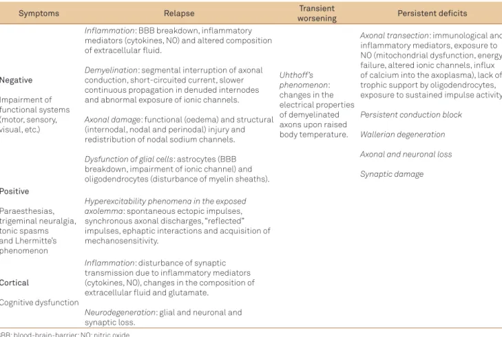
Table 1. Mechanisms that impair axonal conduction and produce clinical manifestations in multiple sclerosis.
Symptoms Relapse Transient
worsening Persistent deficits
Negative
Impairment of functional systems (motor, sensory, visual, etc.)
Inflammation: BBB breakdown, inlammatory mediators (cytokines, NO) and altered composition of extracellular luid.
Demyelination: segmental interruption of axonal conduction, short-circuited current, slower continuous propagation in denuded internodes and abnormal exposure of ionic channels.
Axonal damage: functional (oedema) and structural (internodal, nodal and perinodal) injury and redistribution of nodal sodium channels.
Dysfunction of glial cells: astrocytes (BBB breakdown, impairment of ionic channel) and oligodendrocytes (disturbance of myelin sheaths).
Uhthoff’s phenomenon: changes in the electrical properties of demyelinated axons upon raised body temperature.
Axonal transection: immunological and inlammatory mediators, exposure to NO (mitochondrial dysfunction, energy failure, altered ionic channels, inlux of calcium into the axoplasma), lack of trophic support by oligodendrocytes, exposure to sustained impulse activity.
Persistent conduction block
Wallerian degeneration
Axonal and neuronal loss
Synaptic damage
Positive
Paraesthesias, trigeminal neuralgia, tonic spasms and Lhermitte’s phenomenon
Hyperexcitability phenomena in the exposed axolemma: spontaneous ectopic impulses, synchronous axonal discharges, “relected” impulses, ephaptic interactions and acquisition of mechanosensitivity.
Cortical
Cognitive dysfunction
Inflammation: disturbance of synaptic transmission due to inlammatory mediators (cytokines, NO), changes in the composition of extracellular luid and glutamate.
Neurodegeneration: glial and neuronal and synaptic loss.
to low by continuous propagation. Conduction failure may also be caused by partial myelin thinning along the inter-node and may be even more striking upon myelin loss in the paranodal region14.
Axonal damage
he occurrence of axonal damage in MS is an old obser-vation described by Charcot23, which has been revisited and irmly supported in the last years4. he spectrum of axonal damage may vary from a functional and temporary efect as a result of oedema during relapses to ibre transaction provok-ing persistent neurological sequels.
Marked axonal damage may be found in early disease stages and acute axonal injury correlates with inlamma-tory cells. During a new relapse, the direct harmful stimuli to the axons include axon-speciic antibodies plus comple-ment, CD8 T+ cells perforin, myelin-speciic CD4 T+ cells, NO, matrix metalloproteinases and glutamate24,25. Besides sufering the efects of myelin damage in demyelinated por-tions, axons may be also directly injured in the nodal and perinodal regions, the latter including the paranodes and the juxtaparanodes26. hese nodal and perinodal domains may be impaired by efects of inlammation and axonal swelling, leading to direct and early axonal loss, even in the absence of demyelination. Nodal widening is suicient to block con-duction. Some authors suggest that the axoglial junction in the paranodes, which comprise the axonal adhesion proteins Caspr and contactin, is an area of particular vulnerability dur-ing the initial stages of tissue damage and emphasise the pos-sibility of axoglial antigens being targets for autoimmunity in MS27 and acting like inductors of demyelination. In addition, there is a higher axonal vulnerability due to damaged myelin and demyelination, because when the axon is devoid of my-elin there is a redistribution of the nodal sodium channels that might manipulate the functions and survival of axons.
he mechanisms of axonal transection and permanent loss that provoke persistent neurological deicits will be later described in this article.
Dysfunction of glial cells
In addition, there is evidence that dysfunction of glial cells might inluence the conduction properties of axons. Hence, astrocytic changes not only disturb the integrity of the BBB but also may impair the organization of the ionic channels along the axonal membrane, and oligodendrocytic injury dis-turbs the trophic role to the myelin sheaths, further harming the axoglial junctions in paranodes14.
Mechanisms of transient symptomatic worsening
Apart from relapses, most people with MS experi-ence transient worsening of usual clinical signs and symp-toms, which afect daily life activities, and their complains are sometimes diicult to distinguish from relapses, unless
patients are seen by an experienced neurologist. he daily routine practice in a MS Clinic teaches us that even mildly disabled MS patients may report temporary diiculties in usual motor tasks, as for instance walking or housekeeping, yet sometimes are not able to describe very well the duration of those episodes.
his temporary worsening of function is known since Wilhelm Uhthof ’s descriptions that symptoms can aggra-vate upon heating and improve with body cooling28; namely, after a hot bath or strong exercise, MS patients with a his-tory of optic neuritis reported reversible and stereotyped alterations in vision29. Since then, the heat sensitivity in MS is unanimously recognized and the transient worsening of neurological signs and symptoms consequent to raised body temperature in MS patients is denominated Uhthof ’s phe-nomenon29. his phenomenon led to deine the diagnostic “hot bath test” for patients suspected to have MS30.
Clinically, the impairment explained by Uhthof ’s phe-nomenon is felt in physical and/or neuropsychological func-tions and may occur after passive heat exposure (high am-bient temperatures) and/or after patient’s exercise; stressful situations leading to increased body temperature may also account for some cases31. Transient neurological worsening has also been described in relation to temperature varia-tions due to circadian rhythms as the menstrual cycles32. In a general way, clinical manifestations of MS, including fatigue, worsen with heating and ameliorate with cooling, which is explained by the restoration of conduction in demyelinated axons and decrease in nitric oxide (NO) production induced by decrease in the temperature14. hat is why, in the clinical setting, MS patients are usually advised to avoid situations that might increase the body temperature.
he physiopathological basis for the Uhthof ’s phenom-enon has also been attributed to demyelination and conse-quent reduction in axonal cross-sectional area, thereby de-creasing the conduction velocity, and to loss of internodal conduction, with a predisposition to conduction slowing and block (Table 1). he warming might change the electrical properties of the demyelinated axon and block of conduction ensues thorough an increase in the rate of recovery processes (potassium channel activation and sodium channel inactiva-tion), which surpass the action potential, generating process-es (sodium channel activation)31.
Mechanisms of positive symptoms
he mechanisms underlying these curious and some-times disabling symptoms are based in hyperexcitability phe-nomena arising in the exposed axolemma in response to de-myelination, that can be induced by mechanical stimulation, and somehow vary according to the type of symptomatology (Table 1). Briely, the demyelinated axolemma may generate spontaneous ectopic impulses, the axons may depolarise to-gether in synchronous discharges and ephaptic interactions may occur between adjacent ibres12. Ephapse is deined as a point of lateral contact (other than a synapse) between nerve ibres across which impulses are conducted directly through the nerve membranes.
Regarding paraesthesias, there is evidence that some days after demyelination the axons may acquire the property of generating spontaneous activity in the form of trains of spuri-ous impulses, that may last hours in the absence of stimulus and assume two patterns, either regularly spaced impulses at frequencies of 10–50 Hz related to slow inlux of sodium cur-rent in demyelinated regions, or bursts of impulses explained by inward potassium current12. Tingling sensations may be explained by ectopic activity, and ephaptic transmission be-tween physically adjacent ibres and triggered sensations may as well result from prolonged bursts of ectopic impulses provoked by the passage of normal impulses in demyelinated axon and the production of “relected” impulses that travel in the opposite direction from sites of demyelination12. he re-port of symptoms evoked by body movements that induce a physical distortion of nerve ibres suggests that demyelinat-ed axons may become mechanosensitive. Among movement induced sensations, perhaps the most widely known posi-tive manifestation in MS is the abnormal and disagreeable electric schock-like sensation that some patients feel in the midline trunk upon neck lexion, sometimes irradiating to the limbs, known as Lhermitte’s phenomenon33 that has been related to MS plaques located in the dorsal columns of cervi-cal cord. Movement induced sensations are attributed to the possibility of demyelinated axolemma acquiring ion channel characteristics resembling those of mechanoreceptors and being able to generate ectopic discharges in response to me-chanical distortion34.
Mechanisms of cortical symptoms
MS has been traditionally considered a white matter dis-ease, yet there is a large body of evidence from histopatho-logical and magnetic resonance imaging (MRI) indings that demyelinating lesions may also involve the grey matter struc-tures, mainly the cerebral cortex, but also deep grey nuclei35. However, symptoms thoroughly ascribed to grey matter le-sions are uncommonly reported.
Regarding cortical dysfunction, the most prevailing mani-festation is cognitive impairment, reported to occur in 40– 60% MS patients36, whereas aphasia, hemianopsia or epi-lepsy, are not frequently observed. Cortical demyelination
has been found in 90% of MS autopsy cases, can be detect-ed in early MS and prdetect-edominates in late progressive forms. Cortical lesions are common in chronic MS and may extend over several gyri; regarding location, they can be contiguous with the subcortical white matter (type I, leukocortical), re-stricted to the cortex (type II, intracortical) or extend from the pial surface to cortical layers III–IV (type III, subpial)37. he diagnosis of cortical lesions requires special MRI tech-niques and is indirectly given by measuring the degree of cor-tical atrophy, which is a major correlate of disability and cog-nitive impairment38.
he mechanisms that lead to grey matter pathology are unclear, yet it is plausible that inlammatory and demyelinat-ing changes are both implied (Table 1). Cortical lesions show more neurodegenerative changes, namely glial, neuronal and synaptic loss39 than inlammatory indings. Several fac-tors associated with inlammation can also disturb synaptic transmission, including cytokines — interleukin 1, interleu-kin 2, TNF alpha — and especially NO. In addition, changes in the composition of extracellular luid due to BBB disrup-tion may also afect synaptic funcdisrup-tion, as well as glutamate and other neurotransmitters liberated by activated microglia and leukocytes40.
Mechanisms of neurological recovery in the remitting phase
In most MS patients, the disease evolves, at least in the earlier phases, with remitting periods. he clinical recovery may be total or partial and remissions are expected to be longer in patients treated with DMD1. Various mechanisms underpin the recovery of functions, acting in diferent as-pects and levels of myelin and axonal impairment (Table 2); however, the CNS repair has a dual face, so that some pa-tients experience neurological symptoms related to defects in axonal conduction. Most authors believe that restorative factors vary from patient to patient and also at diferent times in the same patients.
For the sake of simplicity, the mechanisms of repair that explain the re-establishment of axonal conduction, clinically traduced by remission, can be basically grouped in four cate-gories: resolution of inlammation, remyelination, acquisition of internodal excitability in demyelinated axons and adap-tive changes. he irst two processes occur typically in earlier phases, whereas more delayed recovery may relect the use of alternative neuronal pathways or an increase of internodal sodium channels. In addition, there is also the possibility of partial recovery in later progressive phases of MS, the mecha-nisms of which will be described in the next section.
Resolution of inlammation
conduction and myelin damage. herefore, the earlier events are the resolution of oedema, reduction of cell iniltrates around the vessels and dissolution of cytokines, with the consequent relief of functional deicits.
Remyelination
Regarding the restoration of conduction by remyelin-ation, the naturally expected reaction after CNS myelin dam-age, there is evidence that oligodendrocyte progenitor cells migrate to the lesions and diferentiate into premyelinating oligodendrocytes that extend processes to demyelinated axons7. his phenomenon may be active in the early phase of the disease41, where remyelination is extensive; however, as the disease progresses, remyelination is incomplete in chronic white matter lesions and inefective in most cases42. Remyelination was found to occur in about 40% of all MS plaques43, traduced by the formation of uniformly thin my-elin sheaths and shadow plaques. In addition, there is evi-dence that remyelination is more extensive in the cortex than in white matter lesions44 and is still present, but limited, in most chronic MS white matter lesions. Apart from remyelin-ation, it must be emphasised that demyelinated axons placed in ideal conditions might be able to conduct the nervous im-pulse, namely when axonal diameter is small and the inter-node preceding the damaged segment is short, as happens in the axons of the optic nerves and pyramidal tracts, explaining the possibility of recovery from lesions in those pathways14.
Internodal excitability
Demyelinated axons are also able to acquire internod-al excitability in the absence of remyelination, due to the appearance of sodium channels along demyelinated axo-lemma and consequent possibility of a continuous or mi-crosaltatory way of conduction45,46. Notwithstanding, this apparent restoration leads to several defects in the char-acteristics of conduction, which becomes slower and with increased refractory periods, that may be clinically tra-duced by neurological symptoms13. he slower conduction explains the characteristic delay in the latency of evoked
potentials, which has a diagnostic role; the increase in the refractory period diicult the conduction of impulse pairs at physiological frequencies, thus leading to impaired sen-sation and muscle weakness. In demyelinated axons, the re-fractoriness accumulates with repeated activation, so the maximum transmissible frequency becomes progressively poorer during sustained activity; besides, after conduction of impulse trains, intermittent conduction block may ensue because of axolemma hyperpolarization (excessive activity of the sodium pump in response to increased intra-axonal sodium concentration in prior impulse activity); all these situations might explain, for instance, the progressive weak-ness after walking short distances47.
Adaptive changes
Neural plasticity, an outstanding attribute of the nervous system which enables its adaptation to physiological and pathological changes, gives an important contribution to the restoration of functional loss in many neurological diseases. he spectrum of neuroplastic modiications is wide, ranging from large-scale changes in brain areas after an injury to the molecular level underpinning learning, and may be struc-tural (axonal sprouting, dendritic spine changes, formation of new synapses) or functional (brain areas adjacent to the damaged region assume the lost function).
Briely, the adaptive changes of the CNS after injuries are better known since the introduction of functional MRI stud-ies and transcranial magnetic stimulation, and may have a rapid-onset through the activation of other anatomic regions that assume the function of damaged areas, and a long-term duration, relying in the establishment of new neuronal con-nections. In MS patients, functional brain reorganization mainly consists of an increase in the extent of activation of the brain areas used by healthy subjects, as well as the re-cruitment of additional brain areas48. Neural plasticity is a recent and promising research ield in MS, addressed in the literature with studies showing adaptive compensation for motor49,50, visual51, memory and other cognitive tasks52-54, even in the resting state55 and in benign forms56,57.
Table 2. Mechanisms of neural repair and clinical recovery in multiple sclerosis.
Remitting phases Progressive phases
Resolution of inflammation: resolution of oedema, reduction of perivascular cell iniltrates and dissolution of cytokines.
Remyelination: migration of oligodendrocyte progenitor cells to demyelinating lesions and formation of thin myelin sheaths by premyelinating
oligodendrocytes. More extensive in cortical than in white matter lesions.
Internodal excitability: appearance of sodium channels along demyelinated axolemma, continuous or microsaltatory conduction and defective conduction (lower speed, increased refractory periods, intermittent conduction block).
Adaptive changes: anatomical and functional reorganization due to neural plasticity (larger extent of brain areas’ activation, recruitment of additional brain areas; new synapses).
Remyelination: limited and incomplete.
Mechanisms of persistent neurological deficits
Permanent neurological deicits in MS originate after a gradual accumulation of persistent manifestations, particular-ly in later progressive phases, when repair mechanism loose eicacy and are no longer able to restore functions; thereby, the neurodegenerative character of the disease surpasses the inlammatory component. Histopathological studies indicate that chronic MS lesions display demyelination, axonal loss, i-brillary gliosis ad less inlammation than acute lesions14.
Several mechanisms contribute to the irreversible perma-nent neurological disability experienced by MS patients, as loss of axons, dendrites and neurons, persistent block conduction and synaptic damage (Table 1). Actually, in parallel to the axo-nal damage occurring in acute, inlammatory demyelinating lesions in a proportional magnitude to the intensity of inlam-mation, axons sufer a slow degenerative process in chronic in-active MS plaques14. As expected, irreversible axonal and neu-ronal loss is the most fearful pathological situation occurring in MS patients since it is correlated with disability.
he mechanisms of progressive axonal damage and tran-section in chronic phases are various and not completely un-derstood, involving immunological factors and exposure to inlammatory mediators as previously described, as well as lack of trophic support by oligodendroglia resulting from a disturbed axon-glia interaction, and exposure to sustained impulse activity13. In fact, axonal degeneration seems to de-pend of impulse activity, especially when previously demy-elinated portions are exposed to NO, which is liberated in high amounts in MS lesions. In this situation, the conduction block becomes permanent. It is assumed that axons subject-ed to sustainsubject-ed impulse activity have increassubject-ed metabolic needs, however their metabolic capacity is low due to mito-chondrial inhibition by NO, as well as altered ionic homeosta-sis leading to calcium accumulation13. In its turn, mitochon-drial dysfunction and energy failure impair the exchanges
between sodium and calcium, with entrance of calcium into the axoplasma, inducing persistent axonal damage. Upon transection there is Wallerian degeneration and deaferentia-tion. Persistent conduction block may also appear in some axons despite the possibility of acquiring internodal excit-ability mentioned above, mechanism that may not occur in all demyelinated axons. Some factors contribute to abnormal conduction in those axons, as the inability to conduct at usu-al body temperature consequent to biophysicusu-al changes and altered composition of the extracellular luid13. It deserves to be mentioned that Wallerian degeneration has been also evi-denced in normal appearing white matter in acute MS58.
Mechanisms of possible partial recovery in progressive phases
When MS patients enter later stages with progressive course, especially if the disease was not preceded by relaps-es and remissions, the hope of neurological recovery is usu-ally minute. However, in the clinical practice, some patients are seen to have periods of stability or even symptomatic amelioration in those stages, which has been attributed to compensatory and adaptive CNS changes, since remyelin-ation is limited in chronic phases.
Recent research applying functional MRI techniques evidenced that brain plasticity for visuomotor practice is preserved even in MS patients with high lesion bur-den, due to activation of cognitive systems different from those seen in controls, addressing the possibility of per-sistent adaptive plasticity and repair59. Moreover, there is evidence that cortical reorganization of motor func-tions takes place until late in the course of the disease, with activating mechanisms that vary in different stag-es60. Therefore, the role of comprehensive rehabilitation strategies that might assist neural plasticity in chroni-cally disabled patients must be emphasised.
1. Mendes A, Sá MJ. Classical immunomodulatory therapy in multiple sclerosis: how it acts, how it works. Arq Neuropsiquiatr 2011;69:536-543.
2. Costa D, Sá MJ, Calheiros JM. The effect of social support on the quality of life of patients with multiple sclerosis. Arq Neuropsiquiatr 2012;70:108-113.
3. McDonald WI, Sears TA. Effect of demyelination on conduction in the central nervous system. Nature 1969;221:182-183.
4. Trapp BD, Peterson J, Ransohoff RM, Rudick R, Mörk S, Bö L. Axonal transection in the lesions of multiple sclerosis. N Engl J Med 1998;338:278-285.
5. Smith EJ, Blakemore WF, McDonald WI. Central remyelination restores secure conduction. Nature 1979;280:395-396.
6. Prineas JW, Connell F. Remyelination in multiple sclerosis. Ann Neurol 1979;5:22-31.
7. Chang A, Tourtellotte WW, Rudick R, Trapp BD. Premyelinating oligodendrocytes in chronic lesions of multiple sclerosis. N Engl J Med 2002;346:165-173.
8. Noseworthy JH, Lucchinetti C, Rodriguez M, Weinshenker BG. Multiple sclerosis. N Engl J Med 2000;343:938-952.
9. Aggarwal S, Yurlova L, Simons M. Central nervous system myelin: structure, synthesis and assembly. Trends Cell Biol 2001;21:585-593.
10. Lublin FD, Miller AE. Multiple sclerosis and other inlammatory demyelinating diseases of the central nervous system. In: Bradley WG, Daroff RB, Fenichel GM, Jankovic J (Eds). Neurology in Clinical Practice. 5th edition. Philadelphia: Elsevier; 2008:1584-1585.
11. Rushton WAH. Initiation of the propagated disturbance. Proc Roy Soc Lond B Biol Sci 1937;124:210-243.
12. Smith JK, McDonald WI. The pathophysiology of multiple sclerosis: the mechanisms underlying the production of symptoms and the natural history of the disease. Philos Trans R Soc Lond B Biol Sci 1999;354:1649-1673.
13. Smith KJ, McDonald WI. Mechanisms of symptoms production. In: McDonald WI, Noseworthy JH (Eds). Multiple Sclerosis 2. Philadelphia: Elsevier; 2003:59-74.
14. Smith K, McDonald I, Miller D, Lassmann H. The pathophysiology of multiple sclerosis. In: Compston A, Confavreux C, Lassmann H, et al. (Eds). McAlpine’s Multiple Sclerosis. 4th edition. Philadelphia: Elsevier; 2006:601-659.
15. Peterson JW, Trapp BD. Neuropathology of multiple sclerosis. Neurol Clin 2005;23:107-129.
16. Cotton F, Weiner HL, Jolesz FA, Guttmann CR. MRI contrast uptake in new lesions in relapsing-remitting MS followed at weekly intervals. Neurology 2003;60:640-646.
17. Hu S, Sheng WS, Peterson PK, Chao CC. Differential regulation by cytokines of human astrocyte nitric oxide production. Glia 1995;15:491-494.
18. Abbott NJ. Inlammatory mediators and modulation of blood-brain-barrier permeability. Cell Mol Neurobiol 2000;20:131-147.
19. McDonald WI. The effects of experimental demyelination on conduction in peripheral nerve: a histological and electrophysiological study. II. Electrophysiological observations. Brain 1963;83:501-524.
20. Utzschneider DA, Thio C, Sontheimer H, Ritchie JM, Waxman SG, Kocsis JD. Action potential conduction and sodium channel content in the optic nerve of myelin-deicient rat. Proc Biol Sci 1993;254:245-250.
21. Craner MJ, Newcombe J, Black JA, Hartle C, Cuzner ML, Waxman SG. Molecular changes in neurons in multiple sclerosis: altered axonal expression of Nav1.2 and Nav1.6 sodium channels and Na+/Ca2+ exchanger. Proc Natl Acad Sci USA2004;101:8168-8173.
22. Coman I, Aigrot MS, Seilhean D, et al. Nodal, paranodal and juxtaparanodal axonal proteins during demyelination and remyelination in multiple sclerosis. Brain2006;129:3186-3195.
23. Charcot M. Histologie de la sclerose en plaques. Gaz Hosp 1868;141:554-578.
24. Neumann H, Medana IM, Bauer J, Lassmann H. Cytotoxic T
lymphocytes in autoimmune and degenerative CNS diseases. Trends Neurosci 2002;25:313-319.
25. Mathey EK, Derfuss T, Storch MK, et al. Neurofascin as a novel target for autoantibody-mediated axonal injury. J Exp Med 2007;204:2363-2372.
26. Desmazières A, Sol-Foulon N, Lubetzki C. Changes at the nodal and perinodal axonal domains: a basis for multiple sclerosis pathology? Mult Scler J 2012;18:133-137.
27. Derfuss T, Linington C, Hohlfeld R, Meinl E. Axo-glial antigens as targets in multiple sclerosis: implications for axo nal and grey matter injury. J Mol Med 2010;88:753-761.
28. Uhthoff W. Untersuchungen uber bei del multiplen Herdsklerose vorkommenden Augenstorungen. Arch Psychiatr Nervenkrankh 1890;21:55-116.
29. Selhorst JB, Saul RF. Uhthoff and his symptom. J Neuroophthalmol 1995;15:63-69.
30. Malhotra AS, Goren H. The hot bath test in the diagnosis of multiple sclerosis. JAMA 1981;246:1113-1114.
31. Frohman TC, Davis SL, Frohman EM. Modeling the mechanisms of Uhthoff’s phenomenon in MS patients with internuclear ophthalmoparesis. Ann N Y Acad Sci 2011;233:313-319.
32. Namerow NS. Circadian temperature rhythm and vision in multiple sclerosis. Neurology 1968;18:417-422.
33. Lhermitte J, Bollack J, Nicholas M. Les douleurs à type de décharge électrique consécutives à lexion céphalique dans la sclérose en plaques. Rev Neurol 1924;2:56-62.
34. Nordin M, Nystron B, Wallin U, Hagbarth KE. Ectopic sensory discharges and paresthesiae in patients with disorders of peripheral nerves, dorsal roots and dorsal columns. Pain 1984;20:231-245.
35. Vercellino M, Masera S, Lorenzatti M, et al. Demyelination, inlammation, and neurodegeneration in multiple sclerosis deep gray matter. J Neuropathol Exp Neurol 2009;68:489-502.
36. Rao SM, Leo GJ, Bernardin L, Unverzagt F. Cognitive dysfunction in multiple sclerosis. 1. Frequency, patterns, and prediction. Neurology 1991;41:685-691.
37. Peterson JW, Bö L, Mörk S, Chang A, Trapp BD. Transected neurites, apoptotic neurons, and reduced inlammation in cortical multiple sclerosis lesions. Ann Neurol2001;50:389-400.
38. Fisher E, Lee JC, Nakamura K, Rudick RA. Gray matter atrophy in multiple sclerosis: a longitudinal study. Ann Neurol2008;64:255-265.
39. Wegner C, Esiri MM, Chance SA, Palace J, Matthews PM. Neocortical neuronal, synaptic, and glial loss in multiple sclerosis. Neurology 2006;67:960-967.
40. Piani D, Frey K, Do KQ, Cuénod M, Fontana A. Murine brain macrophages induce NMDA receptor mediated neurotoxicity in vitro by secreting glutamate. Neurosci Lett 1991;133:159-162.
41. Patrikios P, Stadelmann C, Kutzelnigg A, et al. Remyelination is extensive in a subset of multiple sclerosis patients. Brain 2006;129:3165-3172.
42. Goldschmidt T, Antel J, König FB, Brück W, Kuhlmann T. Remyelination capacity of the MS brain decreases with disease chronicity. Neurology 2009;72:1914-1921.
43. Barkhof F, Bruck W, De Groot CJ, et al. Remyelinated lesions in multiple sclerosis: magnetic resonance image appearance. Arch Neurol 2003;60:1073-1081.
44. Albert M, Antel J, Brück W, Stadelmann C. Extensive cortical remyelination in patients with chronic multiple sclerosis. Brain Pathol 2007;17:129-138.
45. Felts PA, Deerink TJ, Ellisman MH, et al. Sodium and potassium channel immunolocalisation in demyelinated and remyelinated central axons. Neuropathol Appl Neurobiol 1998;24:154-155.
46. Moll C, Mourre C, Lazdunski M, Ulrich J. Increase of sodium channels in demyelinated lesions of multiple sclerosis. Brain Res 1991;556:311-316.
47. McDonald WI. Mechanisms of functional loss and recovery in spinal cord damage. Outcome of severe damage to the central nervous system. Ciba Found Symp 1975;23-33.
48. Pantano P, Mainero C, Caramia F. Functional brain reorganization in multiple sclerosis: evidence from fMRI studies. J Neuroimaging 2006;16:104-114.
49. Wegner C, Filippi M, Korteweg T, et al. Relating functional changes during hand movement to clinical parameters in patients with multiple sclerosis in a multi-centre fMRI study. Eur J Neurol 2008;15:113-122.
50. Wang J, Hier DB. Motor reorganization in multiple sclerosis. Neurol Res 2007;29:3-8.
51. Werring DJ, Bullmore ET, Toosy AT, et al. Recovery from optic neuritis is associated with a change in the distribution of cerebral response to visual stimulation: a functional magnetic resonance imaging study. J Neurol Neurosurg Psychiatry 2000;68:441-449.
52. Hulst HE, Schoonheim MM, Roosendaal SD, et al. Functional adaptive changes within the hippocampal memory system of patients with multiple sclerosis. Hum Brain Mapp 2011. doi: 10.1002/hbm.21359.
53. Staffen W, Mair A, Zauner H, et al. Cognitive function and fMRI in patients with multiple sclerosis: evidence for compensatory cortical activation during an attention task. Brain 2002;125:1275-1282.
54. Colorado RA, Shukla K, Zhou Y, Wolinsky JS, Narayana PA. Multi-task functional MRI in multiple sclerosis patients without clinical disability. Neuroimage 2012;59:573-581.
55. Liu Y, Liang P, Duan Y, et al. Brain plasticity in relapsing-remitting multiple sclerosis: evidence from resting-state fMRI. J Neurol Sci 2011;304:127-131.
56. Giorgio A, Portaccio E, Stromillo ML, et al. Cortical functional reorganization and its relationship with brain structural damage in patients with benign multiple sclerosis. Mult Scler 2010;16:1326-1334.
57. Rocca MA, Ceccarelli A, Rodegher M, et al. Preserved brain adaptive properties in patients with benign multiple sclerosis. Neurology 2010;74:142-149.
58. Bjartmar C, Kinkel RP, Kidd G, Rudick RA, Trapp BD. Axonal loss in normal-appearing white matter in a patient with acute MS. Neurology 2001;57:1248-1252.
59. Tomassini V, Johansen-Berg H, Jbabdi S, et al. Relating brain damage to brain plasticity in patients with multiple sclerosis. Neurorehabil Neural Repair 2012;26:581-593.