741
LETTERS
Takayasu’s arteritis and cerebral venous
thrombosis: comorbidity or coincidence?
Arterite de Takayasu e trombose venosa cerebral: comorbidade ou coincidência?
Ricardo de Carvalho Nogueira, Emanoela Faro de Oliveira, Adriana Bastos Conforto, Edson Bor-Seng-Shu, Paulo Puglia, Leandro Tavares Lucato, Paulo Eurípedes Marchiori
Neurology Department of Hospital das Clínicas, Universidade de São Paulo (USP), São Paulo SP, Brazil.
Correspondence: Ricardo de Carvalho Nogueira; Departamento de Neurologia, Hospital das Clínicas, Universidade de São Paulo; Avenida Dr. Enéas de Carvalho Aguiar 255/5131; 05403-000 São Paulo SP - Brasil; E-mail: [email protected]
Conflict of interest: There is no conflict of interest to declare.
Received 15 December 2011; Received in final form 29 March 2012; Accepted 09 April 2012
Takayasu’s Arteritis (TA) is an inflammatory disease that affects large blood vessels. The most common neu-rological features are headache, dizziness, syncope and vi-sual blurring1. Symptoms often start in the second or third
decade of life1,2, and diagnosis is made through clinical and
radiological criteria1.
Cerebral Venous Trombosis (CVT) usually affects young patients, and the most common clinical presenta-tion is severe headache with signals and symptoms of in-tracranial hypertension3.
he aim of this paper was to report an uncommon case with clinical and radiological indings of TA and CVT.
CASE REPORT
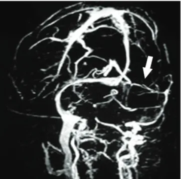
A 39-year old woman complained of leg cramps, weight loss, occipital headache and visual blurring. At the ini-tial assessment, her brachial, femoral, popliteal and tibial pulses were diminished and asymmetric; the neuro-oph-talmic examination revealed papilledema and decreased visual acuity (20/100 bilaterally). Magnetic ressonance im-age (MRI) with magnetic ressonance venography revealed T2 and FLAIR hyperintense areas in the parietal and oc-cipital regions, meningeal thickening and reduced lumen of the left transversal sinus (Fig 1), suggesting chronic ce-rebral venous thrombosis. Computerized Tomography Angiography (CTA) of the aorta and its main branches re-vealed diffuse arterial thickening (Fig 2).
he blood screening disclosed increased values of serum inlammatory markers: Erythrocyte Sedimentation Rate (ESR) of 94 mm/h and C-reactive protein (CRP) of 44.8 mg/dL; the cerebrospinal luid sample had pleocytosis (20 cells/mm3;
64% lymphocyte), and the meningeal biopsy revealed chronic non-speciic inlammatory cell iniltrate. Additional laborato-rial investigation (lupus antibody, anti-cardiolipin antibody, C and S protein, homocystein, Leiden factor V, mutant pro-thrombin gene) excluded other thrombophilia. Treatment with
Fig 1. Magnetic ressonance venography showing narrowed lumen of the left transverse sinus (arrow).
Fig 2. Computerized tomography angiography demonstrates wall thickening of abdominal aorta (arrow) suggesting vasculitis.
LETTERS
anticoagulant medication was initiated, followed by predni-sone. Visual complaints improved, and the serum inlamma-tory markers normalized.
DISCUSSION
he diagnostic criteria for TA are mainly clinical; neverthe-less, laboratory analysis frequently reveals increased ESR and CRP. Although they are not gold standard, CTA and MRI are useful keys to diagnosis1,4. During the acute phase,
inlamma-tory iniltration afects all layers of the arterial wall, whereas at the chronic phase ibrous tissue replaces the damaged layers and arterial stenosis may develop1,2. Diferential diagnoses
in-clude other type of large vessel inlammatory vasculitis1.
CVT is a disease that also affects younger individu-als; however, associated arterial thrombosis is identified in only 10% of the patients5. Whilst blood coagulation
disorders are important causes of CVT, multiple factors may contribute as a precipitant of venous thrombosis5.
Despite all the efforts to find its etiology, it remains un-identified in 20% of the patients3. Our patient fulfilled
four of the six criteria for TA, had radiological imaging suggestive of CVT and was extensively investigated to ex-clude other conditions.
CVT and TA have clinical similarities which are im-portant to be remembered during the diagnostic work up3.
Simultaneous presence of CVT and TA is unexpected and raises the possibility of either causal or casual association between these two conditions.
1. Johnston SL, Lock RJ, Gompels MM. Takayasu arteritis: a review. J Clin Pathol 2002;55:481-486.
2. Vanoli M, Daina E, Salvarani C, et al. Takayasu’s arteritis: a study of 104 Italian patients. Arthritis Rheum 2005;53:100-107.
3. Stam J. Current Concepts: Thrombosis of the Cerebral Veins and Sinuses. N Eng J Med 2005;352:1791-1798.
4. Cantú C, Pineda C, Barinagarrementeria F, et al. Noninvasive cerebrovascular assessment of Takayasu arteritis. Stroke 2000;31:2197-2202.
5. van Gijn J. Cerebral venous thrombosis: pathogenesis, presentation and prognosis. J R Soc Med 2000;93:230-233.