Diagnosis of, surgical
technique for and treatment
results from medullary
lipomas associated with
spinal dysraphism
Experience with 38 patients
José Carlos Lynch1, Juliano Corrêa2, Celestino Pereira3
ABSTRACT
Objective: To observe whether microsurgical removal of medullary lipomas and
untethering of the medulla is a safe and efficient procedure. Method: A retrospective
study was carried out on 38 patients with medullary lipomas associated with spinal dysraphism who underwent operations between January 1986 and January 2008, at the Neurosurgery Department of the Federal Hospital for State Public Servants, in Rio de Janeiro. Results: No deaths occurred in this series, and there was no worsening of motor or bladder function among the patients. Seven individuals presented improvements in their motor deficit. Nine patients presented improvements in bladder function. Three
individuals with trophic lesions achieved wound healing. Conclusion: Microsurgical
removal of medullary lipomas associated with spinal dysraphism proved to be a safe procedure without deaths and with a low morbidity rate, and several patients achieved improvements in their neurological symptoms.
Key words: spinal dysraphism, medullary lipoma of the conus, lumbosacral lipomas, lipomyelomeningoceles, anchored medulla, microsurgery.
Diagnóstico, técnica cirúrgica e resultados nos lipomas medulares associados ao disrafismo vertebral: experiência com 38 pacientes
RESUMO
Objetivo: Observar se a remoção microcirúrgica dos lipomas medulares e a liberação
da medula da tração exercida pelo lipoma é um procedimento seguro e eficaz. Método:
Realizamos estudo retrospectivo de 38 pacientes com lipomas medulares associados ao disrafismo espinhal operados entre janeiro de 1986 a dezembro de 2009 no Serviço de Neurocirurgia do Hospital Federal dos Servidores do Estado do Rio de Janeiro. Resultados:
Nessa série não ocorreu nenhum óbito, ou piora da função motora ou vesical em nenhum paciente. Observamos melhora do défice motor em 7 pacientes. Nove pacientes apresentaram melhora da função vesical. Três indivíduos com lesões tróficas apresentaram
cicatrização das suas feridas. Conclusão: A remoção microcirúrgica dos lipomas
medulares associados ao disrafismo espinhal se mostrou segura, sem nenhum óbito, com baixa morbidade e com melhora dos sintomas neurológicos em vários pacientes.
Palavras-chave: disrafismo espinhal, lipoma de cone medular, lipoma lombo-sacro, lipomielomeningocele, medula ancorada, microcirurgia.
Correspondence José Carlos Lynch
Rua Jardim Botânico 600 / sala 605 22461-000 Rio de Janeiro RJ - Brasil E-mail: [email protected]
Received 26 August 2010 Received in final form 27 May 2011 Accepted 4 April 2011
Several names have been used to describe medullary lipomas of the conus associated with spina biida: lipo-myelomeningoceles, lipomyeloceles, medullary lipoma of the conus or of the ilum terminale and congenital lum-bosacral lipoma. Lipomyelomeningocele is often used as a general term for all lumbosacral lipomas, but these should be distinguished from intramedullary lipoma without spinal dysraphism1-11
. Over the last few years,
several neurosurgeons have reported their experiences with the treatment of lipomyelomeningoceles, in the lit-erature1-16
. Nevertheless, little attention has been paid to
this subject within Brazilian settings, which motivated
us to review and present the cases of this pathological condition treated by our medical team, and to assess the safety and eiciency of microsurgical removal of med-ullary lipomas and the degree of relief from the traction that the lipoma exerts on the medulla.
METHOD
A retrospective study was carried out on 38 consec-utive patients with medullary lipomas associated with spinal dysraphism who underwent surgery between Jan-uary 1986 and JanJan-uary 2008, at the Neurosurgery De-partment of the Federal Hospital for State Public Ser-vants, in Rio de Janeiro. Radiological examinations, patient records, surgical descriptions and, when avail-able, ilming of the surgery, were reviewed, thereby cre-ating a database from which information pertinent to the present study was gathered.
Surgery
All the patients underwent surgery and the same mi-crosurgery technique was used, following these steps: General anesthesia was induced, with endotracheal in-tubation, and the patient was positioned in ventral de-cubitus, resting on the thoracolumbar support. A 4.5× loupe and coaxial lighting were used for the initial steps of the procedure. With a scalpel, a rectilinear midline incision was made, starting one vertebra above and ex-tending to one vertebra below the subcutaneous fat mass, and proceeding around the cutaneous stigma. hroughout the procedure, careful homeostasis was per-formed with bipolar forceps under saline irrigation. Cir-cumferential dissection was performed around the li-poma and/or the lipoibromatous talus. he penetration of the lipoma into the thoracolumbar fascia was viewed (Fig 2B). he fascia was opened along the midline with a scalpel and the paravertebral muscles were carefully disinserted and laterally retracted using a periosteal el-evator. Exposure was maintained using autostatic re-tractors. he lamina and the spinous processes of the upper and lower vertebrae were viewed and the pen-etration of the lipoma or lipomatous talus into the
os-seous failure was observed. Laminectomy of the upper vertebra was usually performed. he normal condition of the dura mater and penetration of the lipoibromatous
Fig 1. Cutaneous stigmata. [A] Dimple, lipoma and hemangioma; [B] Dimple and vestigial tail.
Fig 2. [A] T1-weighted MRI showing penetration of the lipoma into the lumbar fascia. [B] Surgical view of the same patient, re-vealing lipoma penetrating the lumbar fascia.
Fig 3. [A] T2-weighted MRI showing caudal lipoma with lipoi-bromatous talus penetrating the dura mater and adhering to the medullary conus, which was located in an abnormally low posi-tion (arrows). [B] Surgical view of a subcutaneous lipoma and lipo-ibromatous talus penetrating the dura mater (arrows).
talus were identiied (Figs 3B and 4B). At this moment, a surgical microscope was introduced and the remaining surgical procedures were performed under magniica-tion that varied from 10 to 16×. he dura mater was sec-tioned along the midline, while bypassing the lipoma or lipoibromatous talus. he free border of the dura mater was sutured to the paravertebral musculature using 4.0 thread. he adhesion of the lipoibromatous talus to the medullary conus, and the relationship with the nerve ra-dices was identiied. Using microsurgery scissors, the li-poma was resected as close as possible to the medul-lary conus, and as much adipose tissue as possible was removed. However, no attempt was made to resect the intramedullary portion of the lipoma, thus leaving the small remainder of the lipoma freely gliding within the canal sac. he arachnoid bands were also sectioned, but care was taken to lyse this adhesion without damaging functional nerve roots. he ilum terminale was identi-ied and sectioned, with the aim of relieving the medul-lary conus. We only used intraoperative electrical stim-ulation to locate functional neural elements in diicult situations. In all the cases, a decrease in axial tension was observed, while some cases presented cranial dis-placement of the conus. he viable radices of the cauda equine were placed in an anterior position and were ei-ther straightly aligned or had the outlet angle inverted. It was essential to preserve them. he dura mater was sutured hermetically using 4.0 thread. Duraplasty with fascia or using artiicial dura mater was commonly per-formed to prevent straightening of the dural sac diameter and adhesions to the medulla. he Valsalva maneuver was performed with the aim of detecting whether any li-quor istula was present, and such cases were sutured. Bi-ological glue was used on the suturing area with the aim of preventing liquor istula. he paravertebral muscula-ture and of the lumbar fascia were then closed in planes,
using absorbable thread. he skin was sutured in a ten-sion-free manner and the stitches were separated with monoilament thread. During the postoperative period, the patient was kept in the ventral decubitus position and in the Trendelenburg position from 3 to 5 days, to pre-vent any appearance of a liquor istula. Prophylactic an-tibiotics were prescribed for 24 hours.
RESULTS
his was a retrospective study, and therefore it had inherent biases and drawbacks. We did not develop any special protocol for analyzing these patients. Nonethe-less, in addition to the examinations performed in the neurosurgery department, the pediatric patients were routinely examined before and after the operation within the pediatric neurology and urology services and the adults, within the neurology department.
here were no deaths in the current series. he 38 pa-tients included 14 men and 24 women, of ages ranging from 4 months to 37 years (average of 14.5 years).
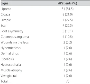
he most frequent sign observed was subcutaneous lumbar lipoma, which was found in 31 cases (81.5%). Genitourinary malformations such as cloaca and du-plication of the genitalia were detected in nine cases (23.6%) and a dimple was seen in seven cases (18.4%). Previous surgical scars, vestigial tails and angiomas were other signs observed in this series (Figs 1 and 2; Table 1). he most frequent symptom was urinary incontinence, which was found in 21 patients (55.2%), followed by motor deficit in 13 (34.2%) and constipation in nine (23.6%). here were only three asymptomatic patients (Table 2).
X-rays identiied spina biida in 29 cases. Magnetic resonance imaging (MRI) clearly showed the lipoma penetrating the rachidian canal and the dura mater, with adhesion to the medullary conus, which was located ab-normally low: below L2 in 16 cases (Figs 3, 4 and 5).
In the current series, we did not observe any wors-ening of motor or bladder function among the patients during the early postoperative period. Seven individ-uals (53.8%) showed improvements in their motor def-icit. Eleven patients (42.3%) showed some improvement in their bladder function, among whom four presented decreases in repetition of urinary tract infections, and seven, decreases in the residual volume. here were three patients with trophic lesions, and they presented healing of chronic ulcerations. Three patients reported tran-sient dysesthesia and numbness in the legs immediately after their operations. Over a mean follow-up period of 7.5 years (range: 2-13 years), we achieved a symptom improvement rate of 55.2%. Stabilization of the symp-toms occurred in 10 patients (26.3%) and some degree of neurological deterioration was noticed in seven indi-viduals (18.4%). he majority of the patients achieved
a satisfactory improvement, which allowed them to re-turn to functional life. Despite the care mentioned above, there were four cases of CSF istula (10.5%), which were treated with prolongation of the ventral decubitus and Trendelenburg positions. Reoperation to close the is-tula was only necessary for one patient (2.6%). In all the other cases, the istulas closed spontaneously, without infectious complications.
Selected case
A 21-year-old patient with lumbosacral lipoma, axial lumbar pain and urinary incontinence previously under-went two supericial interventions to reduce the lipoma at another medical institution. No improvement of the neurological condition was observed, and the lipoma re-gained the original size. At our department, the patient underwent microsurgical resection of the medullary li-poma, with untethering of the medulla, thus achieving better sphincter control and pain relief (Figs 4A and 4B).
DISCUSSION
These patients with lipomyelomeningoceles pre-sented a subcutaneous lipoma in lumbosacral region, usually on the midline. Subcutaneous lipomas develop through closure defects in the thoracolumbar fascia, lamina and dura mater, with adherence to the medullary conus1,4,5,7,10,11 (Figs 2,3,4 and 5). In a 15-mm embryo, the lengths of the medulla and spinal column are the same. From that stage onwards, the spinal column develops towards the caudal end more rapidly than the medulla does, thus causing the medulla to “ascend” towards the encephalon, inside the rachidian canal. he medullary conus usually reaches L3 during the 30th week of preg-nancy and, in adult individuals, its location should not exceed the lower border of L2. If any process such as lipomyelomeningoceles keeps the medulla stuck in one position, thus preventing it from migrating, a gradual longitudinal stretching process that tethers the medulla will take place9,14,16-20. Chapman21 defined three types of lipoma: dorsal, caudal and transitional. The dorsal variant attaches to the dorsal aspect of the conus. he caudal type attaches to the inferior aspect of the conus and the transitional variant is a combination of these two. Lipomas of the terminal ilum are mostly fully en-closed inside the dura and seldom become symptom-atic and require treatment. he great majority of our pa-tients presented with the caudal or transitional variant. We only found three patients with dorsal lipomas and three with lipomas of the ilum.
Yamada et al.20 demonstrated experimentally that the traction exerted on the medulla causes changes to the ox-idative processes of the neuron mitochondria. his might cause paraplegia or urinary incontinence. Patients with
lipomyelomeningoceles are, most often, born neurologi-cally normal. he risk of neurological and/or bladder de-terioration exists at all ages, but particularly during pe-riods of rapid growth3,9-11,13,16. Reviewing the literature, Pierre-Khan et al.6 observed that between 35 and 67% of the patients developed neurological or bladder lesions when not properly treated. Other authors3,7,13-15 have believed that the majority of the patients will present symptoms before they are four years old. The neuro-logical and/or bladder deterioration among these pa-tients is most commonly progressive, but it can appear abruptly during the course of certain events such as nat-ural birth, practicing of physical exercises, sexual inter-course or accidents1,2,6,7,10,16. Several authors who oper-ated on symptomatic patients have stoper-ated that very few of them achieved total regression of their deicits. It is
Table 1. Signs of 38 patients with medullary lipomas.
Signs #Patients (%)
Lipoma 31 (81.5)
Cloaca 8 (21.0)
Dimple 7 (22.5)
Scar 7 (22.5)
Foot asymmetry 5 (13.1)
Cutaneous angioma 4 (10.5)
Wounds on the legs 2 (5.2)
Hypertrichosis 1 (2.6)
Dermal sinus 1 (2.6)
Escoliosis 1 (2.6)
Hydrocephalia 1 (2.6)
Muscle atrophy 1 (2.6)
Vestigial tail 1 (2.6)
Total 70
*Some patients presented more than one sign.
Table 2. Symptoms in 38 patients with medullary lipomas.
Symptoms # Patients (%)
Urinary incontinence 21 (55.2)
Paraparesia 10 (26.3)
Constipation 9 (23.6)
Pain 7 (18.4)
Urinary retention 5 (13.1)
Hypoesthesia 4 (10.5)
Monoparesis 3 (7.8)
Saddle block anesthesia 3 (7.8)
Total 62
harder to achieve recovery of bla dder function than of motor function2-6,11,13. hese characteristics were also ob-served throughout our clinical experience.
In this present group, we achieved a symptom stabili-zation or improvement rate of 81.5%. he aim of surgical treatment is to release the medulla from the traction ex-erted by the lipoma and, in some cases, decompres-sion of the medulla relieves the mass efect. herefore, cosmetic operations that do not untether the medulla are a risk factor for delayed neurological deteriora-tion3,4,5,7,9,10,13-15. his phenomenon occurred in the cases of six patients who had undergone previous surgery at other institutions.
Recently, the treatment of these lesions has generated much controversy. Some physicians have advocated sur-gical treatment for all patients regardless of symptoms, while others have proposed that surgery should be with-held until symptoms develop10.
Kulkarni et al.12 found that the incidence and patterns of neurological deterioration seemed to be very similar, regardless of whether or not early surgery had been per-formed. hese results suggest that conservative treat-ment of asymptomatic patients is a reasonable option. hey observed that, during the follow-up period, deteri-oration was experienced by 33% of the patients who had been conservatively treated, and 46% of those who had undergone prophylactic surgery.
On the other hand, Pang et al.14,15 concluded that total and near-total resection of lipomas and complete recon-struction of the neural placode produced a much better long-term result, with greater likelihood that it would be progression-free, than seen in cases of partial resection and non-surgical treatment. With total resection, pre-operative complications can be successfully treated with low surgical morbidity and a high yield of agreeable long-term postoperative results.
In the present series, all the symptomatic patients un-derwent the microsurgical technique for total/near total lipoma resection, untethering of the medullary cone, lysis of adhesions and sectioning of the ilum terminale, thereby releasing the medulla from any traction, fol-lowed by duraplasty to avoid reanchoring. Some neuro-surgeons have suggested that intraoperative monitoring should be used, such as rectal EMG, urethral EMG, con-tinuous EMG, evoked potential and other types. To date, there has not been any proof that such methods are ef-fective for avoiding injury. In the initial cases of this se-ries, we used intraoperative electrical stimulation to lo-cate functional neural elements, but with progressively increasing experience within our group, we began to use neural stimulation only for diicult situations.
he extent of surgical resection and untethering of the conus was established taking into consideration the
surgeon’s opinion and the postoperative images (MRI and/or CT scans). Over the period (22 years) covered by the present study, there has been remarkable progress in imaging techniques. With the irst patients of this se-ries, assessment was made based on CT scans, which do not allow precise measurement of the tumor residues. In eight cases, postoperative MRI was available for assess-ments, and we observed near total removal (>90%) in ive cases (Figs 5A and 5B), and partial resection in the other three patients. Radiological evaluation of the un-tethering is always diicult to make. We were only able to established ascending of the conus in two individuals with certainty.
Kulkarni et al.12 suggested that it may be diicult to set up close observation of patients in our environment, due to the long distances that usually exist between the patient’s home and the hospital, thus making neurolog-ical surveillance more diicult. In our series, there were even some patients who came from indigenous tribes. herefore, management remains an open question10.
For symptomatic patients, we recommend microsur-gical treatment for all patients, regardless of the symp-toms, in order to protect the neurological function and delay any neurological decline. Our experience with as-ymptomatic patients only consists of three cases, and it is diicult to reach any conclusion with such a small sample. We can conclude that medullary lipomas of the conus associated with spinal dysraphism are a signiicant cause of neurological dysfunction. Early diagnostic procedures should be followed up by pediatricians, urologists or plastic surgeons, thus helping to select the appropriate treatment. Microsurgical treatment can be expected to achieve total/near total resection of the lipoma with re-lief of the traction on the medulla.
Our experience described above, on 38 cases oper-ated on using microsurgical techniques, showed that this is a safe method, with no deaths, low morbidity and sat-isfactory results, comparable to those found in the inter-national literature. For asymptomatic patients, the treat-ment should be decided on an individual basis.
REFERENCES
1. Anderson FM. Occult spinal dysraphism. J Pediatrics 1968;73:163-177. 2. Dubowitz V, Lorber J, Zachary RB. Lipoma of the cauda equine. Arch Dis
Child 1965;40:207-213.
3. Hoffmann HJ, Taecholarn C, Hendrick FB, Humphreys RP. Management of lipomyelomeningoceles: experience at the Hospital for Sick Children. J Neurosurg 1985;62:1-8.
4. Lassman LP, James M. Lumbosacral lipomas: critical survey of 26 cases submitted to laminectomy. J Neurol Neurosurg Psychiatry 1967;30: 174-181.
5. Malis LI. Intramedullary spinal cord tumors. Clin Neurosurg 1977;25: 512-539.
6. Pierre-Kahn A, Lacombe J, Pichon J, et al. Intraspinal lipomas with spina biida: prognosis and treatment in 73 cases. J Neurosurg 1986;65:756-761. 7. Schut L, Bruce DA, Sutton LN. The management of the child with a
8. Flemins KL, Davidson L, Gonzalez-Gomez I, McComb JG. Nondysraphic pe-diatric intramedullary spinal cord lipomas: report of 5 cases. J Neurosurg Pediatr 2010;5:172-178.
9. Rogers HM, Long DM, Chou SN, French LA. Lipomas of the spinal cord and cauda equine. J. Nerosurg 1971;34:349-354.
10. Finn MA, Walker ML. Spinal lipomas: clinical spectrum, embryology, and treatment. Neurosurg Focus 2007;23:1-12.
11. Walker ML.The tethered spinal Cord-walking in the ine line. Neurosurg Focus 2007;23:1-4.
12. Kulkarni AV, Pierre-Kahn A, Zerah M. conservative management of asymp-tomatic spinal lipomas of the conus. Neurosurgery 2004;54:868-875. 13. Blount JP, Scott E. Spinal lipomas. Neurosurg Focus 2001;10:1-13. 14. Pang D, Zovickian J, Oviedo A. Long-term outcome of total and near-total
resection of spinal cord lipomas and radical reconstruction of the neural placode: Part I - surgical technique. Neurosurgery 2009;65:511-529. 15. Pang D, Oviedo A, Zovickian J. Long-term outcome of total and near-total
resection of spinal cord lipomas and radical. Reconstruction of the neural placode: Part II - Outcome analysis and preoperative proiling. Neurosur-gery 2010;66:253-257.
16. Pang D, Wilberger JE Jr. Tethered cord syndrome in adults. J Neurosurg 1982;57:32-47.
17. Lesoin F, Petit H, Destec A, Rousseaux M, Julliot JP, Jomin M. Spinal dys-raphia and elongated spinal cord in adults. Surg Neurol 1984;21:119-124. 18. Barson AJ. The vertebral level of termination of the spinal cord during
normal and abnormal development. J Anat 1970;106:489-497.
19. Pinto FCG, Fontes RBU, Leonhard THC, et al. Anatomic study of the ilum terminale and is correlation with the tethered cord syndrome. Neurosurg 2004;54:868-875.
20. Yamada J, Zinke DE, Sanders D. Pathophysiology of tethered cord syndrome. J Neurosurg 1981;54:494-503.
![Fig 4. [A] T1-weighted MRI showing caudal lipoma penetrating the dura mater, with adherence to the medullary conus, which was located in an abnormally low position, and syringomyelia of the conus](https://thumb-eu.123doks.com/thumbv2/123dok_br/15433248.595282/2.955.518.885.849.998/weighted-showing-penetrating-adherence-medullary-abnormally-position-syringomyelia.webp)
![Fig 5. [A] Sagittal preoperative T1-weighted MRI showing large lipoma tethering the spinal cord](https://thumb-eu.123doks.com/thumbv2/123dok_br/15433248.595282/3.955.68.438.103.346/sagittal-preoperative-weighted-showing-large-lipoma-tethering-spinal.webp)