353
One of the major advantages of bioprostheses is the fact that they have a low incidence of thrombosis 1 as compared with that
of mechanical prostheses, and, therefore, do not require the use of anticoagulant drugs.
In an observational study by Grunkemeier and Rahimtoola 2,
the analysis of the risk of thrombosis showed a significant difference between biological and mechanical prostheses. For the mechanical prostheses in the aortic position, the occurrence of thrombosis varied from 0.05% to 0.25% per year. For the bioprosthesis in the aortic position, the occurrence of thrombosis was 0.03% per year. The mitral prostheses followed a similar pattern, with a predominance of thrombosis occurring with the metallic type (0.28 to 0.62% per year) as compared with that with the biologic type
(Hancock and Carpentier-Edwards 0.02 to 0.07% per year)3.
The diagnosis of prosthetic valve dysfunction is usually confirmed on autopsy or through the surgical inspection of the removed prostheses, but the noninvasive diagnosis has been used with progressive safety in these cases.
Two-dimensional and M-mode echocardiography may be useful instruments for demonstrating obstruction of the bioprosthesis by a thrombus 4,5, the first cases being reported in 1976 6,7. However,
transthoracic echocardiographic images usually do not allow a conclusive approach to thrombus definition due to its imprecision in defining the prosthetic leaflets.
Transesophageal echocardiography (TEE), on the other hand, provides a unique acoustic window for assessing the prosthetic leaflets, particularly in the mitral position, due to the greater proxi-mity of the transducer with the cardiac structures and greater frequency of the crystals used, which results in greater resolution and absence of acoustic interference of the attenuating elements, such as the ribs and lungs, and even the components of the pros-thesis 8. This technique provides accurate images for the definition
of the thickness of the leaflets and their mobility, allowing an a dequate diagnosis of calcifications, stenoses, and ruptures 9-12.
Nonetheless, the descriptions of bioprosthetic mitral valve thrombosis on TEE are rare 13-17.
In regard to the treatment of metallic prosthetic thrombosis, the thrombolytic agent has been efficient in a significant number of cases 18-21, although the use of oral anticoagulants prior to the
thrombolytic agent has also been effective on some occasions, when one is not dealing with an acute hemodynamic decompen-sation 22.
Original Article
Bioprost het ic M it ral Valve Throm bosis. Im port ance
of Transesophageal Echocardiography in t he
Diagnosis and Follow -up Af t er Treat m ent
Adelino Parro Jr, M art a Lancia Carramona, Caio August o Ferreira Amaral,
José Luiz Balt hazar Jacob, José Carlos Nicolau
São José do Rio Pret o, SP - Brazil
Instituto de Moléstias Cardiovasculares - IMC
Mailing address: Adelino Parro Jr. - IMC - Rua Castelo DÁgua, 3030 Cep 15015-210 - São José do Rio Preto, SP, Brazil
E-mail: [email protected] Received: 11/23/00
Accepted: 1/21//02
English version Stela Maris C. e Gandour
Objective
To report the clinical and echocardiographic findings of bio-prosthetic mitral valve thrombosis and the value of transeso-phageal echocardiography (TEE) in its diagnosis and monitoring of thrombolysis.
M ethods
One hundred and eleven patients with mitral bioprostheses underwent TEE, and 4 out of 7 suspected of having a thrombus on these prostheses were included in the study (mean age = 60.2±10.2 years; 2 men). The diagnosis was confirmed with serial TEE and clinical evolution. The morphologic features of the prosthetic leaflets, as well as the presence and characteristics of attached echogenic masses were investigated. The mean gra-dient through the prosthesis and the valvular area were obtained.
Results
The diagnosis of bioprosthetic mitral valve thrombosis was established 48.7±55.2 months after surgery. Two patients had ischemic stroke in the early postoperative period. The mean overall gradient was high (11.4±3 mmHg) and the valvular area reduced (1.24±0.3 cm2). On TEE, echogenic masses on the
left ventricular face of the mitral bioprosthesis suggestive of thrombus were evidenced in all patients. On serial TEE (136±233 days), in 2 patients the thrombus had disappeared and in 2 others it was smaller after treatment, the mean gradient dropped to 6.2±3 mmHg (P = 0.004; 95% CI), and the valvular area increased to 2.07±0.4 (P = NS).
Conclusion
TEE proved to be useful for detecting bioprosthetic mitral valve thrombosis and was effective in monitoring the treatment in all patients.
Key w ords
354
In bioprosthetic thrombosis, therapy with oral anticoagulant agents has been rarely reported, although the results have been promising 17.
The literature has shown that the effect of thrombolytic agents on this type of prosthesis has proved to be an effective alternative 23,24.
This study aims at reporting the echocardiographic findings (particularly those of TEE) in bioprosthetic mitral valve thrombosis and the role played by echocardiography in monitoring therapeutic efficacy.
M ethods
One hundred and eleven patients with mitral bioprostheses who underwent transesophageal echocardiography due to different clinical indications between 1994 and 1998 were retrospectively analyzed. Thrombosis was evidenced in 7 patients. In 4 of these patients (mean age = 60.2±10.2 years; 2 males), the diagnosis of thrombosis could be confirmed through clinical evolution and serial assessment on TEE; therefore, the 4 were included in the study. In the remaining cases, although the transesophageal echocardiographic characteristics were suggestive of thrombus, the clinical evolution did not confirm it, and, therefore, the pa-tients were not included in the study. All papa-tients received intra-venous heparin for the treatment of thrombosis; on one occasion, streptokinase was added.
The number of transesophageal echocardiographies performed and the time interval for their performance were decided by the cardiologist responsible for the case. In this analysis, the last transesophageal echocardiography of each patient was considered for comparison with the initial one. The serial echocardiographies were performed by the same examiner. The mean time between the 2 transesophageal echocardiographies was 136±233 days (11 days 16.2 months). The mitral valve dysfunction was due to the rheumatic cause in all cases.
Two patients had undergone previous surgery for mitral pros-thesis, and none had received a prosthesis in the aortic position. Two patients were using oral anticoagulation prior to the echo-cardiographic diagnosis of thrombosis, and the medication was suspended on the occasion of the surgical procedures (mitral valve re-replacement in 1 case and renal surgery in another). Although the patient undergoing mitral valve re-replacement continued with intravenous heparin, both experienced ischemic stroke in the early postoperative period.
The following devices were used to perform transthoracic echo-cardiography: the Toshiba SSH-140 model; the SIM 7000 model of ESAOTE; and the Acuson XP-10 model. The Toshiba SSH-140
model with biplanar transducer was used for TEE. The measure-ments of the atrial and ventricular cavities were obtained through the left parasternal view, according to previously published recom-mendations 25.
The thickness and mobility of the leaflets of the mitral valve prostheses were analyzed using apical, parasternal, and subcostal mapping, and the presence of echogenic masses in the leaflets was assessed, as was the presence of left atrial thrombus.
The mean gradient through the mitral valve prosthesis was assessed with continuous Doppler using the apical 4-chamber view and the simplified Bernoulli equation 26. The prosthetic mitral
valve area was calculated according to the atrioventricular pressure half-time method reported by Hatle et al 27.
The presence and degree of mitral insufficiency were analyzed with color Doppler 28.
Left ventricular systolic function was assessed through calcu-lation of the ejection fraction according to the method reported by Teichholz et al 29.
The jet of tricuspid insufficiency was used to calculate the systolic pressure of the pulmonary artery adding 10 mmHg to the peak gradient through regurgitation 30.
Transesophageal echocardiography was performed with a 5MHz biplane transducer in the longitudinal and transversal planes, in the high and low esophageal, and gastric positions.
Thickness and mobility of the leaflets were assessed qualita-tively and quantitaqualita-tively; when the measure exceeded 3 mm, the prosthetic leaflet was considered thickened 14.
The presence of echogenic masses in the leaflets was inves-tigated, as was the presence of a thrombus in the left atrium or in its appendage.
The presence and severity of mitral regurgitation were also analyzed on TEE.
After treatment with an anticoagulant or thrombolytic agent, or both, the same parameters were reassessed on transthoracic and transesophageal echocardiography.
The paired Student t test was used for comparison between
pre- and posttreatment parameters.
Results
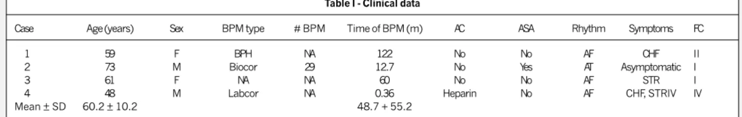
The diagnosis of bioprosthetic mitral valve thrombosis was performed, on average, 48.7±55.2 months after surgery; in 1 patient, it occurred 11 days after surgery (tab. I). On the occasion of the echocardiographic diagnosis of thrombus, 1 patient was in NYHA functional class II congestive heart failure, another in func-tional class IV, and the 2 remaining in funcfunc-tional class I.
Table I - Clinical data
Case Age (years) Sex BPM type # BPM Time of BPM (m) AC ASA Rhythm Symptoms FC
1 59 F BPH NA 122 No No AF CHF II
2 73 M Biocor 29 12.7 No Yes AT Asymptomatic I
3 61 F NA NA 60 No No AF STR I
4 48 M Labcor NA 0.36 Heparin No AF CHF, STRIV IV
Mean ± SD 60.2 ± 10.2 48.7 + 55.2
355
In 1 of these patients in functional class I, obstruction of the mitral bioprosthesis was found on transthoracic echocardiography in a routine assessment of the prosthesis 1 year after surgery, which showed an elevated transprosthetic gradient and leaflets with reduced mobility. The subsequent TEE showed a thrombus on the prosthesis causing the obstruction.
The other patient in functional class I underwent transthoracic echocardiography a few weeks after renal surgery due to the occurrence of stroke in the early postoperative period. The gradient through the prosthesis was increased, and the TEE performed on the same day revealed a thrombus on the mitral prosthesis causing the obstruction.
In the patient with functional class IV congestive heart failure and stroke in the early postoperative period, transthoracic echo-cardiography showed, in addition to the high prosthetic mitral valve gradient, thickened leaflets with reduced mobility, allowing only partial filling through the prosthetic ring on color flow mapping, raising the suspicion of a thrombus. Later, TEE confirmed these findings.
The transprosthetic gradient of the patient in functional class II on transthoracic echocardiography was not very elevated (8.8 mmHg), but as the patients cardiac rhythm was that of atrial fibrillation, cardioversion was considered a possibility in an attempt to improve clinical condition. Prosthetic mitral valve throm-bosis was only detected on routine TEE performed prior to cardio-version to investigate the emboligenic source.
Three patients had atrial fibrillation, and 1 had atrial tachy-cardia. No patient had fever or clinical suspicion of infective endo-carditis.
All patients were treated with intravenous heparin after the diagnosis of thrombosis, streptokinase being added for 1 patient
due to lack of clinical/echocardiographic response to heparin. Oral anticoagulation was administered to all patients after hospital discharge.
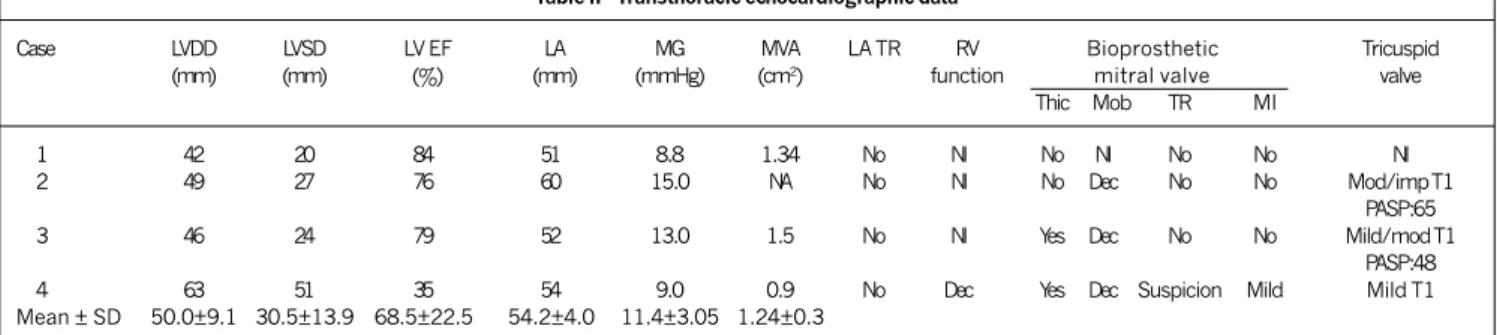
On transthoracic echocardiography, the left atrium ranged from 51 to 60 mm (mean = 55.3±4.1 mm), and left ventricular overall systolic function was decreased only in 1 patient (EF = 35%), who also had a reduced right ventricular overall systolic function.
In 1 patient, transthoracic echocardiography suggested biopros-thetic mitral valve thrombosis, and in all patients the mean trans-prosthetic gradient was elevated, with an overall mean of 11.4± 3 mmHg (tab. II). The valvular area was reduced (1.24±0.3 cm2),
and only 1 patient had mild transprosthetic mitral insufficiency. Three patients had tricuspid valvular incompetence, and the systolic pressure of the pulmonary artery could be measured in 2 of them, being elevated in both (tab. II).
On TEE, the leaflets were thickened and their mobility was reduced in all patients.
Echogenic masses suggestive of thrombi were seen in all patients carpeting the left ventricular face of the prosthetic leaflets, producing an increase in their thickness (5.7 ± 0.9 mm). In 1 patient, thrombi in the left atrial face with a pedunculated aspect were observed, adhered to the atrial margin of the prosthetic leaflet. In 3 patients, the thrombus affected 2 leaflets, and, in 1 patient, the thrombus affected 3 leaflets (tab. III).
A thrombus was observed in the left atrium in 2 patients, and formation of spontaneous contrast was seen in all patients (tab. III). In 3 patients, the recent serial TEE (< 1 month) showed a reduction in size or disappearance of the thrombi (fig. 1); in 1 patient, however, the thrombus only disappeared on the second serial TEE (486 days after the initial study).
Table III - Transesophageal echocardiographic findings
Case LA Bioprosthetic mitral valve
TR SC Thrombus Leaflets Insuf
Type Site Size (mm) # Thic (mm) Mob
1 No Yes Sessile Ventricular face 15 2 Yes 6.0 Dec no
2 Yes Yes Sessile Ventricular face 17 2 Yes 6.7 Dec no
3 No Yes Sessile and Ventricular and 24 3 Yes 4.5 Dec mild
pedunculated atrial face (free margin)
4 Yes Yes Sessile Ventricular face 16 2 Yes 5.7 Dec mild
Mean ± SD 18 ± 4.0 5.7 ± 0.9
SC - spontaneous contrast; Insuf - insufficiency; the other abbreviations are as in table II.
Table II - Transthoracic echocardiographic data
Case LVDD LVSD LV EF LA MG MVA LA TR RV Bioprosthetic Tricuspid
(mm) (mm) (%) (mm) (mmHg) (cm2) function mitral valve valve
Thic Mob TR MI
1 42 20 84 51 8.8 1.34 No Nl No Nl No No Nl
2 49 27 76 60 15.0 NA No Nl No Dec No No Mod/imp T1
PASP:65
3 46 24 79 52 13.0 1.5 No Nl Yes Dec No No Mild/mod T1
PASP:48
4 63 51 35 54 9.0 0.9 No Dec Yes Dec Suspicion Mild Mild T1
Mean ± SD 50.0±9.1 30.5±13.9 68.5±22.5 54.2±4.0 11.4±3.05 1.24±0.3
356
Intravenous heparin was administered followed by anticoagula-tion in all patients, and the mitral transprosthetic gradient subsided in 3 patients, 2 of whom also had disappearance of the thrombus, while the third had a decrease in its size. In the fourth patient, the isolated anticoagulant therapy did not have the effect desired, no modification in the transprosthetic gradient (from 9 to 10 mmHg) occurred, and no significant change in leaflet mobility on transtho-racic echocardiography was observed. Due to clinical instability with persistence of dyspnea on minimal exertion, streptokinase was chosen to be administered at a dosage of 250,000 IU in bolus and 100,000 IU/h as maintenance until the transthoracic echocardio-gram showed a reduction in the transprosthetic gradient, which occurred with 24 hours of infusion (5.6 mmHg). The transesophageal echocardiogram revealed disappearance of the thrombus in the left ventricular face and the presence of a minimum pedunculated mass measuring 3 mm in the margin of the leaflets (atrial face) (fig. 1). On average, the mean mitral transprosthetic gradient after treatment (isolated anticoagulant in 3 patients and streptokinase + anticoagulant in 1 patient) decreased to 6.2±3 mmHg (P = 0.004 versus basal; 95% CI of the difference 3.1 to 7.3),
and the valvular area increased to 2.07±0.4 cm2 (P = NS).
Of the 2 patients, in whom pulmonary arterial systolic pressure could be calculated based on the tricuspid regurgitation jet, regression was observed in 1 (32 mmHg), who also had a reduction in the mean transmitral gradient (7.4 mmHg) in this short
follow-up period (tab. IV). The other patient initially showed a poor response to therapy, evidenced by maintenance of the same di-mensions of the thrombus 1 week after the first serial TEE and no effective reduction in the mean transmitral gradient (11 mmHg) and in the pulmonary arterial systolic pressure (60 mmHg) on transthoracic echocardiography on the occasion. Only on the more recent second serial transesophageal echocardiogram (486 days of evolution tab. IV), the thrombus completely disappeared and a reduction in the mean transmitral gradient (9.5 mmHg) was observed, although the pulmonary pressure levels were maintained (65 mmHg). The left atrial thrombus in the 2 present cases disappeared.
Discussion
Prosthetic mitral valve dysfunction usually occurs due to rupture or calcification of the leaflets, culminating in valvular insufficiency or stenosis 31,32. Bioprosthetic mitral valve thrombosis is rare 2,3.
Thrombosis of the Hancock bioprosthesis was reported in 2/561 (0.3%) patients followed up for a mean period of 2.3 (0.1-7.3) years 33.
The low frequency of bioprosthetic thrombosis 1 is one of the
major advantages of the bioprostheses as compared with the mechanical type, making the use of oral anticoagulants unnecessary in the long run in the first case 33.
However, the real prevalence of thrombosis in patients with mitral bioprosthesis is unknown. Thiene et al 34 reported that,
analyzing the pathologic findings in 50 Hancock bioprostheses placed in the mitral position, thrombus in the leaflets was found in 5 cases (10%), similar to the findings in other reports 35. These
data suggest that the prevalence of bioprosthetic mitral valve thrombosis could be more representative.
The frequency of occurrence of mitral bioprosthetic thrombo-sis on TEE has been controversial in the literature. Kandheria et al 13 reported only 1 patient among 21 (4.7%) with mitral
bio-prosthetic thrombosis, and Daniel et al 14 reported a thrombus in
1 out of 113 (0.8%) patients. On the other hand, Oliver et al 17,
studying 161 (9.3%) patients with mitral bioprosthesis of the porcine type with signs of prosthetic dysfunction assessed on TEE, reported 15 patients with thrombosis, a frequency similar to that of the pathological findings previously reported.
In our study, 7 (6.3%) patients out of 111 had images sugges-tive of bioprosthetic mitral valve thrombosis, 4 of whom were confirmed by clinical response and on serial TEE. In this study, all
Table IV - Echocardiographic data on serial evolution after treatment
2ndTEE 2nd TTE
Case Time (days) LA TR Bioprosthetic mitral valve thrombosis MG (mmHg) MVA (cm2) Tricuspid
Type Site # Size (mm)
1 16 No No - 0 - 2. 3 2. 5 Nl
2 486 No No - 0 - 9.5 1.9 Mod/imp T1
PASP:65
3 22 No Sessile and Ventricular face, 2 11 7.4 1.6 Mild T1
pedunculated free margin PASP:32
4 25 No pedunculated Atrial face, 1 3 5.6 2.3 Nl
free margin
Mean ± SD 136 ± 233 6.2 ± 3 2.07 ± 0.4
Abbreviation as in the previous tables.
Fig. 1 - Transesophageal images of the patient 3 (superior) and 4 (inferior) before (left) and after treatment (right); the arrows point out the site of the thrombus on the leaflets of the mitral prosthesis, which decreased after treatment. VE - left ventricle; AE - left atrium; AAE - left atrial appendage; AO - aorta.
AE
VE
AE
VE
AE AE
AAE AAE
VE
357
patients with a bioprosthesis in the mitral position undergoing TEE were analyzed, including those who were investigated for diseases other than the suspicion of prosthetic dysfunction, diffe-rently from the study by Oliver et al 17.
Additional factors, such as age, sex, time of cardiac surgery, position of the valve, type of bioprosthesis, atrial cardiac rhythm, anticoagulant therapy, left atrial dimension, and left ventricular systolic/diastolic function, may influence in the formation of the thrombus 17. In addition, situations of low transvalvular flow through
the bioprosthesis, requiring circulatory assistance with centrifuge pumps 15, may propitiate the formation of a thrombus in the early
postoperative period.
In the present study, the left atrium was enlarged in all pa-tients, the ventricular function was decreased in 1, all patients had atrial arrhythmia (3 had atrial fibrillation and 1 had atrial tachycardia), and only 1 patient was receiving anticoagulation (IV heparin) due to early postoperative period. The mean patients ages were also advanced. The summation of these factors may have contributed to a greater prevalence of prosthetic mitral valve thrombosis in the present series.
It is important to note the significant role of atrial arrhythmias, mainly atrial fibrillation, in the pathophysiology of atrial thrombus formation, and this could be involved in triggering prosthetic throm-bosis. Several studies 36-38 have shown that the dysfunction of the
atrial appendage present in certain arrhythmias, such as atrial fibrillation or flutter, is more related to the formation of spontaneous contrast or intracavitary thrombus, and with a greater occurrence of embolic events. Mugge et al 36 reported that the
filling/emp-tying velocity of the left atrial appendage (reflecting the left atrial appendage function) was decreased (< 25 cm/s) in some patients with nonrheumatic atrial fibrillation, similarly to the group that had rheumatic atrial fibrillation. More embolic phenomena (5/10 patients) were observed in the group with low velocity as compared with the group with higher velocity (1/19 patients; P < 0.05). A greater occurrence of left atrial appendage thrombosis (30 vs 0%) and spontaneous contrast (80 vs 5%) were also observed.
In the present study, 2 patients had left atrial thrombosis, and all had spontaneous contrast. The left atrial appendage velocity was decreased in 1 patient (0.15 m/s), normal in another (0.46 m/s), and not available in 2 patients, making any conclusion about the significant role played by these data in the pathogenesis of prosthetic thrombosis risky.
The fact that thrombi are found on the ventricular and not on the atrial face of the prostheses is also relevant, and seems related to low flow, and areas of flow stagnation and turbulence through the prostheses. In an experimental study analyzing the characte-ristics of the flow through the different types of prostheses in an in vitro model, Schoephoerster et al 39 showed that, in
biopros-theses, the region close to the periphery of the jet was relatively stagnant, and that the shear stress had greater stress in the margins of the jet with the greatest velocity. Yoganathan et al 40
correlated thrombus formation and tissue growth in valves with a single bascule disc with areas of stagnation and low shear rate in the region with the smallest valvular orifice. Some clinical studies also showed that the thrombus may occupy the sinus of the leaflet, either in the mitral 15,16 or in the aortic 16 position, and
this fact could also be observed in pathological studies 34. The
preservation of both mitral leaflets may cause relative prosthetic
stenosis, and mild abnormalities in blood rheology and local tur-bulence caused by the remaining leaflets might have a thrombo-genic effect 41.
In this study, the thrombi were located in the ventricular face of the prostheses (sinuses of the leaflets) in all patients, corrobo-rating the reports about the site of origin of the thrombi in previous studies.
The period of greater propensity for thrombus development seems not to be well established. Oliver et al 17 reported that
70% of the patients studied had bioprosthetic mitral valve throm-bosis after an 80-month follow-up of valvular replacement, early thrombosis (5 months) occurring in only 1 case. On the other hand, Hagley et al 15 reported the occurrence of bioprosthetic
mitral valve thrombosis early in the postoperative period related to conditions of low transvalvular flow. In the present study, 1 patient had a thrombus on a mitral prosthesis still in the early postoperative period associated with an episode of stroke. It is worth noting that this patient, although being on intravenous heparin therapy, had multiple factors contributing to thrombus formation, such as enlargement of the left atrium, a reduction in the left ventricular systolic function and atrial fibrillation. The patient evolved with NYHA functional class IV congestive heart failure, being discharged from the intensive care unit only after thrombolytic therapy, when thrombus resolution and a reduction in the transprosthetic gradient were obtained.
It is worth noting that in another patient the finding of the prosthetic mitral valve thrombus was occasional. The thrombus was suspected on transthoracic echocardiography, which showed a high gradient in an asymptomatic patient, the definitive diagno-sis being established on TEE.
Treatment with thrombolytic agents for thrombosis on pros-theses of the biological type has rarely been reported 23,24. The
use of thrombolytic agents has been reported for prostheses in the aortic position with favorable results and complete resolution of the thrombus. The rationale for this is similar to that of their use for mechanical prostheses, and, in our study, this treatment was used for thrombosis of a prosthesis in the mitral position. Our decision was based on the clinical instability of the patient and on nonregression of the initial echocardiographic parameters. The patients symptoms improved, and echocardiographic resolution of the mass on the bioprosthesis was observed on TEE with no complications resulting from the thrombolytic treatment.
However, it should be emphasized that the use of thromboly-tic agents is not free of complications 24,42, especially when
consi-dering patients in the early postoperative period. One study 24
reported the occurrence of hemorrhage of the nose and of the venous accesses, which were clinically controlled.
Therefore, the use of thrombolytic agents should be carefully pondered, being basically restricted to those cases in which the clinical and surgical alternatives have been exhausted due the occurrence of other morbid factors that may impair the patients evolution 42. It is important to recall that the use of a thrombolytic
agent does not prevent subsequent reinterventions, when indicated, and that the patient may be referred for surgery when in more stable clinical condition.
In regard to the effect of oral anticoagulants/heparin in throm-bus resolution, Oliver et al 17 and Waksmonski et al 22 showed it to
358
observed) and effective alternative with improvement of the func-tional class in all patients during follow-up. In their study, the treatment with oral anticoagulants was effective in 3 patients, and, 1 patient (receiving intravenous heparin and oral anticoagu-lation) required the additional thrombolytic treatment for thrombus resolution.
In regard to pulmonary pressure levels, one should recall that, in processes of mitral valvular obstruction, the degree of pulmonary arterial hypertension would be theoretically related to the severity of the stenosis 43. However, some patients with significant mitral
stenosis may not develop pulmonary arterial hypertension. The left atrial pressure elevated by valvular obstruction initially increases pulmonary pressure passively with no significant elevation in pul-monary resistance. Chronically, a significant increase in left atrial pressure causes elevation in pulmonary arterial pressure due to an increase in pulmonary vascular resistance, secondary to arteriolar spasm or obstructive vascular changes. Some authors have shown a striking variability among patients in regard to the degree of pulmonary vascular reactivity in response to a chronic elevation in left atrial pressure 43, but the factors determining this variability
remain undefined.
On the other hand, pulmonary arterial hypertension may regress following valvular clearing, which was shown in patients undergoing procedures for mitral valvular dilation. Prediction of this regression could be related to some clinical and echocardiographic factors, such as age 44, and to the immediate result of dilation 45. The
initial reduction in pulmonary arterial pressure seems more related to a reduction in pulmonary capillary pressure in the period follo-wing percutaneous valvuloplasty, while the additional reduction throughout the first week seems more related to the decrease in pulmonary resistance 46.
Although the present study does not assess the specific case of interventional valvular clearance, but of thrombotic clearance, the evolution of pulmonary arterial systolic pressure could be follo-wed up in 2 cases. By the time control TEE (22 days) was perfor-med, only 1 patient showed regression of the pulmonary arterial systolic pressure after treatment (32 mmHg), accompanied by a reduction in the transprosthetic gradient (7.4 mmHg) and an increase in the valvular area (1.6 cm2). In the other patient,
pulmonary arterial systolic pressure remained elevated (65 mmHg) on control TEE (486 days). In this case, the initial response to anticoagulant therapy did not produce the effect desired, because of the permanence of the thrombus on the prosthesis and the insignificant changes in the pulmonary arterial systolic pressure levels (60 mmHg) and in the transprosthetic gradient (11 mmHg) on the 28th day. This poor initial response to anticoagulant treat-ment could have contributed to a chronic change in pulmonary vascular resistance. In addition, the patients more advanced age could have influenced the response of pulmonary arterial systolic pressure to therapy.
Finally, we emphasize the exceptional role played by TEE in assessing therapy efficacy, because of the demonstration of com-plete or partial resolution of the thrombus, a fact also reported by
Oliver et al 17. In their study, in 6 cases, transesophageal
echo-cardiography showed the complete disappearance of the throm-bus after treatment with oral anticoagulants, and, in 2 cases, a significant reduction in the size of the masses was observed. In our study, disappearance of the masses was observed in 2 cases and a reduction in the thrombotic dimensions in the other 2.
The time of the examinations was oriented by the clinician responsible for the patient, and, therefore, the degree of resolution of the thrombi after treatment may have been influenced by it. This may be corroborated by the observation that in 1 patient, the transe-sophageal echocardiogram after 1 week showed no alteration in the size of the thrombus on the mitral bioprosthesis, which only disappeared on the last TEE, considered a control (486 days).
The definitive diagnosis of bioprosthetic thrombosis was established only by the response to the treatment instituted, and was not confirmed by any other methodology. Other abnormalities that could produce images of thickening and masses in the leaflets, such as lipid degeneration, vegetation, fibrocalcification, or hema-tomas, were excluded based on clinical and evolution findings.
Therefore, the favorable results after treatment, with size reduction or disappearance of the masses, allow inferring that those may have corresponded to thrombotic material, an opinion also shared by other authors 17.
The use of other methodologies, such as tissue Doppler, may have contributed to a more definitive determination of the throm-bus 47,48. In the study by Bartel et al 48, all patients with a thrombus
in the left atrial appendage and 75% of the patients with an intra-ventricular thrombus had a coherent leaflet mobility, showing a small phase difference in relation to the adjacent tissue, due to an attenuated oscillation on tissue Doppler. In addition, tissue Doppler also allowed a more instantaneous identification of the structures investigated as compared with that provided by M mode or 2-dimensional echocardiography 48. This study suggests that
throm-bosis with a mitral valve prosthesis, mainly in patients not using anticoagulants, may be more prevalent than previously reported.
Some authors 49,50 emphasize that due to the greater risk of
thromboembolism in the first 3 months after biological replace-ment of mitral valve, oral anticoagulation is frequently recom-mended. Patients with thrombogenic risk factors, such as atrial fibrillation, enlargement of the left atrium, left ventricular dys-function, previous thromboembolic episodes and hypercoagulability conditions, may have a greater risk of thrombotic obstruction of the prosthesis, and, in the absence of contraindications, these patients could be maintained under anticoagulant therapy.
The diagnosis of prosthetic thrombosis should be suspected in a setting of cerebral or peripheral ischemic findings, progressive symp-toms of congestive heart failure or abnormal transprosthetic gradients, in prostheses with no significant signs of degeneration on transthoracic echocardiography.
359
1.Bloomfield P, Wheatley DJ, Prescott RJ, Miller HC. Twelve-year comparison of a Bjork-Shiley mechanical heart valve with porcine bioprostheses. N Engl J Med 1991; 324: 5739.
2. Grunkemeier GL, Rahimtoola SH. Artificial heart valves. Annu Rev Med 1990; 41: 251. 3.McGrath LB, Fernandez J, Laub GW, Anderson WA, Bailey BM, Chen C. Periope-rative events in patients with failed mechanical and bioprosthetic valves. Ann Thorac Surg 1995; 60: S475-8.
4.Chandraratna PAN, San Pedro SR. Echocardiographic features of the normal and malfunctioning porcine xenograft valve. Am Heart J 1978; 95: 548-54. 5.Alam M, Lakier JB, Pickard SD, Goldstein S. Echocardiographic evaluation of
por-cine bioprosthetic valves: experience with 309 normal and 59 dysfunctioning valves. Am J Cardiol 1983; 52: 309-15.
6.Bloch WN, Felner JM, Wickliffe C, Symbas PN, Schlant RC. Echocardiogram of the porcine aortic bioprosthesis in the mitral positon. Am J Cardiol 1976; 38: 293-8. 7.Horowitz MS, Goodman DJ, Hanckock EW, Popp RL. Noninvasive diagnosis of com-plications of the mitral bioprosthesis. J Thorac Cardiovasc Surg 1976; 71: 450-7. 8.Seward JB, Khandheria BK, Oh JK, et al. Transesophageal echocardiography: te-chnique, anatomic correlations, implementation, and clinical aplications. Mayo Clin Proc 1998; 63: 649-80.
9.Scott PJ, Ettles DF, Wharton GA, William GJ. The value of transesophageal echo-cardiography in the investigation of acute prosthetic valve dysfunction. Clin Cardiol 1990; 13: 541-4.
10.Herreras CJ, Chaudhry FA, DeFrino PF, et al. Value and limitations of transesopha-geal echocardiography in evaluating prosthetic or bioprosthetic valve dysfunction. Am J Cardiol 1992; 69: 697-9.
11.Alam M, Serwin JB, Rosman HS, Polanco GA, Sun IS, Silverman NA. Transeso-phageal echocardiographic features of normal and dysfunctioning bioprosthetic valves. Am Heart J 1991; 121: 1149-55.
12.Chaudhry FA, Herrera C, DeFrino PF, Mehlman DJ, Zabalgoitia M. Pathlogic and angiographic correlations of transesophageal echocardiography in prosthetic heart valve dysfunction. Am Heart J 1991; 122: 1057-64.
13.Khandheria BK, Seward JB, Oh JK, et al. Value and limitations of transesophageal echocardiography in assessment of mitral valve prosthesis. Circulation 1991; 83: 1956-68.
14.Daniel WG, Mügge A, Grote J, et al. Comparison of transthoracic and transeso-phageal echocardiography for detection of abnormalities of prosthetic and bio-prosthetic valves in the mitral and aortic positions. Am J Cardiol 1993; 71: 210-5. 15.Hagley MT, Lopez-Candales A, Phillips KJ, Daily BB, Kouchoukos NT. Thrombosis of mitral valve bioprostheses in patients requiring circulatory assistance. Ann Thorac Surg, 1995; 60: 1814-6.
16.Capodilupo RC, Plehn JF. Detection of thrombotic cuspal obstruction of an aortic bioprosthesis with transesophageal echocardiography. J Am Soc Echocardiogr 1997; 10: 685.
17.Oliver JM, Gallego P, Gonzalez A, Dominguez FJ, Gamallo C, Mesa JM. Bioprosthe-tic mitral valve thrombosis: Clinical profile, transesophageal echocardiography features, and follow-up after anticoagulant therapy. J Am Soc Echocardiogr 1996; 9: 691-9.
18.Ledain LD, Ohayon JP, Colle JP Lorient-Roudaut FM, Roudaut RP, Besse PM. Acute thrombotic obstruction with disc valve prostheses: diagnostic considerations and fibrinolytic treatment. J Am Coll Cardiol 1986; 7: 743-51.
19.Kurzrock S, Singh AK, Most AS, Williams DO. Thrombolytic theraphy for prosthe-tic cardiac valve thrombosis. J Am Coll Cardiol 1987; 9: 592-8.
20.McKay CR. Prosthetic valve thrombosis: what can be done with regard to treat-ment? Circulation 1993; 87: 294-6.
21.Nicolau JC, Braile DM, Araújo JD, et al. Terapêutica fibrinolítica em doenças não coronarianas. Arq Bras Cardiol 1991; 56: 451-6.
22.Waksmonski CA, Dye R, Riley M, Berman A. Resolution of prosthetic valve ste nosis (presumed thrombosis) without thrombolytic therapy or surgical intervention (letter). Am J Med 1990; 89: 593.
23.Diamant M, Jaarsma W, Morshuis WJ. Thrombolytic therapy for thrombosis of an aortic bioprosthetic valve. N Engl J Med 1994; 330: 1906-7.
24.Adamick RD, Gleckel LC, Graver LM. Acute thrombosis of an aortic bioprosthetic valve: Transthoracic and transesophageal echocardiographic findings. Am Heart J 1991; 122: 241-2.
25.Sahn DJ, DeMaria A, Kisslo J, Weyman A. Recommendations regarding quantita-tion in M-mode echocardiography: results of a survey of echocardiographic mea-surements. Circulation 1978; 58: 1072-83.
26.Hatle L, BrubaKK AO, Tromsdal A, Angelsen B. Noninvasive assessment of pres-sure drop in mitral stenosis by Doppler ultrasound. Br Heart J 1978; 40: 131-40.
References
27.Hatle L, Angelsen B, Tromsdal A. Noninvasive assessment of atrioventricular pres-sure half-time by Doppler ultrasound. Circulation 1979; 60: 1096-104. 28.Helmcke F, Nanda NC, Hsiung MC, et al. Color Doppler assessment of mitral
regur-gitation with orthogonal planes. Circulation 1987; 75: 175-83.
29.Teichholz LE, Kreulen T, Herman MV, Gorlin R. Problems in echocardiographic de-terminations: echocardiographic-angiographic correlations in the presence or absence of asynergy. Am J Carcdiol 1976; 37: 7-11.
30.Currie PJ, Seward JB, Chan K, Fyfe DA, Hagler DJ, Mair DD. Continuous wave Doppler determination of right ventricular pressure: a simultaneous Doppler-ca-theterization study in 127 patients. J Am Coll Cardiol, 1985; 6: 750-6. 31.Gallo I, Ruiz B, Nistal F, Duran CMG. Degeneration in porcine bioprosthetic
car-diac valves: incidence of primary tissue failures among 938 bioprosthesis at risk. Am J Cardiol 1984; 53:1061-5.
32.Teoh KH, Ivanov J, Weisel R, Daniel LB, Darcel IC, Rakewiski H. Clinical and Dop-pler echocardiographic evaluation of bioprosthetic valve failure after 10 years. Circulation 1990; 82(suppl): IV110-6.
33.Oyer PE, Stinson EB, Reitz BA, Miller C, Rossiter SJ, Shumway NE. Long-term eva-luation of the porcine xenograft bioprosthesis. J Thorac Cardiov Surg 1979; 78: 343-50.
34.Thiene G, Bortolotti U, Panizzon G, Milano A, Galluci V. Pathological substrates of thrombus formation after valve replacement with the Hancock bioprosthesis. J Thorac Cardiov Surg 1980; 414-23.
35.Zeien LB, Klatt EC. Cardiac valve prostheses at autopsy. Arch Pathol Lab Med 1990; 114: 933-7.
36.Mugge A, Kuhn H, Nikutta P, Grote J, Lopez JA, Daniel WG. Assessment of left atrial appendage function by biplane transesophageal echocardiography in pa-tients with nonrheumatic atrial fibrillation: identification of a subgroup of papa-tients at increased embolic risk J Am Coll Cardiol 1994; 23: 599.
37.Pollick C, Taylor D. Assessment of left atrial appendage function by transesopha-geal echocardiography. Implications for the development of thrombus. Circulation 1991; 84: 223-31.
38.Fatkin D, Kelly RP, Feneley MP. Relations between left atrial appendage blood flow velocity, spontaneous echocardiographic contrast and thromboembolic risk in vivo. J Am Coll Cardiol 1994; 23: 961-9.
39.Schoephoerster RT, Chandran KB. Velocity and turbulence measurements past mi-tral valve prostheses in a model left ventricle. J Biomech 1991; 24: 549-62. 40.Yoganathan AP, Corcoran NH, Harrison GC, Carl JR. The Bjork-Shiley aortic
pros-thesis, flow characteristics, thrombus formation and tissue overgrowth. Cir-culation 1978; 53: 70-5.
41.Korkolis DP, Passik CS, Marshalko SJ, Koullias GJ. Early bioprosthetic mitral valve pseudostenosis after complete preservation of the native mitral apparatus. Ann Thorac Surg 2002 Nov;74(5):1689-91.
42.Graver LM, Gelber PM, Tyras DH. The risks and benefits of thrombolytic therapy in acute aortic and mitral prosthetic valve dysfunction: report of a case and a re-view of the literature. Ann Thorac Surg 1988; 46: 85-8.
43.Otto CM, Davis KB, Reid CL, et al. Relation between pulmonary artery pressure and mitral stenosis severity in patientrs undergoing balloon mitral commissuroto-my. Am J Cardiol 1993; 71: 874-8.
44.Cardaioli P, Zennaro M, Ramondo A, et al. Regression of pulmonary hypertension in mitral stenosis: an echo-hemodynamic study in patients who underwent mitral balloon valvuloplasty. G Ital Cardiol 1994; 24: 381-9.
45.Fawzy ME, Mimish L, Sivanandam V, et al. Immediate and long-term effect of mi-tral balloon valvotomy on severe pulmonary hypertension in patients with mimi-tral stenosis. Am Heart J 1996; 131: 89-93.
46.Dev V, Shrivastava S. Time course of changes in pulmonary vascurar resistance and the mechanism of regression of pulmonary arterial hypertension after balloon mitral valvuloplasty. Am J Cardiol 1991; 67: 439-42.
47.Kerut EK. Novel Application of Tissue Doppler Imaging: A Preliminary Observa-tional Echocardiography 1998; 15: 553-62.
48.Bartel T, Muller S, Nesser HJ, Mohlenkamp S, Bruch C, Erbel R. Usefulness of mo-tion patterns identified by tissue Doppler echocardiography for diagnosing various cardiac masses, particularly valvular vegetations. Am J Cardiol 1999; 84: 1428-33. 49.Bonow RO, Carabello B, de Leon AC, et al. Guidelines for the Management of Pa-tients With Valvular Heart Disease: Executive Summary A Report of the American College of Cardiology/American Heart Association Task Force on Practice Guidelines (Committee on Management of Patients With Valvular Heart Disease). Circulation 1998; 98: 1949-84.