1. Laboratory of Human M ovement Biodynamics – School of Health Sci-ences – Vale do Paraíba University – UNIVAP – São José dos Campos, SP – Brazil, Laboratory of Physiology and Pharmacodynamics – Re-search and Development II Institute – IP&D II – Vale do Paraíba Univer-sity – São José dos Campos, SP – Brazil.
2. Biomedical Engineering Group (GENEBIO) – Center of Research and Development in Electrical Engineering – M inas Gerais Federal Univer-sity – Belo Horizonte, M G, Brazil.
3. Department of Pharmacology – Institute of Biomedical Sciences – I. São Paulo University – Cidade Universitária – Butantã – São Paulo, SP – Brazil.
Received in 5/4/05. Final version received in 15/9/05. Approved in 19/9/05.
Correspondence to: Silvia Regina Ribeiro, Research and Development II Institute, Vale do Paraíba University – UNIVAP, Av. Shishima Hifumi, 2.911, Urbanova – 12244-000 – São José dos Campos, SP. Phone: (12) 3947-1000 (2032). E-mail: sribeiro@univap.br
Effects of different strengths in the judo fights,
muscular electrical activity and biomechanical
parameters in elite athletes
Silvia Regina Ribeiro1, Carlos Julio Tierra-Criollo2 and Rodrigo Álvaro Brandão Lopes M artins3
O
RIGINALA
RTICLEKeyw ords: Electromyography. Enzymes. Fatigue. Torque. Financial support: UNIVAP.
ENGLISH VERSION
ABSTRACT
The sportive training causes neuromuscular adaptations and metabolic alterations aiming the competition performance. In judo competitions, the number of fights to w hat athletes are submit-ted, as w ell as their respective endurance and intervals are ran-domized, and these factors may influence the aimed training per-formance. This study investigated the hypothesis that different fight endurances, 90s, 180s, and 300s could influence the enzy-matic and muscular electrical activity, as w ell as the torque peak production. Before and after each fighting, a blood sampling w as collected from each athlete. After and before each fight, they per-formed five dynamic contractions (90o/s) using an isokinetic Dyna-mometer (Biodex System 3). Simultaneously, it w as recorded the electromyographic signal of the agonist, antagonist and synergis-tic muscles of the movement assessed. It w as observed no alter-ations in the torque. The AST and ALT enzymes presented an in-creasing activity in the 90 sec. (p = 0.0033/p = 0.00059), 180 sec. (p = 0.0044/p = 0.0033), and in the 300 sec. (p = 0.0044/p = 0.0033) fights. It w as verified an increase (p = 0.0180) in the CK activity after the 300 sec. fight. LDH decreased after the 90 sec. fight (p = 0.0392). Upon the intermuscular analysis, it w as observed an in-crease in the electromyographic signal of the agonist muscle after the 90 sec. fight (p = 0.005), an increase of the antagonist muscle in the 180 sec. fight (p = 0.0129), and a decrease (p = 0.0137) in the activity of the agonist muscle in the 300 sec. fight. It w as observed that the strength in the 300 sec. fight might reduced the injuries in the muscular tissue characterized by a raise in the plas-matic CK, although the injury w as not sufficient to detect the fa-tigue through the isokinetic dynamometry. It can be concluded that the proposed protocol w as sufficient to the enzymatic and electromyographic alteration, suggesting metabolic and neural ad-aptations from stress caused by the judo fights.
INTRODUCTION
The physical training has as main target to maintain the sportive performance in competitive periods. The plasticity of the muscu-lar skeletal tissue allow s its adaptation to several states upon func-tional demands(1) that along w ith the training corresponds to the increase in the tolerance to the exercise, provoking adaptation pro-cesses in the mechanical, metabolic and electrophysiological fea-tures(2), according to the specificities of each modality. The fatigue is characterized by the inability to keep the torque, and it is a lim-iting factor to the performance in competitions.
The measurement of the variables that indicates the fatigue in sports has been strongly investigated, and among them, the plas-matic serum levels of the muscular enzymes used as an indicator for the injury state and/or damage in the muscular tissue after training periods(3,4), or indicators of the exercise’s intensities(5,6).
The alterations in the electrical activity of the muscular fibers submitted to isometric contractions in the fatigue protocols have been investigated(7-9). But dynamic contractions protocols, mainly in the sportive area, need to be further studied(10-12) w hen the aim is to investigate electromyographic adaptations of the sportive ges-ture to specific trainings(13-17), the performance(18), and the quanti-tative analysis of the muscular fatigue(17).
Despite the fatigue is a shared experience in the sportive area, the process involved in such mechanism still demands further stud-ies due to the divergent results found mainly as to the analysis of the electromyographic signals(13,19).
Before anything, the performance of the sportive gesture de-pends on the strength training. A strength optimization aims the activation of the agonist muscles w ith or w ithout low antagonist co-activations(14,20,21), as to spare unnecessary energetic expendi-tures. Thus, the muscular co-activation suggests debility in the muscular activation performance to accomplish the motor action(7) that suggests damages in the functional performance(17).
In a judo competition, the number of fights to w hat athletes are submitted and their respective endurances and intervals are ran-domized factors that may influence the aimed performance in ev-ery fight(22).
It is generally accepted that the increase in the strength pro-duction is due to neural factors, and this supports the hypothesis that it occurs adaptations in the electrical muscular activity upon physical strengths. In the literature, it is found that these alter-ations in the fatigue status appear in divergent w ays. On the other hand, the relationship betw een the increases in the activity of the plasmatic muscular enzyme related to the increase in the strength caused by the exercises or to the muscular injury status has long been established.
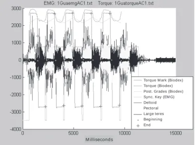
Fig. 1 – Simultaneous collection of the electromyographic signals using an EM G System do Brazil electromyography as w ell as the torque peak using the Biodex System 3 isokinetic dynamometer
Fig. 2 – Recording of a simultaneous collection of the electromyographic signals and the torque of the anterior deltoid, large teres, and the upper pectoral muscles synchronized in the time domain through a key placed on the dynamometer arm and activated on the beginning of the move-ment
Torque M ark (Biodex) Torque (Biodex) Post. Grades (Biodex) Sync. Key (EM G) Deltoid Pectoral Large teres Beginning End
M illiseconds
muscular injury. Those same strengths could change the elec-tromyographic activity of the assessed muscles, and this alter-ation is characterized by a muscular co-activalter-ation that w ould com-promise the production of the strength in the upper limbs of the athletes.
The increase in the enzymatic levels into the plasma could change the electrical activity of the muscle, and maybe those bio-chemical parameters w ould have a correlation to the indicators of the electrical signals of the muscular fatigue, and this could char-acterize a deficient status of the athletes’ fitness for the competi-tion due to a lack of or excessive training.
The aim of this study w as to verify if different judo fighting en-durances, respectively of 90s, 180s, and 300s could change the enzymatic metabolism as w ell as the athletes’ electromyographic activity, thus damaging the torque maintenance, w hich is an indi-cator of the muscular fatigue.
M ATERIALS AND M ETHODS
Subjects
Tw elve 22 ± 4.5 years male elite judo athletes w ho voluntarily signed the consent term to the research. This study w as approved by the UNIVAP Research Ethics Committee under the protocol no. A017/2003/CEP.
Fight protocol
All subjects w ere divided in pairs, according to their body w eight (kg) measured on a Fillizola scale. Three 90s, 180s, and 300s en-durance fights w ere performed betw een the pairs at the follow ing hours: 8am, 1pm, and 6pm in alternate days, w ith a 72 hours inter-val. Athletes w ere orally encouraged by the coach to fight at max-imal intensity. All subjects w ere assessed immediately before and after each fight.
EM G protocol
An 8-channel EM G System do Brasil electromyography w as used, and the EM G signals w ere collected from the follow ing muscles: anterior deltoid, large pectoral, and large teres muscle, respectively: agonist, antagonist, and synergistic of the movement. A local (trichotomia) and the local asepsis on the skin made w ith cotton, alcohol, and gel. A ground electrode w as placed on the non-dominant arm, and bipolar surface electrodes w ere fixed in the medium portion of muscles. The interval betw een the signal takes w as 30sec. from the order of execution of the movement.
Isokinetic dynamometer protocol
In order to measure the torque peak, it w as used a Biodex Sys-tem 3 isokinetic dynamometer. All individuals remained seat on the chair w ith the rotation angle aligned to the glenohumeral re-gion. The individuals performed five flexion movements and of shoulder extension at a 90o/sec. velocity, thus optimizing the col-lection of the electromyographic signal for dynamic protocols (see Gerdle(18)).
The movement started w ith the dominant arm extended anteri-or to the body in a 90o angle from the fundamental anatomic posi-tioning, follow ed by a shoulder flexion up to reach a 70o. Prior to the trial, an adaptation to the equipment w as performed along three days. The motor action w as based on the judo’s sleeve grip fre-quently performed by an athlete. As to the equipments’ synchro-nization, a key connected to the dynamometer arm w as used as a timer device to the beginning of the movement and this allow ed the simultaneous collection of the electromyographic signals and of the dynamometer (figure 1).
TABLE 1
Determination of the torque peak (TP/ ) before (AL) and after (DL) the 90sec., 180sec. and 300sec. fights, performed in the isokinetic test performed at 90os
n = 12 AL DL AL DL AL DL
PT (N.m-1) 70.54 ± 26.8 70.94 ± 29.11 65.7 ± 24.57 71.77 ± 27.10 66.55 ± 25.33 64,36 ± 28.32
TABLE 2
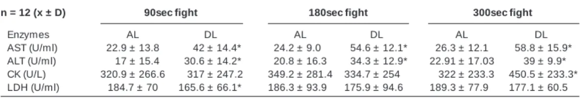
Activity of the enzymes: aminotransferase aspartate (AST), aminotransferase alanine (ALT), creatinekinase (CK), and dehydrogenase lactate (LDH) before (AL) and after (DL) the 90sec 180sec, and 300sec fights n = 12 (x ± D) 90sec fight 180sec fight 300sec fight
Enzymes AL DL AL DL AL DL
AST (U/ml) 22.9 ± 13.8 .* 042 ± 14.4* 24.2 ± 9.00 *54.6 ± 12.1* 26.3 ± 12.1 *58.8 ± 15.9* ALT (U/ml) 0,17± 15.4 *30.6 ± 14.2* 20.8 ± 16.3 *34.3 ± 12.9* 22.91 ± 17.03 * ,39 ± 9.9* CK (U/L) 320.9 ± 266.6 ,0317 ± 247.2 349.2 ± 281.4 334.7 ± 254,* ,0322 ± 233.3 *450.5 ± 233.3* LDH (U/ml) 184.7 ± 70 165.6 ± 66.1* 186.3 ± 93.90 175.9 ± 94.6* 189.3 ± 77.90 177.1 ± 60.50
* M inimum significance level set w as α = 0.05. After the 90sec fight, it w as verified an increase in the AST (p = 0.0059) and in the ALT (p = 0.0033), and a decrease in the LDH (p = 0.0392). After the 180sec fight, it w as observed an increase in the AST (p = 0.0033) and in the ALT (p = 0.0044). After the 300sec fight it w as verified an increase in the AST (p = 0.0033), in the ALT (p = 0.0044), and in the CK (p = 0.0180).
Signal processing protocol
Signals w ere processed using M ATLAB M ath Works 6.1 soft-w are. The rough data soft-w ere collected at a 15sec. interval, and at 2 KHz sampling frequency, w hich w as filtered through a 20 Hz high-pass filter and a 250 Hz low -high-pass filter, and rectified by the F.F.T. – Fast Fourier Transformation. In order to attain the same time res-olution in the EM G (0,05 ms) signals as w ell as in the torque (10 ms), the torque signal w as interpolated using the Cubic Spline at 0,05 ms intervals. The first phase of each concentric contraction w as analyzed from the beginning of the movement.
The EM G signals w ere normalized by the basic line, and they w ere analyzed in the time domain through the total calculation of the absolute EM G signal value (iEM G) for each contraction using the below show ed equation, w here T is the endurance of the con-traction, and f(t) is the electromyographic signal.
Protocol to the blood sampling collection and biochemical dosage
It w as collected 10 ml of the individual’s blood using a dispos-able syringe and needle. All sampling w ere immediately centri-fuged in a 4oC JOUAN CR3I centrifuge. The activity of the LDH, CK, AST, and ALT enzymes w ere verified using a HITACHI UV-2001 spectrophotometer and the Analisa Diagnostica kit through the colorimetric method.
Statistical analysis of data
To the statistical analysis of the data, the non-parametric WIL-COXON test for paired data w as used w ith a significance level of
α = 0.05.
RESULTS
No difference w as found in the torque peak (TP) before and after different fight endurances, as it can be seen in the values show n on table 1.
Upon the AST and ALT activities, it w as verified an increase related to the increase in the 90sec, 180sec, and 300sec endur-ance fights, as it can be observed in the values show n on table 2 and figure 3.
It w as observed a decrease in the LDH only after the 90sec fight, as it can be observe on table 2, and an increase in the CK activity after the 300sec fight, according to figure 3c.
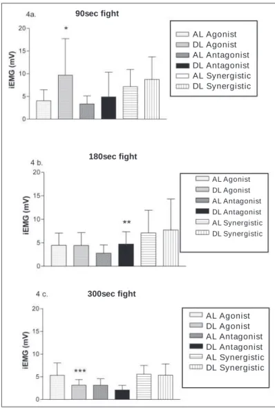
It w as observed an increase in the iEM G (mV) signal after every muscle in the 90sec fight. But such increase w as significant (p = 0.005) in the agonist muscle as it can be seen in table 3.
After the 180sec fight, the agonist muscle did not present any alteration, keeping the resting values, but it w as observed an in-crease (p = 0.0129) in the iEM G (mV) signal in the antagonist
mus-Fig. 3 – 3a) Activity of the transferase alanine enzyme (ALT), and 3b) transaminase aspartate (ALT) and creatinine kinase (CK) before and after the 90sec. 180sec., and 300sec. fight
ALT x Fights
Time (sec)
Time (sec)
Time (sec) CK x Fights
90sec AL 90sec DL 180sec AL 180sec DL 300sec AL 300sec DL
90sec AL 90sec DL 180sec AL 180sec DL 300sec AL 300sec DL
90sec AL 90sec DL 180sec AL 180sec DL 300sec AL 300sec DL
cle, and an increase, although non-significant, in the synergistic muscle.
In the 300sec fight, it w as verified a decrease in the activity of the agonist muscle (p = 0.0137), and a decrease (p = 0,05), al-though non-significant, in the activation of the antagonist muscle, and the synergistic muscle kept its mean resting values.
DISCUSSION
The mean values seen on table 1 suggest that the stressing fights to w hich individuals w ere submitted w as not enough to change the torque peak of muscles, and/or the resting time be-tw een the strength and the signal collection posed a sufficient recovering time to the ability to generate strength in the analyzed muscles.
The main biomechanical phenomena w ere verified in the simul-taneous analysis of the muscles involved in the movement, w hich w as already observed by other authors(23).
Despite de 90sec fight w as not sufficient to decrease the torque, it caused neural adaptations observed in the increasing iEM G sig-nals of the agonist muscle, and this may represent an adaptation dynamics observed in other studies using fatigue protocols(18).
Opposed to Kellis(17), it could be verified in the 180sec fight an increase in the amplitude of the iEM G signal of the antagonist muscle, suggesting the muscle has an important role for the sta-bility of the joint as a motor controlling of the regulation, and to the protection of the joints in a fatigue process(24). An explanation for such increase can be attributed to a central nervous system attempt to compensate the muscular fatigue through the increase in the number of motor units available(25).
If the antagonist muscle is able to increase the strength upon the accomplishment of repetitive strengths, it could represent an additional load to attend the agonist m uscle and keeping the strength, contributing to the resulting decrease in the involved joint and in protecting it, and this must be a parameter to be con-sidered w hen it is aimed the training performance in sportive ges-tures.
On the other hand, such co-activation suggests debility in the activation performance of the muscles to the accomplishment of the motor action, and this could pose a damage to the functional performance(24). The optimization of the strength training aiming the athlete’s performance has as purpose the activation of the agonist muscles w ith or w ithout low antagonist co-activation(16,22). The neuromuscular adaptations related to the endurance of fights suggest that it w as necessary a higher energetic demand to per-form the same strength observed w ith the AST and ALT activity regulated by the energetic demand necessary to the strengths.
Even w ith a predominance of the glycolytic w ay of the judo fights, the muscular proteins, the alanine and the glutamine w ere recruited to the ATP synthesis, suggesting in this study that the AST and ALT w ere biomarkers to the intensity level of the exer-cise(6).
The increasing iEM G (mV) of the agonist muscle after the first fight, as w ell as the co-activation after the second fight can both be explained by the dynamics of the neuromuscular adaptation to perform the w ork follow ing a gradual increase in the energetic demand characterized by the ASL and ALT activities, as it can be seen in figures 3a and 3b.
Analyzing the LDH, it w as verified a decrease in every fight pro-tocol, but it w as significant only after the 90sec fight, and this can be explained by the potentialized capability of the aerobic resis-tance that training athletes are submitted to. It is generally ac-cepted that the endurance training and the reduced intensity cause an accentuated reduction in the LDH activity(26,27). Thus, the regu-lating factor to the increasing LDH is the pyruvate and NADH of-fering. M aybe the individual’s training condition made the level of the substrates did not exceed the PDH capability to metabolize the pyruvate and/or the alpha-glycerophosphate system to supply the energetic demands(28). As the LDH is a regulating enzyme, maybe a negative modulator that could reduce the cytosolic NADH w ould cause such inhibition.
TABLE 3
Integral activity of the electromyographic signal (iEM G) in the agonist, antagonist, and synergistic muscles of the muscular (puxada de manga) action in the judo, simulated in the isokinetic dynamometer before (AL) and after (DL) the 90sec, 180sec, and 300sec fight n = 12 (X ± DP) 90s fight 180sec fight 300sec fight
iEM G (mV) AL DL AL DL AL DL
Agonist 4.04 ± 2.42 * 09.7 ± 8.02* 4.46 ± 2.61 4.45 ± 2.72* * 5.34 ± 2.73 3.16 ± 1.23* * *
Antagonist 3.35 ± 1.77 4.89 ± 5.45 2.79 ± 1.75 4.72 ± 2.64* * 3.14 ± 1.45 2.10 ± 1.03* * * Synergistic 7.14 ± 3.80 8.74 ± 5,00 07.1 ± 4.85 7.72 ± 6.61* * 5.55 ± 1.96 5.38 ± 2.44* * *
* The minimum significance level established w as α = 0.05. After the 90sec fight, it w as verified an increase in the iEM G signal of the agonist muscle (p = 0.005); after the 180sec fight, it w as observed an increase in the iEM G signal of the antagonist muscle (p = 0.0129), and after the 300sec fight it w as verified a decrease in the iEM G signal in the agonist muscle (p = 0.0137).
Fig. 4 – 4a) iEM G (mV) activity of the agonist, antagonist, and synergistic muscles of the movement assessed before and after the 90sec fight. 4b) iEM G (mV) activity of the agonist, antagonist and synergistic muscles of the movement assessed before and after the 180sec fight. 4c) iEM G (mV) activity of the agonist, antagonist, and synergistic muscles of the move-ment assessed before and after the 300sec fight.
90sec fight
180sec fight
300sec fight
AL Agonist DL Agonist AL Antagonist DL Antagonist AL Synergistic DL Synergistic
AL Agonist DL Agonist AL Antagonist DL Antagonist AL Synergistic DL Synergistic
In the 180sec. and 300sec. fights, the potentialized increase of the lactate due to the intermitting feature of the judo fight maybe has been enough to not allow a significant decrease in the LDH activity(29).
In the 300sec. fight, the iEM G (mV) signals presented a decrease in the agonist and antagonist activity, and at the same time, it kept the synergistic activation as the resting values.
The decreased activation of the agonist muscle may suggest a deficient motor action, combined w ith the synergistic actuation that should be stabilizing the movement instead of acting as pri-mary motor.
These data show the importance of studies aiming the syner-gistic activity of the movement, as to the effective strength train-ing, besides the activation of the agonist muscle and the reduc-tion in the co-activareduc-tion, an optimizareduc-tion in the synergistic muscle w ould be able to exert an important role in the increase of the torque system produced on the joints(12).
It w as verified that the 300sec fight protocol caused physiolog-ical adaptations w ith the increasing CK, and this damaged the motor units and/or the spreading of the muscular electric signal.
The lack of alteration in the CK activity in the first tw o fights can be explained by the inter-individual variations expressed by the high values of the standard deviation, making difficult to analyze the alterations caused by the exercise.
The increase in the CK after the 300sec fights can be a result of the intermittent submaximal strength(6) to w hat individuals w ere submitted, w ith an increase in the temperature and in the meta-bolic rate and a predominant eccentric contraction, thus increas-ing the release of that enzyme(30). The intensity of the exercise and its endurance are directly related variables to the alterations in the plasmatic CK concentrations(30). While the strength increas-es, the sarcolemma’s porosity and/or the rupture of the membrane provokes the passage of such cellular proteins to the plasma, and this is a phenomenon observed in this study after the 300sec fight. The decrease in the iEM G of the primary motor muscle of the movement associated to the increase in the plasmatic CK during the 300sec fight suggests that the strength w as sufficient to cause an increase in the permeability and/or micro-injuries in the sarco-lemma, and this provoked an alteration in the polarity, damaging the activation and spreading of the electrical signals, resulting in a decrease in the electromyographic signal amplitude.
Such phenomenon can result from the presence of metabolic by-products caused by the exercise, provoking an alteration in the membrane’s potential through the decrease in the pH caused by the acidification resulting from the injured muscular fiber, w hat could lead to damages to the cellular activation(31).
The fights athletes w ere submitted to in this study constitute a limiting factor, because despite the stimuli given by the coach and every official rules of the competition w ere follow ed, they w ere simulated to the accomplishment of this trial. So, athletes w ould present different and individual physiological responses in an offi-cial competition.
Despite of the increase w hich w as verified in the strength in-tensities through the AST and ALT activity, the mechanics of the muscular actions used in different fighting endurances is another aspect to be considered, once the fights are different as to their actions, limiting the functional assessment of the motor actions used by athletes in the fights.
The adaptations in the EM G signals observed in this study evi-dence the need of a simultaneous analysis as to the intermuscular coordination. The results of the AST and ALT activity related to the respective fight endurances confirm the utilization of such enzymes as strength intensity biomarkers, indicating a dynamics in the met-abolic adaptation to the gradually increased strengths.
The alterations in the electrical and biochemical parameters in the proposed protocol suggest that the intensity of the strength is related to the alteration in the electrical activity of the skeletal
muscular tissue, and the effects of the 300sec fight provoked an increase in the CK activity, and a decrease in the electrical activity of the agonist muscle. This suggests that the decrease in the am-plitude of the primary motor muscle signal is associated to possi-ble micro-injuries in the muscular membrane that damage the ac-tivation and spreading of the electromyographic signal.
CONCLUSION
It can be concluded that the stress of the 90sec, 180sec, and 300sec fights, despite of being unable to change the torque capa-bility w as stimuli sufficient to cause alterations in the enzymatic and muscular electrical activity that can be damaging to the ath-letes’ performance.
All the authors declared there is not any potential conflict of inter-ests regarding this article.
REFERENCES
1. M ujica I, Padilla S. M uscular characteristics of the training in humans. M ed Sci Sports Exerc 2000;33:1297-3.
2. Coyle EF. Detraining and retention of training-induced adaptations. M ed Sci Sports Exerc 1990;2:1-5.
3. Apple FS, Hellsten Y, Clarkson PM . Early detection of skeletal muscle injury by assay creatine kinase M M isoforms in serum after acute exercise. Clin Chem 1998;32:41-4.
4. Volfinger L, Lassorourd V, M ichaux JM , Braun JP, Tiurtain PL. Kinetic evaluation of muscle damage during exercise by calculation of amount of creatine kinase released. Am J Physiol 1994;266:434-1.
5. M eulen Van Der JH, Kuipers H, Drukker J. Relationship betw een exercise-induced muscle damage and enzyme release in rats. The American Physiological Society 1991;161:999-4.
6. Lijnen P, Hespel P, Fagard R, Lysens R, Van Den Eynde E, Goris M , et al. Indica-tors of cell breakdow n in plasma of men during and after a marathon race. J Sports M ed 1988;9:108-13.
7. M asuda T, Tizuca T, Zhe JY, et al. Influence of contraction force and speed on muscle fiber conduction velocity during dynamic voluntary exercise. J Electromyo-gr Kinesiol 2001;1:85-4.
8. Perry SR, Housh TJ, Weir JP, Johnson GO, Bull A, Ebersole KT. M ean pow er frequency and amplitude of the mechanomyographic and electromyographic sig-nals during incremental cycle ergometry. J Electromyogr Kinesiol 2001;11:299-5.
9. Gabriel DA, Basford JR, Kai-Nan AN. Neural adaptations to fatigue: implications for muscle strength and training. M ed Sci Sports Exerc 2001;33:1354-69.
10. Hakkinen K, Kramer WJ, New ton RU, Alen M . Changes in electromyographic activity muscle fibre and force production characteristics during heavy resistance/ pow er strength training in middle-aged and older men and w omen. Scandinavian Physiological Society 2001;171:51-62.
11. Kell RT, Bell G, Quinney A. M uscle skeletal fitness, health outcomes and quality of life. Sports M ed 2001;12:863-73.
12. Ross A, Leveritt M , Riek S. Neural Influences on sprint running. Sports M ed 2001;31:409-25.
13. Haw ley JA, Stepto NK. Adaptations to training in endurance cyclists. Implications for performance. Sports M ed 2001;31:511-0.
14. Clarys JP, Cabri J. Electromyography and study of sports movements: a review. J Sports Sci 1993;11:379-48.
15. Kazumi M , Tadashi M , Tsugutake S, M itsuharu I, Shigeru K. Changes in surface EM G parameters during static and dynamic fatiguing contractions. J Electromyo-gr Kinesiol 1999;9:39-6.
16. Vollestad NK. M easurement of human muscle fatigue. J Neurosci M ethods 1997; 74:219-7.
17. Kellis E. The effects of fatigue on the resultant joint moment, agonist and antag-onist electromyographic activity at different angles during dynamic knee exten-sion efforts. J Electromyogr Kinesiol 1999;9:191-9.
18. Gerdle B, Larsson B, Karlsson S. Criterion validation of surface EM G variables as fatigue indicators using peak torque. J Electromyogr Kinesiol 2000;10:225-2.
19. Lamontagne A, Richards CL, M alouin F. Coactivation during gait as an adaptive behavior after stroke. J Electromyogr Kinesiol 2000;10:407-5.
20. Hautier CA, Arsac LM , Deghdegh K. Influence of fatigue on EM G/force ratio and co-contraction in cycling. M ed Sci Sports Exerc 2000;32:839-43.
22. Franchini E. Características fisiológicas em testes laboratoriais e resposta da con-centração de lactato sanguíneo de três lutas em judocas das classes juvenil-A, Júnior e Sênior. Revista Paulista de Educação Física 1998;12:5-16.
23. Berger W, Trippl M , Discher M , Dietz V. Influence of subjects height on the stabi-lization of posture. Acta Otolaryngol 1992;30:112-22.
24. Van Handel PJ, Watson P, Troup J, Plyley M . Effects of treadmill running on oxida-tive capacity of regenerated skeletal muscle. Clin J Sport M ed 1981;2:92-6. 25. Spriet LL, Heigenhairser GJF. Regulation of Pyruvate Dehydrogenase (PDH)
ac-tivity in human skeletal muscle during exercise. Exerc Sport Sci Rev 2002;30: 91-5.
26. Almada A, M itchell T, Earnest C. Impact of chronic creatine supplementation on serum enzyme concentrations. FASEB J 1996;10:45-67.
27. Noakes TD. Effect of exercise on serum activities in humans. Sports M ed 1987; 4:245-67.
28. Bourdon L, Stieglitz P, Pouzeratte N, Curé M . Effect of incubation temperature on the creatine kinase release from in vitro rat skeletal muscle preparation. J Therm Biol 1996;21:109-13.
29. Hortobagyi T, Denahan D. Variability in creatine kinase: methodological, exercise and clinically related factors. Int J Sports M ed 1989;10:69-80.
30. Janssen GM , Kuipers H, Willems GM , Does RJ, Janssen, EP, Geurten P. Plasma activity of muscle enzymes. Quantification of skeletal muscle damage and rela-tionship w ith metabolic variables. J Sports M ed 1989;3:160-8.