Rev Bras Ter Intensiva. 2013;25(4):345-347
Aortic luminal thrombus and intramural
hematoma after cardiopulmonary resuscitation
CASE REPORT
INTRODUCTION
Cardiopulmonary resuscitation (CPR) is a life saving procedure. However, it can be associated with thoracic and especially skeletal injuries, although direct cardiac damage is rare.(1-6) A few cases of acute intramural
aortic hematoma leading to fatal aortic rupture have been described.(4,7) We
describe a case of aortic hematoma combined with a loating intraluminal thrombus associated with manual CPR.
CASE REPORT
A 92-year old male experienced cardiac arrest due to ventricular ibrillation. Witnesses immediately initiated manual CPR, relayed by a medical team upon arrival on site. Circulation was restored after 20 minutes. Electrocardiogram showed an anterior ST segment elevation myocardial infarction suggesting an occlusion of the left anterior descending artery. Hypothermia was initiated during transfer to the hospital. he patient was admitted to the emergency room in cardiogenic shock, requiring immediate
David Fagnoul1, Antoine Herpain1, Jean-Louis
Vincent1, Daniel De Backer1
1. Department of Intensive Care, Erasme University Hospital, Université Libre de Bruxelles - Brussels,
Belgium. We describe the case of a patient
with an intramural hematoma and loating thrombus after cardiopulmonary resuscitation. he 92-year old man had a cardiac arrest due to ventricular ibrillation and witnesses immediately initiated manual cardiopulmonary resuscitation. Transesophageal echocardiography was performed immediately on hospital admission because the patient was in cardiogenic shock. In addition to an akinetic anterior wall, examination of the descending thoracic aorta demonstrated an intramural hematoma and a loating intra-aortic thrombus at a distance of 40cm from the dental arch. here was
Conflicts of interest: None.
Submitted on October 29, 2013 Accepted on November 11, 2013
Corresponding author: Daniel De Backer
Department of Intensive Care Erasme University Hospital Route de Lennik 808 B-1070 Brussels, Belgium E-mail: [email protected]
Trombo aórtico intraluminal e hematoma intramural após
manobra de ressuscitação cardiopulmonar
ABSTRACT
Keywords: Cardiopulmonary resuscitation/ complications; Hematoma/etiology; Thrombosis/ etiology; Echocardiography; Anticoagulants/ therapeutic use; Case reports
no aortic dissection. he thrombus was attributed to aortic compression during cardiopulmonary resuscitation. Although the aortic thrombus and intramural hematoma were not associated with any complications in this patient, insertion of an intra-aortic balloon may have led to aortic rupture or embolic events. Transesophageal echocardiography should be performed, when available, prior to insertion of an intra-aortic balloon for counterpulsation in patients who have undergone cardiopulmonary resuscitation.
346 Fagnoul D, Herpain A, Vincent JL, De Backer D
Rev Bras Ter Intensiva. 2013;25(4):345-347
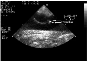
administration of norepinephrine. Transesophageal echocardiography was performed immediately to evaluate cardiac function. In addition to an akinetic anterior wall, examination of the aorta, before possible insertion of an aortic counterpulsation balloon in this elderly patient, showed an intramural hematoma and a loating intraluminal thrombus in the descending aorta, at a distance of 40cm from the dental arch (Figures 1 and 2, videos available in electronic supplement). here was no aortic dissection. he thrombus was attributed to aortic compression during CPR. Of note, there were no other signs of thoracic trauma (no rib nor sternum fracture). Shortly after admission, a coronary angiogram was performed using radial arterial access and occlusion of the left main coronary artery was treated by endovascular stenting. In view of the advanced age of the patient, presence of an intra-aortic hematoma(8,9) and persistent cardiogenic shock, stenting
of the aorta was not performed. he patient was treated with unfractionated heparin and clopidogrel. A second transesophageal echocardiography performed 2 days later showed that the intra-aortic thrombus had disappeared but the intramural hematoma persisted. here was no evidence of systemic embolization. he patient made a good neurologic recovery but died of severe cardiac failure on the 9th day. An autopsy could
not be performed due to relative refusal.
Figure 1 - Transverse view of descending aorta. An intramural hematoma and intraluminal clot are visualized at 40cm from the dental arch.
Figure 2 - Longitudinal view of descending aorta. This view shows the floating aspect of the clot.
DISCUSSION
Aortic thrombus may occur spontaneously in patients with a normal aorta or in the presence of atherosclerotic plaques. It can also occur in aortic trauma.(10) In this
patient, the thrombus may have been related to aortic trauma induced by chest compressions during CPR.
Manual compression CPR can result in signiicant thoracic trauma with multiple rib fractures, bilateral pulmonary contusions, lacerations of intercostal arteries, myocardial laceration and hemopericardium.(1-5) Acute
intramural aortic hematomas, which rarely lead to fatal aortic rupture,(4,7) have been described as an uncommon
complication of CPR. To the best of our knowledge, there is no report of endoluminal thrombus (combined with intramural hematoma) associated with CPR in the literature.
Given the paucity of reports in the literature, it is diicult to identify factors associated with CPR-induced aortic lesions. he use of mechanical devices may be involved in some cases(4) although they do not increase
the incidence of serious complications.(2) Of note,
Aortic luminal thrombus and intramural hematoma after cardiopulmonary resuscitation 347
Rev Bras Ter Intensiva. 2013;25(4):345-347
CPR is performed by bystanders rather than trained personnel.(6) Nevertheless, age alone should not be used
as a reason for not performing CPR, because old patients also beneit from CPR, especially when associated with neuroprotective measures as in this patient.(11)
Optimal treatment for such aortic lesions is debated. Mobile aortic thrombi are often associated with thromboembolic events.(12) Heparin remains the
irst-line therapy for mobile aortic thrombi,(13,14) and
thrombectomy through direct aortotomy and aortic stenting can also be considered.(13,15) Usually, post-CPR
aortic lesions are diagnosed at autopsy.(4) Although
the aortic thrombus and intramural hematoma were not associated with any complications in this patient, insertion of an intra-aortic balloon may have resulted in aortic rupture or embolic events.
CONCLUSION
Transesophageal echocardiography should be performed, when available, prior to insertion of intra-aortic balloon counterpulsation in patients who have undergone cardiopulmonary resuscitation.
Descrevemos o caso de um paciente com hematoma intramu-ral e trombo lutuante após ressuscitação cardiopulmonar. Esse homem, de 92 anos de idade, teve uma parada cardíaca causada por ibrilação atrial e testemunhas iniciaram imediatamente ma-nobras manuais de ressuscitação cardiopulmonar. Ao ser admitido no hospital, o paciente apresentava-se em choque cardiogênico, sendo, então, imediatamente submetido a ecocardiograia tran-sesofágica. Além de uma parede anterior acinética, o exame da aorta torácica descendente mostrou um hematoma intramural e um trombo intra-aórtico lutuante a uma distância de 40cm do arco dental. Não havia dissecção da aorta. O trombo foi atribuído
à compressão aórtica durante a ressuscitação cardiopulmonar. Embora o trombo aórtico e o hematoma intramural não tenham se associado a qualquer complicação nesse paciente, a inserção de um balão intra-aórtico poderia ter levado a uma ruptura da aorta ou a eventos embólicos. Recomenda-se a realização de ecocardio-graia transesofágica, quando disponível, antes da inserção de um balão intra-aórtico de contrapulsação em pacientes submetidos à ressuscitação cardiopulmonar.
RESUMO
Descritores: Ressuscitação cardiopulmonar/complicações;
Hematoma/etiologia; Trombose/etiologia; Ecocardiograia; Anticoagulantes/uso terapêutico; Relatos de casos
REFERENCES
1. Hoke RS, Chamberlain D. Skeletal chest injuries secondary to cardiopulmonary resuscitation. Resuscitation. 2004;63(3):327-38. 2. Lafuente-Lafuente C, Melero-Bascones M. Active chest
compression-decompression for cardiopulmonary resuscitation. Cochrane Database Syst Rev. 2004;(2):CD002751. Update of Cochrane Database Syst Rev. 2002;(3):CD002751.
3. Rabl W, Baubin M, Broinger G, Scheithauer R. Serious complications from active compression-decompression cardiopulmonary resuscitation. Int J Legal Med. 1996;109(2):84-9.
4. Englund E, Kongstad PC. Active compression-decompression CPR necessitates follow-up post mortem. Resuscitation. 2006;68(1):161-2. 5. Smekal D, Johansson J, Huzevka T, Rubertsson S. No difference in autopsy
detected injuries in cardiac arrest patients treated with manual chest compressions compared with mechanical compressions with the LUCAS device--a pilot study. Resuscitation. 2009;80(10):1104-7.
6. Kim MJ, Park YS, Kim SW, Yoon YS, Lee KR, Lim TH, et al. Chest injury following cardiopulmonary resuscitation: a prospective computed tomography evaluation. Resuscitation. 2013;84(3):361-4.
7. Bjork RJ, Snyder BD, Campion BC, Loewenson RB. Medical complications of cardiopulmonary arrest. Arch Intern Med. 1982;142(3):500-3. 8. Maraj R, Rerkpattanapipat P, Jacobs LE, Makornwattana P, Kotler MN.
Meta-analysis of 143 reported cases of aortic intramural hematoma. Am J Cardiol. 2000;86(6):664-8.
9. Evangelista A, Dominguez R, Sebastia C, Salas A, Permanyer-Miralda G, Avegliano G, et al. Long-term follow-up of aortic intramural hematoma: predictors of outcome. Circulation. 2003;108(5):583-9.
10. Cogert G, Siegel RJ. Giant floating aortic thrombus: a rare finding on transesophageal echocardiography. Am J Cardiol. 2007;99(5):739-40. 11. Mosier J, Itty A, Sanders A, Mohler J, Wendel C, Poulsen J, et al.
Cardiocerebral resuscitation is associated with improved survival and neurologic outcome from out-of-hospital cardiac arrest in elders. Acad Emerg Med. 2010;17(3):269-75.
12. Piffaretti G, Tozzi M, Mariscalco G, Bacuzzi A, Lomazzi C, Rivolta N, et al. Mobile thrombus of the thoracic aorta: management and treatment review. Vasc Endovascular Surg. 2008;42(5):405-11.
13. Moriwaki Y, Sugiyama M, Tahara Y, Iwashita M, Kosuge T, Harunari N, et al. Complications of bystander cardiopulmonary resuscitation for unconscious patients without cardiopulmonary arrest. J Emerg Trauma Shock. 2012;5(1):3-6.
14. Bowdish ME, Weaver FA, Liebman HA, Rowe VL, Hood DB. Anticoagulation is an effective treatment for aortic mural thrombi. J Vasc Surg. 2002;36(4):713-9.