Reclassifying the spectrum of septic patients using
lactate: severe sepsis, cryptic shock, vasoplegic
shock and dysoxic shock
INTRODUCTION
Sepsis remains a major challenge to public health, even after years of study
and progression in the understanding of the condition.(1-3) In recent years, the
incidence of sepsis has been increasing, and the associated mortality remains
high, with great variability between countries and continents.(3-6) To better
stratify sepsis, serum lactate levels have been used worldwide,(7-12) and the
current literature demonstrates good results for the use of serum lactate as a prognostic measure, as well as for therapeutic decisions and clinical classiication
for inclusion in randomized studies and benchmarking.(9-11,13-16)
Otavio Tavares Ranzani1,2, Mariana Barbosa Monteiro1, Elaine Maria Ferreira3, Sergio Ricardo Santos1, Flavia Ribeiro Machado3,4, Danilo Teixeira Noritomi1, on behalf of “Grupo de Cuidados Críticos Amil”
1. Intensive Care Unit, Hospital Paulistano - São Paulo (SP), Brazil.
2. Intensive Care Unit, Discipline of Clinical Emergency, Hospital das Clínicas, Universidade de São Paulo - USP - São Paulo (SP), Brazil. 3. Latin American Sepsis Institute - São Paulo (SP), Brazil.
4. Discipline of Anesthesiology, Pain and Intensive Care, Escola Paulista de Medicina, Universidade Federal de São Paulo - UNIFESP - São Paulo (SP), Brazil.
Objective: he current deinition of severe sepsis and septic shock includes a heterogeneous proile of patients. Although the prognostic value of hyperlactatemia is well established, hyperlactatemia is observed in patients with and without shock. he present study aimed to compare the prognosis of septic patients by stratifying them according to two factors: hyperlactatemia and persistent hypotension.
Methods: he present study is a
secondary analysis of an observational study conducted in ten hospitals in Brazil (Rede Amil - SP). Septic patients with initial lactate measurements in the irst 6 hours of diagnosis were included and divided into 4 groups according to hyperlactatemia (lactate >4mmol/L) and persistent hypotension: (1) severe sepsis (without both criteria); (2) cryptic shock (hyperlactatemia without persistent hypotension); (3) vasoplegic shock (persistent hypotension without hyperlactatemia); and (4) dysoxic shock (both criteria).
Conflicts of Interest: None.
Submitted on October 12, 2013 Accepted on November 28, 2013
Corresponding Author: Otavio Tavares Ranzani
Departamento de Cuidados Críticos - Rede AMIL Rua Martiniano de Carvalho, 741
Zip code: 01321-001 - São Paulo (SP), Brazil E-mail: [email protected]
Reclassificando o espectro de pacientes sépticos com o uso do
lactato: sepse grave, choque críptico, choque vasoplégico e choque
disóxico
ABSTRACT
Keywords: Infection; Sepsis; Shock; Lactic acid
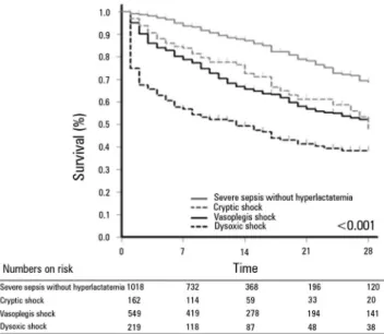
Results: In total, 1,948 patients were analyzed, and the sepsis group represented 52% of the patients, followed by 28% with vasoplegic shock, 12% with dysoxic shock and 8% with cryptic shock. Survival at 28 days difered among the groups (p<0.001). Survival was highest among the severe sepsis group (69%, p<0.001 versus others), similar in the cryptic and vasoplegic shock groups (53%, p=0.39), and lowest in the dysoxic shock group (38%, p<0.001 versus others). In the adjusted analysis, the survival at 28 days remained diferent among the groups (p<0.001) and the dysoxic shock group exhibited the highest hazard ratio (HR=2.99, 95%CI 2.21-4.05).
Conclusion: he deinition of sepsis includes four diferent proiles if we consider the presence of hyperlactatemia. Further studies are needed to better characterize septic patients, to understand the etiology and to design adequate targeted treatments.
he current deinition of severe sepsis requires the presence of organ dysfunction associated with infection, and lactatemia is included as a variable.(7) Septic shock is deined by the presence of sepsis associated with persistent hypotension after adequate volume replacement and the
need for vasoactive drugs.(7) However, septic patients
classiied as being in severe sepsis or septic shock exhibit great variability with respect to phenotype, clinical
outcomes, and prognosis(7,17-21) he two patient proiles of
sepsis are classic septic shock and cryptic shock, which is characterized as severe sepsis associated with serum lactate levels above 4mmol/L. Two studies have reported that there is no diference in the mortality of patients with these two sepsis diagnoses.(19,21) Recently, two other studies reclassiied patients with classic septic shock as dysoxic shock patients if the patients exhibited hyperlactatemia and as vasoplegic shock patients if the patients exhibited
persistent hypotension without hyperlactatemia.(17,20)
Patients with vasoplegic shock exhibited better outcomes compared to patients with dysoxic shock.
Few studies have addressed this topic in the current literature, and the topic is of fundamental importance when managing and classifying sepsis. Furthermore, no study has compared the new groups among themselves. hus, the present study aimed to compare patients with severe sepsis without hypoperfusion and patients with cryptic shock, vasoplegic shock and dysoxic shock. Secondarily, we aimed to assess whether intermediate initial values of lactate have a role in the prognosis of patients with sepsis.
METHODS
he present study constitutes a post-hoc analysis of a retrospective, multicenter, observational cohort study conducted by analyzing a prospectively collected
database.(22) Patients admitted to ten hospitals of the
Rede Amil from May 2010 to January 2012 in São Paulo
were included. Of these hospitals, one specializes in heart diseases and the remaining nine are general hospitals, providing 1,650 beds in total, 191 of which are located in intensive care units (ICU).
he database was built in partnership with the Latin
America Sepsis Institute (LASI).(23) Data were collected
using the electronic form provided by LASI based on the
Surviving Sepsis Campaign (SSC) bundles.(24) In each
hospital, a nurse was responsible for including data in the database. hese data were prospectively collected using a data collection sheet designed speciically for the present study that was completed by the healthcare team from
the time sepsis was diagnosed until the irst 24 hours of resuscitation. To collect data, the nurse also reviewed the charts. hroughout the collection, storage, and analysis of data, the patient's privacy was maintained, and all cases were identiied only by an identiication number. he Research Ethics Committee of the reference hospital, the
Hospital Pró-Cardíaco, approved the retrospective analysis and the publication of data on behalf of the entire network (protocol number 104,931), so a signed informed consent was not necessary.
he inclusion criteria consisted of patients diagnosed with severe sepsis or septic shock according to the sepsis
consensus conference deinitions(7,25) in all areas of the
hospital (emergency room, ward and ICU), and only the irst sepsis episode was included. he exclusion criteria consisted of patients under 18 years, receiving palliative care or those who refused intensive care. Patients whose initial lactate levels were not measured within the 6 irst hours of diagnosis were excluded from the present study. his cutof value for inclusion was chosen because it is the recommended time window for resuscitation therapeutic approaches.(7)
Definitions
Severe sepsis was deined by the presence of two or more signs of systemic inlammatory response syndrome resulting from a proven or suspected infectious process and at least one organ dysfunction associated with sepsis. Septic shock was considered when the hypotension associated with sepsis was refractory to adequate volume replacement with the subsequent need for vasopressors. he following were considered to be organ dysfunctions: hypotension (systolic blood pressure <90mmHg or mean arterial pressure <65mmHg or decrease >40mmHg in the systolic pressure); bilateral iniltrates on chest thorax radiograph and arterial oxygen pressure/fraction
of inspired oxygen ratio (PaO2/FiO2) ≤300 or the need
for supplemental oxygen to maintain oxygen saturation >90% (excluding the prior need for oxygen); total serum bilirubin >2mg/dL; urine output ≤0.5mL/kg/h for more than 2 hours or creatinine >2mg/dL; platelet count
≤100x109/L, international normalized ratio >1.5 or
activated partial thromboplastin time >60 seconds; and
serum lactate ≥2mmol/L.(7)
resuscitation strategy of these patients, according to the
SSC.(7) he presence of cryptic shock was considered
when patients exhibited severe sepsis criteria and
systemic hypoperfusion.(19) Patients with septic shock
criteria without systemic hypoperfusion were considered
vasoplegic shock patients,(20) and the presence of dysoxic
shock was considered when patients exhibited septic shock criteria and systemic hypoperfusion.(20)
To classify the patients in the four groups deined above, we performed a retrospective classiication using two variables already speciied in our database: persistent hypotension despite adequate volume expansion and the initial serum lactate level.
Demographic data (age and gender), clinical characteristics (temperature, heart and respiratory rate, systemic blood pressure, consciousness level, and chills) and laboratory data (blood glucose levels, blood lactate levels, and leukocyte counts) were collected at diagnosis. he severity scores were also collected at diagnosis (Acute Physiologic and Chronic Health Evaluation (APACHE
II) and Sequential Organ Failure Assessment (SOFA),(26)
as were the setting of sepsis diagnosis (ward, emergency room, or ICU), the site of infection and the compliance with measures for the resuscitation of septic patients. As the primary outcome, we evaluated the survival at 28 days after the sepsis diagnosis. As a secondary outcome, we evaluated the hospital mortality and the length of stay in the ICU and in the hospital.
Statistical analysis
To analyze the data distribution, a visual analysis of histograms and the Kolmogorov-Smirnov or the Shapiro-Wilk test, when appropriate, were performed. For continuous variables, data were presented as the mean and standard deviation if they exhibited a normal distribution or as the median and interquartile range if they did not exhibit a normal distribution. For categorical variables, data were presented as percentages.
According to the criteria deined above, patients were divided into four groups: (1) severe sepsis; (2) cryptic shock; (3) vasoplegic shock; and (4) dysoxic shock. he continuous variables were compared among the four groups using analysis of variance (ANOVA) for those with a normal distribution and the Kruskal-Wallis test for those without a normal distribution. For post-hoc comparisons, the correction proposed by Bonferroni was used. he categorical variables were compared using the chi-square test or, when appropriate, the Fisher's exact test or.
To analyze the survival at 28 days among the 4 groups, we used the Kaplan-Meier method. he probability of survival between the groups was analyzed by the log-rank test; for multiple post-hoc comparisons, we used the Holm-Sidak method. We evaluated the efect of each group on the survival at 28 days using the Cox regression model, both unadjusted and adjusted. For the adjusted model, we inserted pre-speciied variables based on the current literature using the enter method. We created two models: one that considers age, APACHE II, and SOFA in Model A, and one that considers age, APACHE II, SOFA, early use of antibiotics, local of diagnosis, and source hospital in Model B. We calculated the hazard ratio (HR) and its respective conidence interval (95%CI) for each group, and the severe sepsis group was used as a reference. For the Cox regression models, we tested second order interactions between age, APACHE II, and SOFA. To analyze the continuous variable lactate and the hospital mortality outcomes in patients with severe sepsis and septic shock, a nonlinear locally weighted function called Locally Weighted Scatterplot Smoothing (LOESS) was adjusted.
A two-tailed p value ≤0.05 was considered signiicant. he analyses and graphs were created using the programs Statistical Package for the Social Sciences (SPSS) version 19.0 (SPSS Inc, Chicago, IL), SigmaPlot 12.0 (Systat Software, San Jose, CA), and R v3.0.2 (R Development
Core Team 2013).(27)
RESULTS
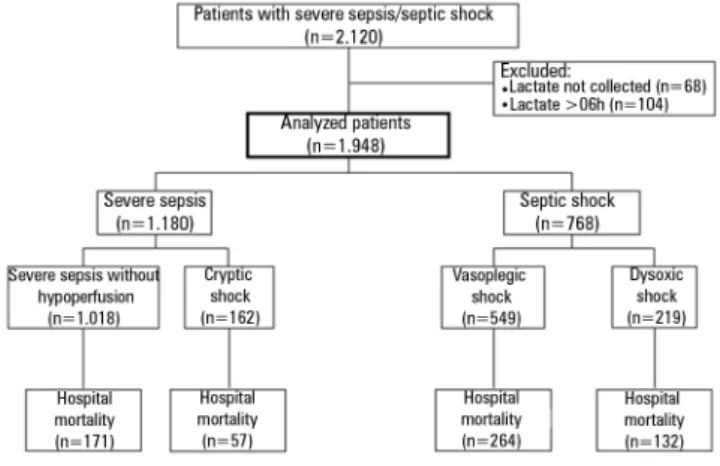
Within May 2010 and January 2012, there were 2,120 patients with severe sepsis or septic shock. Lactate collection was not performed in 68 patients and in 104 patients the irst collection occurred more than 6 hours after diagnosis. hus, 1,948 patients were analyzed and 172 (8%) patients were excluded (Figure 1). he average time between diagnosis and lactate collection was 19 (5-45) minutes.
here were 1,180 (61%) initial diagnoses of severe sepsis and 768 (39%) of septic shock. Among patients with severe sepsis, 1,018 (86%) exhibited no hypoperfusion criteria, whereas 162 (14%) were classiied as cryptic shock patients. Vasoplegic shock was present in 549 (72%) patients with an initial diagnosis of septic shock, whereas dysoxic shock was diagnosed in 219 (28%) patients (Table 1).
APACHE II, and SOFA (Model A, Table 2), the survival diference among the 4 groups persisted (p<0.001), and the dysoxic shock group exhibited increased adjusted risk (Beta=1.095, HR=2.99, 95% CI=2.21-4.05; p<0.001). he results were similar for model B (Table 2). None of the interactions tested were signiicant, so they were not included in the models. Figure 3 presents the adjusted survival curves of the four groups from models A and B. he hospital mortality was 624 (32%), ranging from 16.8% (95% CI=14.4-19.1) for patients with severe sepsis without hyperlactatemia to 60.3% (95% CI=53.9-67.1) for patients with dysoxic shock.
Figure 4 presents the relationship between the continuous values of lactate levels and hospital mortality in patients with a diagnosis of severe sepsis and septic shock separately. Interestingly, the risk of death begins to increase signiicantly in both groups when the lactate level exceeds 2.0mmol/L. Furthermore, the risk of death is greater among patients with shock.
DISCUSSION
Our results conirm that patients with a clinical diagnosis of severe sepsis or septic shock exhibit variable presentations and outcomes. We demonstrated that the percentage of patients with severe sepsis and hypoperfusion (cryptic shock) at diagnosis is relatively low. Similarly, among patients with septic shock, the number of patients with hypoperfusion (dysoxic shock) is also low. Moreover, the survival at 28 days difered among the groups; survival was lowest in the dysoxic shock group, but similar between patients with cryptic shock and vasoplegic shock, although the prognostic scores difered between the latter two groups. We also demonstrated that among patients with sepsis, the risk of death increases signiicantly when the initial lactate values are greater than 2mmol/L.
he initial serum lactate level is accepted as a prognostic marker and as a method for evaluating tissue perfusion in several populations of critically ill patients.(10,11,15,28) In both retrospective and prospective studies performed with patients with suspected infection, lactate levels exhibited prognostic value irrespective of the number of organ dysfunctions.(8) Moreover, the initial lactate values are often used for screening, as they are the trigger for the beginning of sepsis resuscitation measures. In the present study, patients with classic severe sepsis and septic
shock(3) were divided into new groups according to the
serum lactate level. hus, we add to the current literature the comparison between the four phenotypes of septic patients, given that previous studies analyzed patients with Figure 1 - Study diagram.
was higher when compared with those with severe sepsis without hypoperfusion and those with cryptic shock. In total, 47% of the patients were male and the majority of the patients were diagnosed in the emergency room. he vasoplegic shock group exhibited a diferent pattern and was less often diagnosed in the emergency room when compared with both the severe sepsis without hypoperfusion group and the cryptic shock group. he most frequent site of infection was the lung, followed by the urinary tract and the abdominal tract (Table 1).
At diagnosis, the prevalence of fever, hypothermia, tachycardia, and leukopenia difered among the groups (p<0.001). Fever, hypothermia, and leukopenia were more frequent in the group with severe sepsis without hypoperfusion when compared with the dysoxic shock group. Among patients with severe sepsis, arterial hypotension responsive to volume was present in 49% of patients without hypoperfusion and in 38% of patients with hypoperfusion, respectively. he severity scores of the patients at diagnosis also difered; the APACHE II score ranged from 15±6 in patients with severe sepsis without hypoperfusion to 24±8 in patients with dysoxic shock (p<0.001), with a signiicantly progressive increase among the four groups (Table 1). With respect to the SOFA score, patients with dysoxic shock exhibited higher scores (10 [8-13]), with no signiicant diference between patients with severe sepsis without hypoperfusion and patients with cryptic shock (3 [2-5] versus 4 [2-6]; p=0.20, respectively).
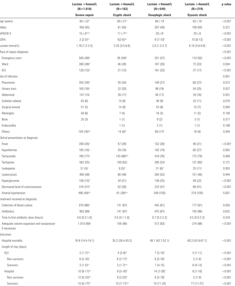
Table 1 - General characteristics of the sample according to the groups
Severe sepsis Septic shock
Lactate <4mmol/L (N=1.018)
Lactate >4mmol/L (N=162)
Lactate <4mmol/L (N=549)
Lactate >4mmol/L (N=219)
p value
Severe sepsis Cryptic shock Vasoplegic shock Dysoxic shock
Age (years) 60±22b 60±21d 66±18 63±18 <0.001
Males 458 (45) 81 (50) 267 (49) 109 (50) 0.321
APACHE II 15±6a,b,c 17±7d,e 20±8f 24±8 <0.001
SOFA 3 [2-5]b,c 4[2-6]d,e 9 [7-10]f 10 [8-13] <0.001
Lactate (mmol/L) 1.78 [1.2-2.5] 5.25 [4.5-6.6] 2.0 [1.3-2.7] 6.10 [4.8-8.8] <0.001
Place of sepsis diagnosis <0.001
Emergency room 500 (49)b 95 (59)d 201 (37) 110 (50) <0.001
Ward 390 (38)b 46 (28) 167 (30) 72 (33) 0.004
ICU 128 (13)b 21 (13) 181 (33) 37 (17) <0.001
Site of infection 0.001
Pneumonia 350 (34)b 55 (34) 149 (27) 60 (27) 0.012
Urinary tract 165 (16)c 32 (20) 99 (18) 54 (25) 0.027
Abdominal 147 (14) 28 (17) 94 (17) 34 (16) 0.501
Catheter-related 63 (6) 10 (6) 48 (9) 23 (11) 0.070
Surgical wound 51 (5) 14 (9) 43 (8) 15 (7) 0.084
Meningitis 58 (6) 7 (4) 16 (3) 11 (5) 0.100
Bone 25 (3) 1 (1) 9 (2) 3 (1) 0.317
Endocarditis - 1 (1) 2 (1) 1 (1) 0.188
Others 159 (16)a,c 14 (9)d 89 (17)e 18 (9) 0.004
Clinical presentation at diagnosis
Fever 359 (35)c 47 (29) 152 (28) 46 (21) <0.001
Hypothermia 165 (16)c 29 (18) 102 (19) 60 (27) 0.002
Tachycardia 780 (77)a 143 (88)d,e 416 (76) 172 (79) 0.006
Tachypnea 562 (55) 100 (62) 290 (53) 131 (60) 0.121
Leukopenia 51 (5)c 8 (5)e 31 (6)f 25 (11) 0.003
Leukocytosis 490 (48) 80 (49) 284 (52) 101 (46) 0.444
Hyperglycemia 156 (15)b 34 (21) 138 (25) 49 (22) <0.001
Decreased level of consciousness 316 (31)b 62 (38) 224 (41) 89 (41) <0.001
Arterial hypotension 496 (49)b,c 61 (38)d,e 549 (100) 219 (100) 0.001
Treatment received at diagnosis
Collection of blood culture 870 (86)c 141 (87) 445 (81) 177 (81) 0.050
Antibiotics 903 (89) 141 (87) 475 (87) 193 (88) 0.632
Time to first antibiotic dose (hours) 0.6 [0.2-1.6] 0.5 [0.1-1.8] 0.7 [0.2-2.3] 0.5 [0.0-2.3] 0.234
Adequate volume expansion and vasopressor if necessary
1.010 (99)c 156 (96) 512 (93) 214 (98) <0.001
Outcomes
Hospital mortality 16.8 (14.4-19.1) 35.2 (28.4-43.2) 48.1 (43.7-52.1) 60.3 (53.9-67.1) <0.001
Length of stay (days)
ICU 3 [1-7]d,e 4 [2-9]d 7 [3-15]f 4 [1-11] <0.001
Non-survivors 8 [3-16]c 6 [2-17]e 8 [3-16]f 2 [1-8] <0.001
Survivors 3 [1-5]b,c 3 [1-7]d,e 7 [4-15] 8 [4-13] <0.001
Hospital 10 [6-17]b,c 9 [5-18]d 14 [7-28]f 8 [1-18] <0.001
Non-survivors 12 [6-23]b,c 9 [3-22]e 9 [3-19]f 2 [1-9] <0.001
Survivors 10 [6-17]b,c 10 [7-17]d,e 19 [11-33] 17 [11-27] <0.001
Figure 2 - Survival curve 28 days after the sepsis diagnosis. A significant difference is observed among the four sepsis phenotypes (log-rank test, p<0.001). In post-hoc comparisons, the survival was different among the four groups, except for the comparison between the cryptic shock group and the vasoplegic shock group (p=0.387).
septic shock only with respect to the presence or absence
of hypoperfusion(17,18,20) or compared cryptic shock with
septic shock without considering the presence or absence
of hyperlactatemia in patients with septic shock.(19,21)
Among patients with persistent hypotension, we observed that 72% did not exhibit hyperlactatemia, which is higher than has been reported in previous studies (31% and 50%),(17,18,20) although these prior studies involved smaller sample sizes and focused on emergency room patients. Among patients with severe sepsis, only 14% exhibited hyperlactatemia, consistent with the previously described range of 8 to 25%.(8,21)
Occult hypoperfusion has been reported in the literature both in septic patients and in other proiles of critically ill patients.(21,29) he fact that occult hypoperfusion exists and is not diagnosed is unsurprising. he imbalance between the supply and consumption of oxygen is a characteristic of
Table 2 - Survival at 28 days according to the study groups
Univariate analysis Model A Model B
Beta HR (CI95%) p value Beta HR (CI95%) p value Beta HR (CI95%) p value
Severe sepsis without hyperlactatemia Ref. 1 <0.001 Ref. 1 <0.001 Ref. 1 <0.001
Cryptic shock 0.792 2.21 (1.59-3.06) <0.001 0.712 2.01 (1.47-2.83) <0.001 0.768 2.16 (1.54-3.01) <0.001
Vasoplegic shock 0.919 2.51 (2.03-3.10) <0.001 0.597 1.82 (1.41-2.34) <0.001 0.618 1.86 (1.43-2.40) <0.001
Dysoxic shock 1.522 4.58 (3.59-5.84) <0.001 1.095 2.99 (2.21-4.05) <0.001 1.171 3.23 (2.37-4.39) <0.001
Model A - adjusted for age, APACHE II, and total SOFA. Model B - adjusted for age, APACHE II, SOFA total, place of diagnosis, use of adequate antibiotic treatment within the period (1 hour/3 hours), and the source hospital.
shock conditions, and other markers such as blood pressure, heart rate, are urine output exhibit a low sensitivity of
detecting the presence of shock.(21) hus, even when vital
signs are normal, the serum lactate may increase through other mechanisms. his condition is called cryptic shock and is associated with evidence of hypoperfusion, despite the ability of the organism to maintain normal blood pressure with compensatory mechanisms. Moreover, patients with hypotension refractory to volume may also exhibit varying levels of serum lactate, and the relationship between these levels and other clinical parameters is unclear. Hernandez et al.(18) reported that the presence or absence of hyperlactatemia in patients with septic shock did not correlate with age, comorbidities, source of sepsis, or macrohemodynamic parameters, including cardiac output. Conversely, patients with normal levels of lactate exhibited better microcirculation parameters and exhibited a microcirculatory low close to normal, as evaluated using
images of the sublingual microcirculation.(18) Wacharasint
et al.(30) also evaluated the importance of intermediate
lactate values (cut-of for the Q4 quartile ≥4.4mmol/L) in patients with septic shock. In the original cohort (VASST study), the mean arterial pressure did not diferentiate the lactate groups, but the heart rate and central venous pressure was able to diferentiate among the groups. In the validation cohort, the heart rate was similar between the
groups.(30) In 2013, Sterling et al. analyzed 247 patients
with septic shock, dividing them in vasoplegic shock and dysoxic shock, reasserting previous indings that reported
no clinical manifestation diference between the groups.(20)
However, in sepsis, tissue hypoxia is not the only
factor involved in increasing lactate levels.(31) here are
Figure 3 - Adjusted survival curves 28 days after the sepsis diagnosis. Model A is adjusted for age, APACHE II, and SOFA, and model B is adjusted for age, APACHE II, SOFA, early use of antibiotics, place of sepsis diagnosis, and source hospital. In both models, the group with dysoxic shock exhibited a higher hazard ratio when compared with the group with severe sepsis without hyperlactatemia.
Figure 4 - Role of intermediate lactate values in patients with severe sepsis and septic shock. An important increase in the risk of death can be observed from the 2 mmol/L value in both groups, although patients with septic shock exhibit a higher risk of death. The risk of death was adjusted by a nonlinear locally weighted function called Locally Weighted Scatterplot Smoothing (LOESS). The gray area corresponds to a confidence interval of 95%.
associated with sepsis. Moreover, lactate clearance is also decreased in septic patients, and increased lactate levels are a result of its decreased metabolism.(32) Additionally, there may be exacerbations of the glycolytic pathway mediated by the use of adrenergic drugs,(11,33) i.e., patients using adrenaline or high doses of noradrenaline may exhibit hyperlactatemia. In the present study, due to the early collection of our samples, this potential cause of increased
lactate levels is not likely. Because hyperlactatemia may not relect reduced tissue supply with hypoperfusion, the prognostic value of increased lactate levels may represent the intrinsic severity of the patient's condition.
hypoperfusion to the dysoxic shock group, except for SOFA in patients with severe sepsis. he survival at 28 days, both in the univariate and in the adjusted analysis, difered among the four groups. Patients with cryptic shock and vasoplegic shock exhibit similar risks when compared with the group with severe sepsis without hypoperfusion, although patients with vasoplegic shock exhibited higher scores on both scales used in the study. Patients with cryptic shock and vasoplegic shock difered, however, from patients with dysoxic shock for whom the survival was shorter, which suggests greater severity. To our knowledge, this fact is infrequently reported in the literature and may contribute to a better understanding of the disease. Previously, Puskarich et al.(19) reported that the mortality associated with cryptic shock is similar to that associated with the classically deined septic shock (21% versus
19%). Hernandez et al.(17) also observed longer survival
times in patients with vasoplegic shock when compared with patients with dysoxic shock (92.3% versus 57.1%).
Guided by the study of Rivers et al.,(34) the current
guidelines recommend similar treatment for patients with septic shock and severe sepsis with hypoperfusion using the criterion of initial serum lactate greater than 4mmol/L. Some recent studies demonstrated that even intermediate levels of lactate are associated with an increased risk of unfavorable outcomes in patients with sepsis, suggesting that therapeutic strategies should be established for this group. Indeed, a recent study(13) that used an initial lactate value of 3 mmol/L reported that management guided by lactate clearance and by central venous oxygen saturation
is equivalent. In our study, we observed an increased risk of death in patients with sepsis with lactate values greater than 2mmol/L, suggesting that patients with severe sepsis and lactate levels lower than the currently established cutof must be further studied to develop better therapeutic treatments for this patient population.(30,35)
Our study had limitations that should be emphasized. he present was an observational and retrospective study; thus, there is no information on the etiological diagnosis, inlammatory response, and other important hemodynamic variables of patients with sepsis, such as central venous oxygen saturation and other markers of hypoperfusion, that would better support the indings observed. In addition, we have no data on variables that can interfere with the lactate values, such as the presence of liver disease, the dose of vasoactive drugs, or enzyme deiciencies, which could allow subgroup analyses. he inding of an increased risk of death with lactate values greater than 2mmol/L also suggests that the use of diferent values to deine hyperlactatemia could inluence the reported results.
CONCLUSION
In a multicenter study of septic patients, we demonstrated that there are at least four diferent phenotypes within the two current classic sepsis classiications. he diferentiation between the groups is of fundamental epidemiological importance for a possible adaptation of the targeted treatments and can inform better patient selection in future studies.
Objetivo: A deinição atual de sepse grave e choque séptico inclui um peril heterogêneo de pacientes. Embora o valor prognóstico de hiperlactatemia seja bem estabelecido, ela está presente em pacientes com ou sem choque. Nosso objetivo foi comparar o prognóstico de pacientes sépticos estratiicando-os segundo dois fatores: hiperlactatemia e hipotensão persistente.
Métodos: Este estudo é uma análise secundária de um
estudo observacional conduzido em dez hospitais no Brasil (Rede Amil - SP). Pacientes sépticos com valor inicial de lactato das primeiras 6 horas do diagnóstico foram incluídos e divididos em 4 grupos segundo hiperlactatemia (lactato>4mmol/L) e hipotensão persistente: (1) sepse grave (sem ambos os critérios); (2) choque críptico (hiperlactatemia sem hipotensão persistente); (3) choque vasoplégico (hipotensão persistente sem hiperlactatemia); e (4) choque disóxico (ambos os critérios).
Resultados: Foram analisados 1.948 pacientes, e o grupo sepse grave constituiu 52% dos pacientes, seguido por 28% com choque vasoplégico, 12% choque disóxico e 8% com choque críptico. A sobrevida em 28 dias foi diferente entre os grupos (p<0,001), sendo maior para o grupo sepse grave (69%; p<0,001
versus outros), semelhante entre choque críptico e vasoplégico (53%; p=0,39) e menor para choque disóxico (38%; p<0,001
versus outros). Em análise ajustada, a sobrevida em 28 dias permaneceu diferente entre os grupos (p<0,001), sendo a maior razão de risco para o grupo choque disóxico (HR=2,99; IC95% 2,21-4,05).
Conclusão: A deinição de pacientes com sepse inclui quatro diferentes peris, se considerarmos a presença de hiperlactatemia. Novos estudos são necessários para melhor caracterizar pacientes sépticos e gerar conhecimento epidemiológico, além de possível adequação de tratamentos dirigidos.
RESUMO
REFERENCES
1. Silva E, Pedro Mde A, Sogayar AC, Mohovic T, Silva CL, Janiszewski M, Cal RG, de Sousa EF, Abe TP, de Andrade J, de Matos JD, Rezende E, Assunção M, Avezum A, Rocha PC, de Matos GF, Bento AM, Corrêa AD, Vieira PC, Knobel E; Brazilian Sepsis Epidemiological Study. Brazilian Sepsis Epidemiological Study (BASES study). Crit Care. 2004;8(4):R251-60. 2. Silva E, Akamine N, Salomao R, Townsend SR, Dellinger RP, Levy M.
Surviving sepsis campaign: a project to change sepsis trajectory. Endocr Metab Immune Disord Drug Targets. 2006;6(2):217-22.
3. Dellinger RP, Levy MM, Rhodes A, Annane D, Gerlach H, Opal SM, Sevransky JE, Sprung CL, Douglas IS, Jaeschke R, Osborn TM, Nunnally ME, Townsend SR, Reinhart K, Kleinpell RM, Angus DC, Deutschman CS, Machado FR, Rubenfeld GD, Webb S, Beale RJ, Vincent JL, Moreno R; Surviving Sepsis Campaign Guidelines Committee including The Pediatric Subgroup. Surviving Sepsis Campaign: international guidelines for management of severe sepsis and septic shock, 2012. Intensive Care Med. 2013;39(2):165-228.
4. Phua J, Koh Y, Du B, Tang YQ, Divatia JV, Tan CC, Gomersall CD, Faruq MO, Shrestha BR, Gia Binh N, Arabi YM, Salahuddin N, Wahyuprajitno B, Tu ML, Wahab AY, Hameed AA, Nishimura M, Procyshyn M, Chan YH; MOSAICS Study Group. Management of severe sepsis in patients admitted to Asian intensive care units: prospective cohort study. BMJ. 2011;342:d3245. 5. Sogayar AM, Machado FR, Rea-Neto A, Dornas A, Grion CM, Lobo SM,
Tura BR, Silva CL, Cal RG, Beer I, Michels V, Safi J, Kayath M, Silva E; Costs Study Group - Latin American Sepsis Institute. A multicentre, prospective study to evaluate costs of septic patients in Brazilian intensive care units. Pharmacoeconomics. 2008;26(5):425-34.
6. Teles JM, Silva E, Westphal G, Filho RC, Machado FR. Surviving sepsis campaign in Brazil. Shock. 2008;30 Suppl 1:47-52. Review.
7. Dellinger RP, Levy MM, Rhodes A, Annane D, Gerlach H, Opal SM, Sevransky JE, Sprung CL, Douglas IS, Jaeschke R, Osborn TM, Nunnally ME, Townsend SR, Reinhart K, Kleinpell RM, Angus DC, Deutschman CS, Machado FR, Rubenfeld GD, Webb S, Beale RJ, Vincent JL, Moreno R; Surviving Sepsis Campaign Guidelines Committee including The Pediatric Subgroup. Surviving Sepsis Campaign: international guidelines for management of severe sepsis and septic shock, 2012. Intensive Care Med. 2013;39(2):165-228.
8. Mikkelsen ME, Miltiades AN, Gaieski DF, Goyal M, Fuchs BD, Shah CV, et al. Serum lactate is associated with mortality in severe sepsis independent of organ failure and shock. Crit Care Med. 2009;37(5):1670-7.
9. Bakker J, Gris P, Coffernils M, Kahn RJ, Vincent JL. Serial blood lactate levels can predict the development of multiple organ failure following septic shock. Am J Surg. 1996;171(2):221-6.
10. Bakker J, Jansen TC. Don't take vitals, take a lactate. Intensive Care Med. 2007;33(11):1863-5.
11. Bakker J, Nijsten MW, Jansen TC. Clinical use of lactate monitoring in critically ill patients. Ann Intensive Care. 2013;3(1):12.
12. Machado FR, Salomão R, Rigato O, Ferreira EM, Schettino G, Mohovic T, et al. Late recognition and illness severity are determinants of early death in severe septic patients. Clinics (Sao Paulo). 2013;68(5):586-91. 13. Jansen TC, van Bommel J, Schoonderbeek FJ, Sleeswijk Visser SJ, van
der Klooster JM, Lima AP, Willemsen SP, Bakker J; LACTATE study group. Early lactate-guided therapy in intensive care unit patients: a multicenter, open-label, randomized controlled trial. Am J Respir Crit Care Med. 2010;182(6):752-61.
14. Maciel AT, Park M. Unmeasured anions account for most of the metabolic acidosis in patients with hyperlactatemia. Clinics (Sao Paulo). 2007;62(1):55-62.
15. Noritomi DT, Soriano FG, Kellum JA, Cappi SB, Biselli PJ, Libório AB, et al. Metabolic acidosis in patients with severe sepsis and septic shock: a longitudinal quantitative study. Crit Care Med. 2009;37(10):2733-9.
16. Park M, Taniguchi LU, Noritomi DT, Libório AB, Maciel AT, Cruz-Neto LM. Clinical utility of standard base excess in the diagnosis and interpretation of metabolic acidosis in critically ill patients. Braz J Med Biol Res. 2008;41(3):241-9.
17. Hernandez G, Castro R, Romero C, de la Hoz C, Angulo D, Aranguiz I, et al. Persistent sepsis-induced hypotension without hyperlactatemia: is it really septic shock? J Crit Care. 2011;26(4):435.e9-14.
18. Hernandez G, Bruhn A, Castro R, Pedreros C, Rovegno M, Kattan E, et al. Persistent Sepsis-Induced Hypotension without Hyperlactatemia: A Distinct Clinical and Physiological Profile within the Spectrum of Septic Shock. Crit Care Res Pract. 2012;2012:536852.
19. Puskarich MA, Trzeciak S, Shapiro NI, Heffner AC, Kline JA, Jones AE; Emergency Medicine Shock Research Network (EMSHOCKNET). Outcomes of patients undergoing early sepsis resuscitation for cryptic shock compared with overt shock. Resuscitation. 2011;82(10):1289-93. 20. Sterling SA, Puskarich MA, Shapiro NI, Trzeciak S, Kline JA, Summers RL,
Jones AE; Emergency Medicine Shock Research Network (EMSHOCKNET). Characteristics and outcomes of patients with vasoplegic versus tissue dysoxic septic shock. Shock. 2013;40(1):11-4.
21. Howell MD, Donnino M, Clardy P, Talmor D, Shapiro NI. Occult hypoperfusion and mortality in patients with suspected infection. Intensive Care Med. 2007;33(11):1892-9.
22. Noritomi DT, Ranzani OT, Monteiro MB, Ferreira EM, Santos SR, Leibel F, et al. Implementation of a multifaceted sepsis education program in an emerging country setting: clinical outcomes and cost-effectiveness in a long-term follow-up study. Intensive Care Med. 2013 Oct 22. [Epub ahead of print].
23. Latin American Sepsis Institute [Internet]. [cited 2012 feb 13]. Available from: http://www.sepsisnet.org/
24. Surviving Sepsis Campaign [Internet]. [cited 2012.Feb 13]. Available from: http://www.survivingsepsis.org.
25. Bone RC, Balk RA, Cerra FB, Dellinger RP, Fein AM, Knaus WA, et al. Definitions for sepsis and organ failure and guidelines for the use of innovative therapies in sepsis. The ACCP/SCCM Consensus Conference Committee. American College of Chest Physicians/Society of Critical Care Medicine. Chest. 1992;101(6):1644-55. Review.
26. Ranzani OT, Battaini LC, Moraes CE, Prada LF, Pinaffi JV, Giannini FP, et al. Outcomes and organ dysfunctions of critically ill patients with systemic lupus erythematosus and other systemic rheumatic diseases. Braz J Med Biol Res. 2011;44(11):1184-93.
27. R Development Core Team. R: a language and environment for statistical computing. Viena, Austria: R Foundation for Statistical Computing; 2013. 28. Maciel AT, Noritomi DT, Park M. Metabolic acidosis in sepsis. Endocr
Metab Immune Disord Drug Targets. 2010;10(3):252-7. Review. 29. Park M, Maciel AT, Noritomi DT, Brunialti MK, Salomão R, Schettino GP,
et al. Is persistent hypotension after transient cardiogenic shock associated with an inflammatory response? Braz J Med Biol Res. 2008;41(8):648-56. 30. Wacharasint P, Nakada TA, Boyd JH, Russell JA, Walley KR. Normal-range blood lactate concentration in septic shock is prognostic and predictive. Shock. 2012;38(1):4-10.
31. Gladden LB. Lactate metabolism: a new paradigm for the third millennium. J Physiol. 2004;558(Pt 1):5-30.
32. Levraut J, Ciebiera JP, Chave S, Rabary O, Jambou P, Carles M, et al. Mild hyperlactatemia in stable septic patients is due to impaired lactate clearance rather than overproduction. Am J Respir Crit Care Med. 1998;157(4 Pt 1):1021-6.
33. Mizock BA, Falk JL. Lactic acidosis in critical illness. Crit Care Med. 1992;20(1):80-93.
34. Rivers E, Nguyen B, Havstad S, Ressler J, Muzzin A, Knoblich B, Peterson E, Tomlanovich M; Early Goal-Directed Therapy Collaborative Group. Early goal-directed therapy in the treatment of severe sepsis and septic shock. N Engl J Med. 2001;345(19):1368-77.