Factors associated with blood oxygen partial pressure
and carbon dioxide partial pressure regulation during
respiratory extracorporeal membrane oxygenation
support: data from a swine model
INTRODUCTION
Despite a worldwide increase in respiratory extracorporeal membrane
oxygenation (ECMO) support,(1,2) studies exploring the physiology of
veno-venous conigurations are still lacking.(3)
he use of respiratory extracorporeal support allows ultra-protective mechanical ventilation, which leads to reduced stress and strain on the lungs and
is associated with better outcomes in ECMO-supported patients.(4) Reductions
Marcelo Park1,2, Pedro Vitale Mendes1,2, Eduardo Leite Vieira Costa1,2, Edzangela Vasconcelos Santos Barbosa2, Adriana Sayuri Hirota2, Luciano Cesar Pontes Azevedo1,2
1. Research and Education Institute, Hospital Sírio-Libanês - São Paulo (SP), Brazil. 2. Intensive Care Unit, Hospital das Clinicas, Faculdade de Medicina, Universidade de São Paulo - São Paulo (SP), Brazil.
Objective: he aim of this study
was to explore the factors associated with blood oxygen partial pressure and carbon dioxide partial pressure.
Methods: he factors associated
with oxygen - and carbon dioxide regulation were investigated in an apneic pig model under veno-venous extracorporeal membrane oxygenation support. A predeined sequence of blood and sweep lows was tested.
Results: Oxygenation was mainly
associated with extracorporeal membrane oxygenation blood low (beta coeicient = 0.036mmHg/mL/min), cardiac output (beta coeicient = -11.970mmHg/L/ min) and pulmonary shunting (beta coeicient = -0.232mmHg/%). Furthermore, the initial oxygen partial pressure and carbon dioxide partial pressure measurements were also associated with oxygenation, with beta coeicients of 0.160 and 0.442mmHg/ mmHg, respectively. Carbon dioxide partial pressure was associated with Conflicts of interest: The authors received
a donation of PLS systems from Maquet Cardiopulmonary of Brazil to perform the experimental research and provide patient support.
Submitted on November 16, 2015 Accepted on January 6, 2016
Corresponding author: Marcelo Park
Avenida Dr. Enéas Carvalho de Aguiar, 255, 6º Andar, Sala 6.040
Zip Code: 05403-010 - São Paulo (SP), Brasil E-mail: [email protected]
Responsible editor: Felipe Dal Pizzol
Fatores associados à regulação da pressão parcial de oxigênio e da
pressão parcial de gás carbônico durante suporte respiratório com
oxigenação por membrana extracorpórea: dados de um modelo
em suínos
ABSTRACT
Keywords: Respiratory distress
syndrome, adult; Respiration, artiicial; Extracorporeal membrane oxygenation; Swine
cardiac output (beta coeicient = 3.578mmHg/L/min), sweep gas low (beta coeicient = -2.635mmHg/L/ min), temperature (beta coeicient = 4.514mmHg/ºC), initial pH (beta coeicient = -66.065mmHg/0.01 unit) and hemoglobin (beta coeicient = 6.635mmHg/g/dL).
Conclusion: In conclusion,
elevations in blood and sweep gas lows in an apneic veno-venous extracorporeal membrane oxygenation model resulted in an increase in oxygen partial pressure and a reduction in carbon dioxide partial pressure 2, respectively. Furthermore, without the possibility of causal inference, oxygen partial pressure was negatively associated with pulmonary shunting and cardiac output, and carbon dioxide partial pressure was positively associated with cardiac output, core temperature and initial hemoglobin.
12 Park M, Mendes PV, Costa EL, Barbosa EV, Hirota AS, Azevedo LC, et al.
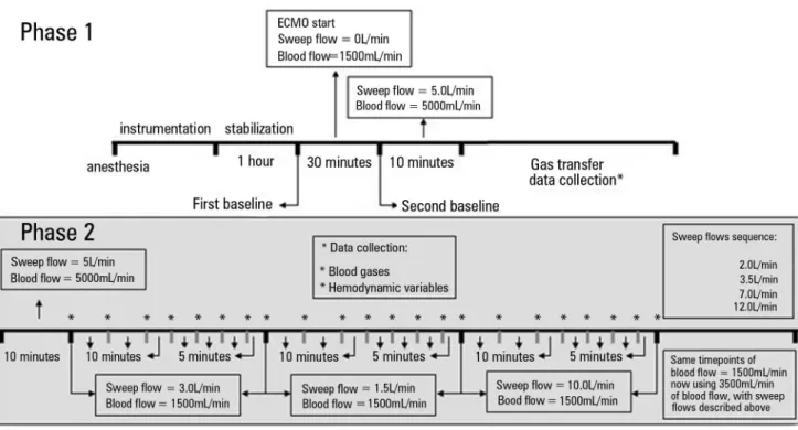
and, subsequently, to 10.0L/min at 50-minute intervals. he blood low was then increased to 3500mL/min, and a sequence of sweep lows of 2.0, 3.5, 7.0 and 12.0L/min of 50 minutes each was applied. he same 50-minute data collection sequence described (for blood low = 1500mL/ min and sweep low = 3.0L/min) was performed for each new tested sweep gas low tested (Figure 1).
he following data were collected every ten minutes: heart rate (HR), mean arterial blood pressure (ABPm), central venous pressure, mean pulmonary artery pressure, pulmonary artery occlusion pressure, cardiac output, core
temperature, peripheral oxygen saturation, end-tidal CO2
(EtCO2), and mixed venous oxygen saturation (SvO2).
Pre- and post-membrane port blood samples from the pulmonary and femoral arteries were collected after ten minutes of each new tested sweep low. Subsequently, a new femoral artery blood sample was collected every ten minutes for up to 30 minutes and every ive minutes for up to 50 minutes at the beginning of each new sweep low tested thereafter. Blood samples were analyzed in a standard radiometer ABL 600 (Radiometer, Copenhagen, Denmark). Biochemical samples were collected from the femoral artery catheter.
Calculations were performed using standard formulas as follows:(13-16)
• Blood oxygen content CbO2 [mL O2/100mL
blood] = 1.36 x Hb x SatbO2 + 0.0031 x PbO2 • Alveolar O2 pressure = capillary O2 partial pressure
= PaO2 = (FiO2 x (694 - 46)) - (PaCO2/0.8)
• Capillary O2 saturation = 100%
• Pulmonary shunt [mL O2/100mL blood] =
(capillary O2 content - arterial O2 content)/
(capillary O2 content - venous O2 content)
• Blood CO2 content [mL/min] = ((1 - ((0.0289
x Hb)/(3.352 - 0.456 x (SatO2 /100) x (8.142
- pH)))) x 2.226 x 0.0307 + (0.00057 x (37 -
temperature)) + (0.00002 x (37 - temperature)2)
x PaCO2 x (1 + 10 (pH - 6.086 + (0.042 x (7.4 - pH)) + ((38
- temperature) x 0.00472 + (0.00139 x (7.4 - pH)))))
Statistical analysis
Data normality was assessed with the Shapiro-Wilk goodness-of-it model. Normal data are presented as the means ± the standard deviations, and non-normal
data are presented as the median and the 25th and 75th
percentiles. Within group comparisons were performed in both airway pressures and the fraction of inspired
oxygen (FiO2) may aggravate already severe hypoxemia and hypercapnia if those blood gas changes are not corrected
by respiratory ECMO support.(5) ECMO parameters
are set based on the results of blood gas analysis, and
impaired oxygenation and carbon dioxide (CO2) removal
are corrected for by increasing the ECMO blood low
and the sweep gas low, respectively.(6) In more severely
ill patients in whom hypoxemia persists,(7) knowledge
of venous-venous ECMO physiology is necessary to better understand the scenario and modify the ECMO parameters accordingly.(8)
With the rationale of improving the knowledge of venous-venous ECMO physiology, the aim of this study was to explore the factors associated with the regulation of
blood oxygen partial pressure (PaO2) and carbon dioxide
partial pressure (PaCO2) during standardized sweep gas
low and ECMO blood low combinations in an apneic swine ECMO-supported model.
METHODS
his study was approved by the Institutional Animal
Research Ethics Committee of the Hospital Sírio Libanês
in São Paulo, Brazil (Protocol CEUA-P-20143), and was performed according to the National Institutes of Health guidelines for the use of experimental animals.
his study is part of an experimental sequence applied to ECMO-supported animals. Other aspects of these
experiments have been published elsewhere.(9,10) he
instrumentation, surgical preparation, sepsis induction, and pulmonary injury were performed as previously described and published in this journal.(11,12)
he animal was maintained in apnea with 10cmH2O
Figure 1 - Timeline of the entire study. The gray ground illustrates the sequence of the current analysis. ECMO - extracorporeal membrane oxygenation.* Data collection.
using Friedman’s test. Multivariate analysis was performed using a backward elimination mixed linear generalized model with the animals as random factors to account for the within-subject correlation among the repeated observations. he Markov chain Monte Carlo procedure with 10,000 simulations to reach the equilibrium of distributions was used to retrieve a ixed probability of each resulting independent variable from the mixed generalized model after backward elimination. Collinearity among the independent variables was tested with the Spearman’s test of correlations in a matrix that included all of the tested variables. Variables with r coeicients > 0.85 were further tested for multicollinearity with the variance inlation factor (VIF). Variables with VIFs < 2.5 were considered appropriate
for the analysis. he pseudo-R2 was calculated for each
model to determine the goodness of it. his calculation was performed with the squared ratio of the Spearman correlation between the itted values of the model and the original values retrieved from the experiment. he R free source statistical package and comprehensive-R archive network (CRAN)-speciic libraries were used to create the graphics and analyze the data.(17)
RESULTS
In four animals, 20-French catheters were used, and a 21-French catheter was used in one animal to drain the blood into the ECMO device. In three animals, 21-French return catheters from the ECMO system were used, and 20-French catheters were used in two. To ensure non-signiicant re-circulation, the pre-membrane oxygen saturation was collected ten minutes after the beginning of each sweep gas low test. he values obtained were 58% [51, 67] at a blood low of 1,500mL/minute and 65% [64; 70] at a blood low of 3,500mL/minute. here was no need to re-position the cannulae.
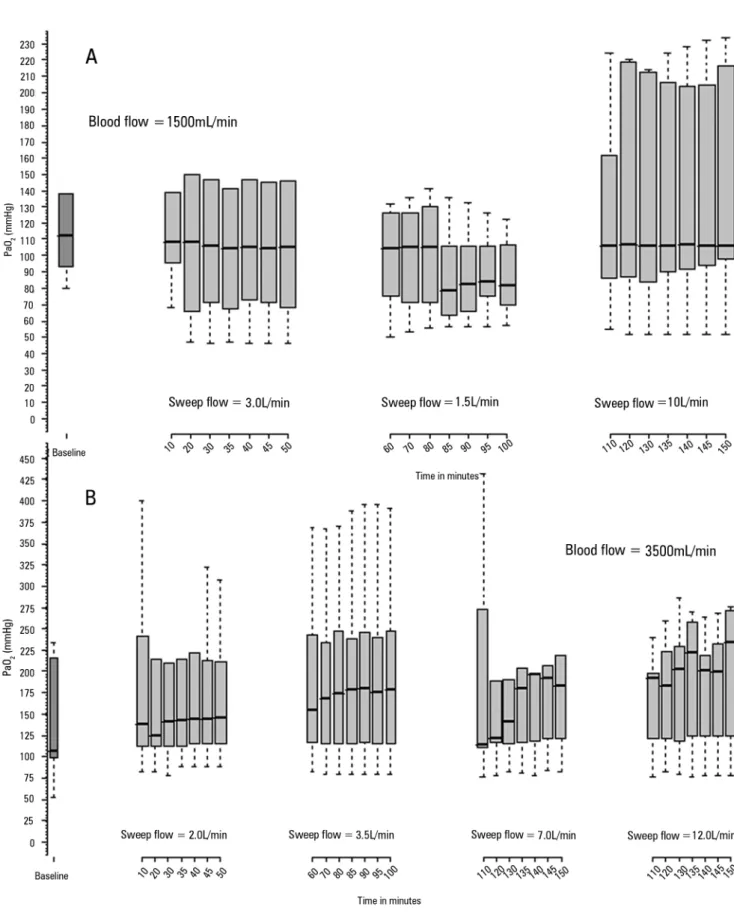
Figure 2 illustrates blood PaO2 behavior during the 50 minutes of observation. Among the other variables tested in the multivariate analysis, the variations in the experimental parameters were as follows: the pulmonary shunting (%) ranged from 45 [29, 66] to 60 [33, 67]; the temperature
(°C) ranged 37.2 [36.8, 38.5] to 38.0 [37.0, 38.1]; the
hemoglobin (g/dL) ranged from 11 [10, 13] to 14 [13, 14]; the cardiac output (L/min) ranged from 6.3 [4.0, 6.7] to 9.0 [4.4, 9.1]; the pH ranged from 7.03 [7.01, 7.16] to
7.43 [7.37, 7.49]; and the blood CO2 content (mL/100mL)
14 Park M, Mendes PV, Costa EL, Barbosa EV, Hirota AS, Azevedo LC, et al.
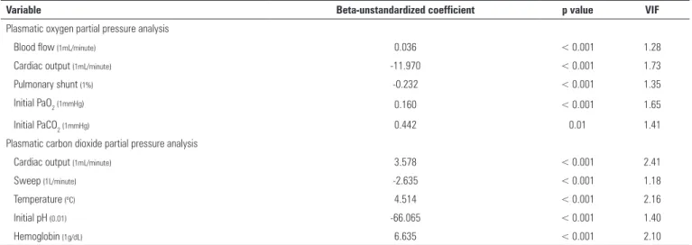
Table 1- Backward elimination multivariate analysis exploring the variables associated with plasma oxygen and carbon dioxide partial pressures during extracorporeal membrane oxygenation support
Variable Beta-unstandardized coefficient p value VIF
Plasmatic oxygen partial pressure analysis
Blood flow (1mL/minute) 0.036 < 0.001 1.28
Cardiac output (1mL/minute) -11.970 < 0.001 1.73
Pulmonary shunt (1%) -0.232 < 0.001 1.35
Initial PaO2 (1mmHg) 0.160 < 0.001 1.65
Initial PaCO2 (1mmHg) 0.442 0.01 1.41
Plasmatic carbon dioxide partial pressure analysis
Cardiac output (1mL/minute) 3.578 < 0.001 2.41
Sweep (1L/minute) -2.635 < 0.001 1.18
Temperature (ºC) 4.514 < 0.001 2.16
Initial pH (0.01) -66.065 < 0.001 1.40
Hemoglobin (1g/dL) 6.635 < 0.001 2.10
This multivariate analysis was performed using a mixed model with backward elimination. The initial dependent variables in the systemic oxygenation analysis were blood flow, cardiac output, pulmonary shunting, initial PaO2, initial PaCO2, temperature and hemoglobin. Temperature and hemoglobin were removed during the backward elimination of the multivariate analysis. The coefficient of determination of the final model (pseudo - R2) was 0.61. The initial dependent variables in the carbon dioxide transfer analysis were sweep flow, cardiac output, temperature, initial pH and hemoglobin. The coefficient of determination of the final model (pseudo - R2) was 0.79. Blood samples were acquired from the pre-membrane port. Beta-unstandardized coefficient - estimated variation in the oxygen transference in mL/min for each unit (the units are cited in the table) variation in the independent variables. VIF - variance inflation factor; PaO2 - partial pressure of oxygen; PaCO2 - partial pressure of carbon dioxide.
Figure 3 - Spider plots based on the data collected from the animals illustrating the PaO2 (Panel A) and PaCO2 percent (Panel B) variations associated with the variations in the main related variables. The respective main related variables were extracted from the multivariate analysis. PaO2 - partial pressure of oxygen; PaCO2 - partial pressure of carbon dioxide.
Table 1 presents the multivariate analysis evaluating
the factors associated with blood PaO2 and PaCO2
after 50 minutes of ECMO blood and sweep gas lows. Figure 3 displays a spider plot quantifying the inluences
of the deviation of each variable extracted from the multivariate analysis on the inal (after 50 minutes) PaO2
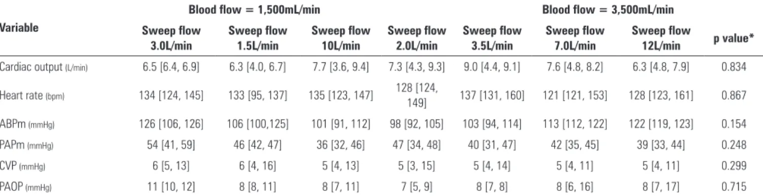
andPaCO2. Table 2 presents the hemodynamic behavior
16 Park M, Mendes PV, Costa EL, Barbosa EV, Hirota AS, Azevedo LC, et al.
Table 2 - Hemodynamic variables after fifty minutes of sweep or blood flow modifications
Variable
Blood flow = 1,500mL/min Blood flow = 3,500mL/min
Sweep flow 3.0L/min
Sweep flow 1.5L/min
Sweep flow 10L/min
Sweep flow 2.0L/min
Sweep flow 3.5L/min
Sweep flow 7.0L/min
Sweep flow
12L/min p value*
Cardiac output (L/min) 6.5 [6.4, 6.9] 6.3 [4.0, 6.7] 7.7 [3.6, 9.4] 7.3 [4.3, 9.3] 9.0 [4.4, 9.1] 7.6 [4.8, 8.2] 6.3 [4.8, 7.9] 0.834
Heart rate (bpm) 134 [124, 145] 133 [95, 137] 135 [123, 147] 128 [124,
149] 137 [131, 160] 121 [121, 153] 128 [123, 161] 0.867
ABPm (mmHg) 126 [106, 126] 106 [100,125] 101 [91, 112] 98 [92, 105] 103 [94, 114] 113 [112, 122] 122 [119, 123] 0.154
PAPm (mmHg) 54 [41, 59] 46 [42, 47] 36 [32, 46] 47 [34, 48] 40 [31, 47] 42 [35, 45] 39 [33, 44] 0.248
CVP (mmHg) 6 [5, 13] 6 [4, 16] 5 [4, 13] 5 [3, 15] 5 [4, 14] 5 [4, 11] 5 [4, 11] 0.299
PAOP (mmHg) 11 [10, 12] 8 [8, 11] 8 [7, 11] 7 [5, 9] 8 [7, 8] 8 [6, 16] 8 [7, 17] 0.715
ABPm - mean arterial blood pressure; PAPm - mean pulmonary arterial blood pressure; CVP - central venous pressure; PAOP - pulmonary artery occlusion pressure. * The p values were obtained based on Friedman’s tests. Post-hoc analyses were not performed due to the large variety of comparisons (varying through the blood flow and sweep gas flows domains).
DISCUSSION
After changing the blood low, the oxygen partial pressure reached equilibrium almost immediately, and variations in the sweep gas low did not improve oxygenation. Oxygenation after 50 minutes was mainly associated with ECMO blood low (beta coeicient = 0.036mmHg/mL/min), cardiac output (beta coeicient = -11.970mmHg/L/min), and pulmonary shunting (beta coeicient = -0.232mmHg/%). Furthermore,
the initial PaO2 and PaCO2 were also associated with
blood oxygenation, with beta coeicients of 0.160 and
0.442mmHg/mmHg of the given gas, respectively. PaCO2
after 50 minutes was associated with cardiac output (beta coeicient = 3.578mmHg/L/min), sweep gas low (beta coeicient = -2.635mmHg/L/min), temperature (beta coeicient = 4.514 mmHg/°C), initial pH (beta coeicient = -66.065mmHg/0.01 unit) and hemoglobin (beta coeicient = 6.635mmHg/g/dL).
Compared with carbon dioxide, the volume of the distribution of oxygen in the body is small. his is in line with our inding of a very fast oxygen equilibrium after a step change in the ECMO settings and contrasts with the
long time required to reach PaCO2 equilibrium; however,
this issue should be the subject of another manuscript due to its complexity.
It is interesting to consider the physiological bases
of the factors associated with the inal arterial PaO2 at
equilibrium. he dependence of arterial oxygenation on ECMO blood low occurred primarily because only a fraction of the cardiac output was oxygenated by the veno-venous ECMO. he remaining fraction of the venous return crossed the vena cava native bed straight to the heart without gas exchange and thus shunted the
ECMO circuit.(8) With higher ECMO blood lows for a
given venous return, this fraction increases, which leads to increased oxygen delivery by the return cannula. his increased delivery occurs despite the fall in the PaO2 of the return cannula that results from the limited difusibility
of oxygen through the lung membrane.(18) Additionally,
the lungs, however sick, provide an additive oxygenation efect (in series with the ECMO circuit). he delivery of highly oxygenated blood to the lungs (from the ECMO circuit) may worsen their ventilation perfusion mismatch by impeding hypoxic vasoconstriction. Despite these considerations, our inding that arterial oxygenation increases with increasing ECMO blood low implies that the fall in oxygen content and the worsening of hypoxic vasoconstriction are ofset by the increase in blood low. Notably, the reverse of the above described efect, i.e., the reduction of cardiac output (for example, due to the use of beta-blockers) has been described to optimize peripheral
oxygenation during veno-venous ECMO support.(19)
he pre-ECMO PaCO2 value was also related to
oxygenation, most likely due to modulation of hemoglobin-oxygen ainity.(20) Finally, in an intuitive manner, the lower
pre-ECMO PaO2 is also associated with greater oxygenation. In a recent study, Messai et al. demonstrated that peripheral oxygen saturation can be predicted based on ECMO blood low, cardiac output, after-membrane oxygen saturation,
and pulmonary artery oxygen saturation.(3) hese indings
are very similar to those of the current study; however, the importance of the pulmonary shunt was highlighted in this experiment.
he following ive variables were associated with the
PaCO2. (1) he cardiac output is the main determinant
of the local tissue low and therefore is a modulator of
tissue CO2 transport to the core circulation and to the
lungs.(21) Increased cardiac output results in enhanced
lungs. In contrast, an elevation of the PaCO2, regardless of the cause, initiates a sympathetic autonomic response and, consequently, an elevation in cardiac output.(22) In this
experiment, it is diicult to ascribe cause and consequence.
(2) Sweep low is an intuitive PaCO2 modulator once the
membrane is ventilated by the gas low.(6) (3) Temperature
is an aerobic metabolism regulator that leads to increased
or decreased CO2 production.(23) (4) Initial blood pH
is also a PaCO2 modulator because it can disturb the
transportation, storage, and production of CO2.(23-25) (5)
Hemoglobin is a PaCO2 regulator because an increased
hemoglobin level represents increased CO2, resulting in a lower partial pressure of CO2 in the plasma.(25)
In a bedside practical approach, taking PaO2 and
PaCO2 together, ECMO blood low and sweep gas low are
important variables for ECMO support.(6) Furthermore,
in special clinical situations, such as persistent hypoxemia and/or hypercapnia, patient temperature and cardiac output are variables that exhibit strong interactions and
strong efects on oxygenation and CO2 removal, which
can be modulated by the care team. he hemoglobin level is a particularly important variable in ECMO-supported patients. Higher hemoglobin levels are associated with
increased oxygen transport,(26) and according to our
indings, associated with lower blood PaCO2. Currently,
some ECMO groups allow hemoglobin levels as low as
7g/dL.(27) However, experienced groups keep hemoglobin
levels above 10g/dL, which results in high survival rates for severely hypoxemic patients.(7)
he hemodynamics of animals exhibited low sweep lows that were associated with increased pulmonary artery pressure and reduced cardiac output. In addition to these indings, a higher mean systemic arterial pressure was also observed, which likely resulted from sympathetic activation.
In this manuscript, we described the associations of
some variables with the oxygenation and inal PaCO2 of
the ECMO-supported patient. However, we would like to stress that other classical variables, such as the sweep gas low FiO2, subject weight and type and surface area of the oxygenator, are also associated with gas transfer and the consequent inal blood gases.(27)
his study has some limitations. First, the low number of animals used may have resulted in type II errors that attenuated the validity of our indings regarding the lack of associations. Notably, this limitation would not afect our positive indings. Second, these results are based on an animal model with a physiology diferent from that of humans.
CONCLUSION
In conclusion, elevations in the blood and sweep gas lows in an apneic veno-venous extracorporeal membrane oxygenation model resulted in increased partial oxygen and reduced partial carbon dioxide pressures. Furthermore, without the possibility of causal inference, the partial pressure of oxygen was negatively associated with pulmonary shunting and cardiac output, and the partial pressure of carbon dioxide was positively associated with cardiac output, core temperature and initial hemoglobin.
Objetivo: Explorar os fatores associados aos níveis sanguí-neos da pressão parcial de oxigênio e da pressão parcial de gás carbônico.
Métodos: Os fatores associados com a regulação do oxigênio e de gás carbônico foram investigados em um modelo com porcos em apneia com suporte de oxigenação por membrana extracorpórea venovenosa. Foi testada uma sequência predeinida de luxos de sangue e gás.
Resultados: A oxigenação associou-se principalmente com o luxo da oxigenação por membrana extracorpórea (coeiciente beta = 0,036mmHg/mL/minuto), débito cardíaco (coeiciente
beta = -11,970mmHg/L/minuto) e shunt pulmonar (coeiciente
beta = -0,232mmHg/%). As mensurações iniciais da pressão parcial de oxigênio e da pressão parcial de gás carbônico também se associaram com oxigenação, com coeicientes beta de 0,160 e 0,442mmHg/mmHg, respectivamente. A pressão parcial
de gás carbônico se associou com débito cardíaco (coeiciente beta = 3,578mmHg/L/minuto), luxo de gás (coeiciente beta = -2,635mmHg/L/minuto), temperatura (coeiciente beta = 4,514mmHg/°C), pH inicial (coeiciente beta = -66,065mmHg/0,01 unidade) e hemoglobina (coeiciente beta = 6,635mmHg/g/dL).
Conclusão: Elevações nos luxos de sangue de gás em um modelo de oxigenação por membrana extracorpórea venovenosa durante apneia resultaram em aumento da pressão parcial de oxigênio e redução da pressão parcial de gás carbônico, respectivamente. Ainda, sem a possibilidade de uma inferência causal, a pressão parcial de oxigênio associou-se negativamente
com o shunt pulmonar e o débito cardíaco, e a pressão parcial
de gás carbônico teve associação positiva com o débito cardíaco, temperatura central e hemoglobina inicial.
RESUMO
18 Park M, Mendes PV, Costa EL, Barbosa EV, Hirota AS, Azevedo LC, et al.
REFERENCES
1. Australia and New Zealand Extracorporeal Membrane Oxygenation (ANZ ECMO) Influenza Investigators, Davies A, Jones D, Bailey M, Beca J, Bellomo R, Blackwell N, et al. Extracorporeal Membrane Oxygenation for 2009 Influenza A(H1N1) Acute Respiratory Distress Syndrome. JAMA. 2009;302(17):1888-95. 2. Pham T, Combes A, Rozé H, Chevret S, Mercat A, Roch A, Mourvillier B,
Ara-Somohano C, Bastien O, Zogheib E, Clavel M, Constan A, Marie Richard JC, Brun-Buisson C, Brochard L; REVA Research Network. Extracorporeal membrane oxygenation for pandemic influenza A(H1N1)-induced acute respiratory distress syndrome: a cohort study and propensity-matched analysis. Am J Respir Crit Care Med. 2013;187(3):276-85.
3. Messaï E, Bouguerra A, Harmelin G, Di Lascio G, Cianchi G, Bonacchi M. A new formula for determining arterial oxygen saturation during venovenous extracorporeal oxygenation. Intensive Care Med. 2013;39(2):327-34. 4. Schmidt M, Stewart C, Bailey M, Nieszkowska A, Kelly J, Murphy L, et al.
Mechanical ventilation management during extracorporeal membrane oxygenation for acute respiratory distress syndrome: a retrospective international multicenter study. Crit Care Med. 2015;43(3):654-64. 5. Peek GJ, Mugford M, Tiruvoipati R, Wilson A, Allen E, Thalanany MM,
Hibbert CL, Truesdale A, Clemens F, Cooper N, Firmin RK, Elbourne D; CESAR trial collaboration. Efficacy and economic assessment of conventional ventilatory support versus extracorporeal membrane oxygenation for severe adult respiratory failure (CESAR): a multicentre randomised controlled trial. Lancet. 2009;374(9698):1351-63.
6. Schmidt M, Tachon G, Devilliers C, Muller G, Hekimian G, Bréchot N, et al. Blood oxygenation and decarboxylation determinants during venovenous ECMO for respiratory failure in adults. Intensive Care Med. 2013;39(5):838-46. 7. Lindén V, Palmér K, Reinhard J, Westman R, Ehrén H, Granholm T, et al.
High survival in adult patients with acute respiratory distress syndrome treated by extracorporeal membrane oxygenation, minimal sedation, and pressure supported ventilation. Intensive Care Med. 2000;26(11):1630-7. 8. Nunes LB, Mendes PV, Hirota AS, Barbosa EV, Maciel AT, Schettino GP, Costa
EL, Azevedo LC, Park M; ECMO Group. Severe hypoxemia during veno-venous extracorporeal membrane oxygenation: exploring the limits of extracorporeal respiratory support. Clinics (Sao Paulo). 2014;69(3):173-8.
9. Park M, Costa EL, Maciel AT, Silva DP, Friedrich N, Barbosa EV, et al. Determinants of oxygen and carbon dioxide transfer during extracorporeal membrane oxygenation in an experimental model of multiple organ dysfunction syndrome. PLoS One. 2013;8(1):e54954.
10. Park M, Costa EL, Maciel AT, Barbosa EV, Hirota AS, Schettino Gde P, et al. Effect of flow rate and temperature on transmembrane blood pressure drop in an extracorporeal artificial lung. Perfusion. 2014;29(6):517-25. 11. Park M, Costa EL, Maciel AT, Hirota AS, Vasconcelos E, Azevedo LC.
Acute hemodynamic, respiratory and metabolic alterations after blood contact with a volume priming and extracorporeal life support circuit: an experimental study. Rev Bras Ter Intensiva. 2012;24(2):137-42.
12. Park M, Mendes PV, Hirota AS, Santos EV, Costa EL, Azevedo LC. Blood flow/pump rotation ratio as an artificial lung performance monitoring tool during extracorporeal respiratory support using centrifugal pumps. Rev Bras Ter Intensiva. 2015;27(2):178-84.
13. da Silva Almeida JR, Machado FS, Schettino GP, Park M, Azevedo LC. Cardiopulmonary effects of matching positive end-expiratory pressure to abdominal pressure in concomitant abdominal hypertension and acute lung injury. J Trauma. 2010;69(2):375-83.
14. Rosário AL, Park M, Brunialti MK, Mendes M, Rapozo M, Fernandes D, et al. SvO(2)-guided resuscitation for experimental septic shock: effects of fluid infusion and dobutamine on hemodynamics, inflammatory response, and cardiovascular oxidative stress. Shock. 2011;36(6):604-12.
15. Siggaard-Andersen O. The van Slyke equation. Scand J Clin Lab Invest Suppl. 1977;146:15-20.
16. Douglas AR, Jones NL, Reed JW. Calculation of whole blood CO2 content. J Appl Physiol (1985). 1988;65(1):473-7.
17. Team RDC. R: A language and environment for statistical computing. Viena, Austria: R Foundation for Statistical Computing; 2009.
18. Chauhan S, Subin S. Extracorporeal membrane oxygenation, an anesthesiologist’s perspective: physiology and principles. Part 1. Ann Card Anaesth. 2011;14(3):218-29.
19. Guarracino F, Zangrillo A, Ruggeri L, Pieri M, Calabrò MG, Landoni G, et al. β-Blockers to optimize peripheral oxygenation during extracorporeal membrane oxygenation: a case series. J Cardiothorac Vasc Anesth. 2012;26(1):58-63.
20. West JB. Hemoglobin O2 affinity and tissue hypoxia. J Appl Physiol (1985). 1989;67(5):2163.
21. Anderson CT, Breen PH. Carbon dioxide kinetics and capnography during critical care. Crit Care. 2000;4(4):207-15. Review.
22. Carvalho CR, Barbas CS, Medeiros DM, Magaldi RB, Lorenzi Filho G, Kairalla RA, et al. Temporal hemodynamic effects of permissive hypercapnia associated with ideal PEEP in ARDS. Am J Respir Crit Care Med. 1997;156(5):1458-66.
23. Vleck D. Measurement of O2 consumption, CO2 production, and water vapor production in a closed system. J Appl Physiol (1985). 1987;62(5):2103-6.
24. Cherniack NS, Longobardo GS. Oxygen and carbon dioxide gas stores of the body. Physiol Rev. 1970;50(2):196-243.
25. Meldrum NU, Roughton FJ. The state of carbon dioxide in blood. J Physiol. 1933;80(2):143-70.
26. Spinelli E, Bartlett RH. Relationship between hemoglobin concentration and extracorporeal blood flow as determinants of oxygen delivery during venovenous extracorporeal membrane oxygenation: a mathematical model. ASAIO J. 2014;60(6):688-93.