Characterization of patients transported with
extracorporeal respiratory and/or cardiovascular
support in the State of São Paulo, Brazil
INTRODUCTION
The use of extracorporeal membrane oxygenation (ECMO) support has
increased in recent years,
(1)especially following the pandemic of influenza A
(H1N1) virus pneumonitis.
(2-4)Although the results of previous randomized
trials in which ECMO was used for respiratory support are inconclusive,
(5,6)new technologies
(7)associated with the application of ultraprotective mechanical
ventilation
(8)have improved survival and the quality of life when ECMO is
used for patients with severe respiratory failure.
(9,10)The high cost of the training and support required for ECMO use may
have a negative economic impact, especially in developing countries.
(11)Ho Yeh Li1, Pedro Vitale Mendes1,4, Livia Maria Garcia Melro1,2, Daniel Joelsons1, Bruno Adler Maccagnan Pinheiro Besen1,4, Eduardo Leite Viera Costa1,3, Adriana Sayuri Hirota1, Edzangela Vasconcelos Santos Barbosa1, Flavia Krepel Foronda1, Luciano Cesar Pontes Azevedo1,3, Thiago Gomes Romano1,4, Marcelo Park1
1. Hospital das Clínicas, Faculdade de Medicina, Universidade de São Paulo - São Paulo (SP), Brazil.
2. Hospital TotalCor - São Paulo (SP), Brazil. 3. Hospital Sírio Libanês - São Paulo (SP), Brazil. 4. Oncological Intensive Care Unit, Hospital São Luiz, Rede D’Or - São Paulo (SP), Brazil.
Objective:
To characterize the
transport of severely ill patients
with extracorporeal respiratory or
cardiovascular support.
Methods:
A series of 18 patients in
the state of São Paulo, Brazil is described.
All patients were consecutively evaluated
by a multidisciplinary team at the
hospital of origin. The patients were
rescued, and extracorporeal membrane
oxygenation support was provided on
site. The patients were then transported
to referral hospitals for extracorporeal
membrane oxygenation support. Data
were retrieved from a prospectively
collected database.
Results:
From 2011 to 2017, 18
patients aged 29 (25 - 31) years with
a SAPS 3 of 84 (68 - 92) and main
primary diagnosis of leptospirosis
and influenza A (H1N1) virus were
transported to three referral hospitals
in São Paulo. A median distance of 39
(15 - 82) km was traveled on each rescue
mission during a period of 360 (308
Conflicts of Interest: None.
Submitted on January 29, 2018 Accepted on April 30, 2018
Corresponding author:
Marcelo Park
Disciplina de Emergências Unidade de Terapia Intensiva Hospital das Clínicas
Avenida Dr. Enéas Carvalho de Aguiar, 255, 6º andar, sala 6.040
Zip code: 05403-000 - São Paulo (SP), Brazil E-mail: [email protected]
Responsible editor: Alexandre Biasi Cavalcanti
Caracterização de pacientes transportados com suporte
respiratório e/ou cardiovascular extracorpóreo no Estado de
São Paulo − Brasil
ABSTRACT
Keywords:
Artificial, respiration;
Respiratory insufficiency; Extracorporeal
membrane oxygenation; Transportation
of patients; Critical illness; Intensive
care units
- 431) min. A median of one (0 - 2)
nurse, three (2 - 3) physicians, and one
(0 - 1) physical therapist was present per
rescue. Seventeen rescues were made by
ambulance, and one rescue was made by
helicopter. The observed complications
were interruption in the energy supply
to the pump in two cases (11%) and
oxygen saturation < 70% in two cases.
Thirteen patients (72%) survived and
were discharged from the hospital.
Among the nonsurvivors, there were
two cases of brain death, two cases of
multiple organ dysfunction syndrome,
and one case of irreversible pulmonary
fibrosis.
Conclusions:
Transportation with
extracorporeal support occurred without
serious complications, and the hospital
survival rate was high.
However, the high cost of the initial installation of the
system is compensated for by its low cost of maintenance
and the good outcomes obtained when ECMO support
is used with adequate staff training, making this therapy
cost-effective in developed countries
(9,12)and potentially
cost-effective in developing countries.
(13)Considering that ensuring the availability of
appropriate staff in health centers with a relatively small
occupancy rate may increase the cost of extracorporeal
support, ECMO-equipped transport to specialized centers
has been made available at an acceptable cost, with high
survival rates and improvement in the quality of life.
(4,9)Considering the importance of transport with ECMO,
the objective of this study was to characterize the transport
performed by our team in the State of São Paulo since
2011.
METHODS
Data were retrieved from a prospectively collected
database. The analysis of the database was assessed and
approved by the Research Ethics Committee of the
Hospital das Clínicas
of the
Faculdade de Medicina
of the
Universidade de São Paulo
(USP) (number 107,443), and
the requirement for informed consent was waived. Data
on each patient were collected as previously described
(14,15)using an online worksheet in the REDCap system.
(16)The contact was made by telephone by a local team
member. Data were stored in an online spreadsheet. The
severity of the patient’s condition was determined, and
the indications and contraindications for extracorporeal
support were analyzed. The indication and contraindication
criteria were previously described by our group.
(17)These
criteria were modified slightly because the initial results
were suboptimal due to occasional problems in the initial
experience.
(14,18-20)The current criteria are described in the
Supplementary Material. Although the contraindication
criteria were restrictive, special situations that generated
doubts were discussed by our group. In cases in which
the indication criteria were fulfilled or in which there
were doubts, the remaining members of the team were
contacted, and the final decision on whether or not to
undertake support was made by the team as a whole.
The rescue team was composed of at least three
professionals, of whom at least two were physicians (the
third professional was a physician, a nurse, or a physical
therapist). All the professionals who formed the team
were trained to operate the system and to engage in open
and direct communication with patients, relatives, and
caregivers.
All the professionals made an initial assessment of the
patients. When there was agreement about the indication,
the two physicians were responsible for cannulation, and
the third professional was responsible for communication
with the patient’s relatives and for equipment assembly,
including priming the system.
Because an adequate transport system was not
available, the requesting center was responsible for
transporting the hospital staff to the requested location by
ambulance or private transport. The team was responsible
for carrying some equipment on the mission, including
an ECMO system, a voltage stabilizer for the ECMO
pump, two infusion pumps, a noninvasive blood pressure
measurement system, and an oximeter. The transport
of these items was confirmed using a checklist before
departure for the mission. The remaining monitoring and
support were provided by the ambulance in charge of the
return transport.
Initial support, initial patient stabilization, and
migration to protective/ultraprotective ventilation were
performed in the presence of all three professionals.
The stepwise technique used in this process was
previously described.
(14)The ECMO system included a
polymethylpentene membrane oxygenator connected
to the following centrifugal pumps: (1) Rotaflow/Jostra
Quadrox-D/Permanent Life Support (PLS;
Maquet
Cardiopulmonary AG, Hirrlingen, Germany), and
(2) a BioPump with campanula and Affinity
TMcircuit
(Medtronic Inc, MN, USA) with a BIOCUBE 6000
membrane (Nipro Ltda, Sorocaba, São Paulo, Brazil).
Ambulances could be used to transport critical patients
provided these vehicles had a mechanical ventilator
capable of delivering at least 10cmH
2O of positive end
expiratory pressure and an inverter with a power of at least
2,000 watts. The latter feature was requested because less
powerful inverters were not able to keep the ECMO pump
working together with the other required devices. Team
workload was reduced during transport by not carrying
the ECMO thermoregulator and by carefully keeping the
ambulance air conditioner off to avoid excessive cooling
of the patient.
Statistical analysis
The data were considered nonparametric because of
the small sample size and are reported as the median [25
th- 75
thpercentile] if quantitative and as the number of
Fisher’s exact test for qualitative data. The confidence
interval of the survivor ratio was calculated according to
the method described by the Association of Public Health
Observatories
(21)using R software for calculations and
graph creation.
(22)RESULTS
The ECMO program was initiated in 2011, and the
transport of ECMO patients began in the same year.
(14)A flowchart of the 28 requests for extracorporeal support
outside the referral hospitals is shown in figure 1S. The
first seven patients in this series were described in another
publication.
(15)During the six years of the program,
18 patients in the state of São Paulo were rescued and
transported with ECMO support by our team. Seventeen
patients received exclusive respiratory support
(veno-venous - VV configuration), and one patient received
respiratory and cardiovascular support (veno-arterial - VA
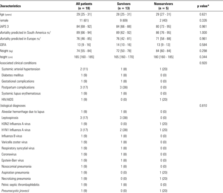
configuration). A profile of the patients is shown in table 1.
The characteristics of the patients shortly before initiation
of the support are shown in table 2. The Respiratory
ECMO Survival Prediction Score (RESP score) and
the tidal volume in pre-ECMO mechanical ventilation
differed significantly in survivors and nonsurvivors. The
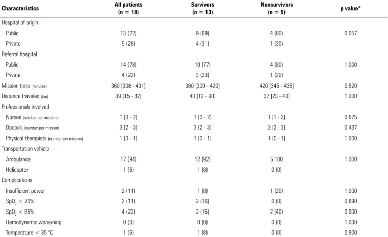
data on the rescue missions and complications during
transport are shown in table 3. The referral hospitals were
Hospital Sírio Libanês
(two patients),
Hospital TotalCor
(two patients), and the
Hospital das Clínicas
of São Paulo
(14 patients).
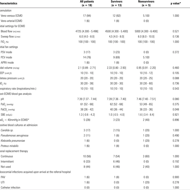
The data on the extracorporeal support are shown
in table 4. Respiratory support was provided using the
femoral-jugular configuration, and veno-arterial support
(one case) was provided using the femoral-femoral
configuration. The venous cannulae were 21 - 22 Fr, and
the arterial cannulae were 16 - 19 Fr. Apart from
veno-arterial cannulation, anticoagulation was started upon
patient arrival at the referral hospital. Five patients did
not use anticoagulation at any time because of pulmonary
hemorrhage (four cases) or the presence of cerebral
vasculitis with hemorrhagic areas (one case). None of the
evaluated patients had a change of itinerary or a change in
the support configuration related to initial cannulation.
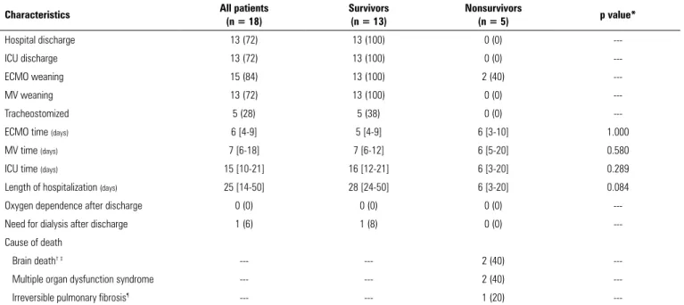
The final results are shown in table 5. The minimum
and maximum duration of support was 3 and 60 days,
respectively. Of the 18 patients, 13 (72%, 95%CI 49 -
88) survived to hospital admission (Figure 2S). Of the
survivors, only one patient needed dialysis after hospital
admission, and none required home oxygen therapy. The
individual patient data are presented in table 1S.
DISCUSSION
In this case series of 18 severe patients transported to
specialized centers with ECMO support in São Paulo,
the rate of complications was low, and hospital survival
was 72%. Of the patients who were discharged from the
hospital, only one needed renal replacement therapy, and
none required home oxygen therapy.
Fewer than 2% of the patients admitted to the intensive
care unit (ICU) suffered from severe respiratory failure.
Of these, fewer than 0.5% were refractory to protective
mechanical ventilation and salvage therapy for hypoxemia
and severe hypercapnia
(23)and sometimes required ECMO
support. The low rate of very severe patients limits the
ability to maintain a team to perform ECMO support
in all ICUs. Therefore, in developed countries, transport
with installed ECMO support was used to reduce the risk
of transportation to specialized centers, and the patient
survival rate was 62% (95%CI 57 - 68%).
(4,15)In our
series, hospital survival was 72% (95%CI 49 - 88%), in
agreement with the data reported in the literature.
(15)These results are attributed to two main causes. The first
is the use of more rigorous inclusion and exclusion criteria,
which resulted in restricting the use of ECMO to highly
selected patients because ECMO support seems to have a
survival benefit with improved quality of life for patients
with few comorbidities and few acute dysfunctions.
(9,24)In addition, the application of rescue therapy, such as
the use of the prone position before ECMO, is essential
whenever possible because this therapy is inexpensive
and there is strong evidence that its use improves patient
survival.
(25)Second, the use of ECMO support can be
optimized by providing adequate training and experience
to the multidisciplinary team
(18)and by the involvement
of professionals who possess comprehensive knowledge
of emergency care and possible complications during
ECMO support.
(26-28)Table 1 - General characteristics of patients transported with extracorporeal membrane oxygenation support
Characteristics All patients
(n = 18)
Survivors (n = 13)
Nonsurvivors
(n = 5) p value*
Age (years) 29 [25 - 31] 28 [25 - 31] 29 [27 - 31] 0.621
Female 11 (61) 9 (69) 2 (40) 0.326
SAPS 3 84 [68 - 92] 84 [66 - 88] 80 [73 - 95] 0.961
Mortality predicted in South America (%)† 89 [66 - 94] 89 [62 - 92] 86 [76 - 95] 1.000
Mortality predicted in Europe (%)† 76 [46 - 85] 76 [42 - 81] 71 [58 - 88] 0.961
SOFA 13 [9 - 16] 14 [10 - 16] 13 [9 - 13] 0.584
Weight (kg) 74 [55 - 84] 72 [50 - 78] 84 [60 - 84] 0.298
Height (cm) 165 [160 - 185] 165 [160 - 170] 180 [160 - 185] 0.344
Associated clinical conditions 0.920
Systemic arterial hypertension 2 (11) 1 (8) 1 (20)
Diabetes mellitus 1 (9) 1 (8) 0 (0)
Gestational complications 1 (9) 1 (8) 0 (0)
Postpartum complications 3 (17) 3 (38) 0 (0)
Systemic lupus erythematosus 1 (9) 1 (8) 0 (0)
HIV/AIDS 1 (9) 0 (0) 1 (20)
Etiological diagnoses 0.810
Alveolar hemorrhage due to lupus 1 (9) 1 (8) 0 (0)
Leptospirosis 3 (17) 3 (38) 0 (0)
H3N2 Influenza A virus 1 (9) 0 (0) 1 (20)
H1N1 Influenza A virus 3 (17) 2 (38) 1 (20)
Influenza B virus 1 (9) 1 (8) 0 (0)
Varicella zoster virus 1 (9) 1 (8) 0 (0)
Respiratory syncytial virus 1 (9) 1 (8) 0 (0)
Coronavirus 1 (9) 1 (8) 0 (0)
Epstein-Barr virus 1 (9) 1 (8) 0 (0)
Nosocomial pneumonia 1 (9) 1 (8) 0 (0)
Aspiration pneumonia 1 (9) 0 (0) 1 (20)
Necrotizing pneumonia 1 (9) 0 (0) 1 (20)
Pelvic septic thrombophlebitis 1 (9) 1 (8) 0 (0)
Pneumocystis jirovecii 1 (9) 0 (0) 1 (20)
SAPS 3 - Simplified Acute Physiology Score 3; SOFA - Sequential Organ Failure Assessment Score. * Value of comparison between survivors and nonsurvivors; † calculation was performed
using the logit of SAPS 3 for South America and Western Europe, respectively. The results are expressed as the median [interquartile 25 - 75] or the number (%).
therefore be more accurate.
(30)The Survival After
Veno-Arterial ECMO Score (SAVE score) was described, but
the effects of using this score were not analyzed because it
was used in only one case.
Another relevant factor in our sample of nonsurvivors
was that the partial pressure decrease in carbon dioxide
(PaCO
2) from pre- to post-ECMO was critical. This
characteristic is known to be related to higher patient
mortality in ECMO.
(31)This factor may have contributed
to the deaths of two patients who progressed to brain
death while in the ICU. This outcome alerted us to the
importance of the careful initiation of extracorporeal
ventilation, especially in hypercapnic patients with gas/
blood flow < 1, to ensure a smaller initial decrease in
PaCO
2.
Table 2 - Respiratory and hemodynamic characteristics of patients who received extracorporeal membrane pre-oxygenation support
Characteristics All patients
(n = 18)
Survivors (n = 13)
Nonsurvivors
(n = 5) p value*
Murray score 3.7 [3.0 - 4.0] 3.5 [3.0 - 4.0] 3.8 [3.0-4.0] 0.448
Mechanical ventilation time (days) 7 [1 - 11] 5 [1 - 8] 8 [7-15] 0.113
ICU time (days) 7 [2 - 11] 5 [1 - 9] 8 [8-15] 0.199
RESP score 0.00 [-2.00 - 2.00] 1.50 [-0.25 - 3.25] -2.00 [-3.00 - -1.00] 0.023
Survival rate (RESP) (%) 50 [40 - 60] 58 [48 - 69] 40 [35 - 45] 0.034
SAVE score 0.00 0.00 ---
---Survival rate (SAVE) (%) 40 40 ---
---Mechanical ventilation
Pressure-controlled mode 14 (78) 10 (77) 4 (80) 1.000
Volume-controlled mode 4 (22) 3 (23) 1 (20)
PEEP (cmH2O) 14 [10 - 18] 12 [10 - 15] 17 [13 - 18] 0.269
FiO2(%) 100 [100 - 100] 100 [100 - 100] 100 [100 - 100] 0.288
Tidal volume (mL/kg) 4 [4 - 6] 5 [4 - 6] 3 [3 - 4] < 0.001
Respiratory rate (ipm) 28 [25 - 35] 28 [25 - 35] 28 [28 - 35] 0.723
Plateau pressure (cmH2O) 33 [30 - 35] 34 [30 - 35] 32 [31 - 35] 0.960
Blood gas analysis
pH 7.27 [7.08 - 7.35] 7.27 [7.09 - 7.35] 7.10 [7.00 - 7.34] 0.622
PaO2(mmHg) 54 [38 - 60] 58 [39 - 65] 50 [45 - 60] 0.882
PaCO2(mmHg) 61 [46 - 90] 53 [42 - 80] 90 [49 - 90] 0.429
SBE (mEq/L) 1.5 [-2.5 - 4.8] 1.0 [-3.0 - 6.0] 2.0 [0.0 - 4.0] 0.805
Lactate (mEq/L) 2.7 [2.2 - 3.9] 2.7 [2.2 - 3.3] 2.7 [2.7 - 4.4] 0.692
P/F ratio (mmHg) 55 [39 - 60] 60 [39 - 65] 50 [45 - 60] 0.657
Salvage therapy
Alveolar recruitment 15 (84) 10 (77) 5.100 0.638
Nitric oxide 2 (11) 0 (0) 2 (40) 0.114
Prone position 12 (67) 7 (54) 5.100 0.193
Curarization 15 (84) 10 (77) 5.100 0.638
TGI 2 (11) 0 (0) 2 (40) 1.000
Corticosteroids 12 (67) 8 (62) 4 (40) 0.852
Hemodynamic support
Noradrenaline 16 (89) 12 (92) 4 (80) 1.000
Vasopressin 4 (22) 1 (8) 3 (60) 0.078
Adrenaline 3 (17) 1 (8) 2 (40) 1.000
Dobutamine 3 (17) 3 (23) 0 (0) 0.638
ICU - intensive care unit; RESP score - Respiratory ECMO Survival Prediction Score; SAVE score - Survival After Veno-Arterial-ECMO Score; PEEP - positive end-expiratory pressure; FiO2 - fraction of inspired oxygen; PaO2 - partial pressure of oxygen; PaCO2 - partial pressure of carbon dioxide; SBE - standard base excess; P/F ratio - PaO2/FiO2 ratio; TGI - tracheal gas insufflation.
Table 3 - Characteristics of missions and transportation
Characteristics All patients (n = 18)
Survivors (n = 13)
Nonsurvivors
(n = 5) p value*
Hospital of origin
Public 13 (72) 9 (69) 4 (80) 0.057
Private 5 (28) 4 (31) 1 (20)
Referral hospital
Public 14 (78) 10 (77) 4 (80) 1.000
Private 4 (22) 3 (23) 1 (20)
Mission time (minutes) 360 [308 - 431] 360 [300 - 420] 420 [345 - 435] 0.520
Distance traveled (km) 39 [15 - 82] 40 [12 - 90] 37 [23 - 40] 1.000
Professionals involved
Nurses (number per mission) 1 [0 - 2] 1 [0 - 2] 1 [1 - 2] 0.675
Doctors (number per mission) 3 [2 - 3] 3 [2 - 3] 2 [2 - 3] 0.437
Physical therapists (number per mission) 1 [0 - 1] 1 [0 - 1] 1 [0 - 1] 1.000
Transportation vehicle
Ambulance 17 (94) 12 (92) 5.100 1.000
Helicopter 1 (6) 1 (8) 0 (0)
Complications
Insufficient power 2 (11) 1 (8) 1 (20) 1.000
SpO2 < 70% 2 (11) 2 (16) 0 (0) 0.890
SpO2 < 85% 4 (22) 2 (16) 2 (40) 0.900
Hemodynamic worsening 0 (0) 0 (0) 0 (0) 1.000
Temperature < 35 °C 1 (6) 1 (8) 0 (0) 0.900
SpO2 - blood oxygen saturation. * Value of comparison between survivors and nonsurvivors. The results are expressed as the number (%) or the median [interquartile 25 - 75].
because of the severity of lung injury, associated with a
cardiac output. Severe hypoxemia may occur during the
acute phase of respiratory support and sometimes needs
to be tolerated by the team;
(32)although this complication
may not directly affect survival or cognitive outcome, it
indicates the severity of the patient’s condition.
(33)Although the sample described in this study does
not provide new data, it represents the first case series
of patients transported in ECMO in Brazil. However, the
results of this study should be viewed with caution for
several reasons. First, because the sample size was small,
it was not possible to perform a multivariate analysis.
Second, the results of the analyses are preliminary and
should not be used to change procedures at the bedside.
Third, generalization of the results reported here to other
centers should be made with caution because the number
of ECMO support cases per year was low (5 - 10). Fourth,
the indications were restricted to a small subset of patients.
Table 4 - Support and complications of patients in the intensive care unit
Characteristics All patients
(n = 18)
Survivors (n = 13)
Nonsurvivors
(n = 5) p value*
Cannulation
Veno-venous ECMO 17 (94) 12 (92) 5.100 1.000
Veno-arterial ECMO 1 (6) 1 (8) 0 (0)
Initial settings for ECMO
Blood flow (mL/min) 4725 [4.300 - 5.498] 4500 [4.300 - 5.400] 5000 [4.300 - 6.400] 0.521
Sweep flow (L/min) 6.0 [4.0 - 8.0] 4,5 [4.0 - 8.0] 6.0 [6.0 - 10.0] 0.136
FiO2(%) 100 [100 - 100] 100 [100 - 100] 100 [100 - 100] 1.000
Initial fan settings
PSV mode 3 (17) 3 (23) 0 (0) 0.372
PCV mode 14 (78) 9 (69) 5.100
APRV mode 1 (6) 1 (8) 0 (0)
Tidal volume (mL/kg) 2.1 [0.89 - 2.71] 2.33 [0.80 - 2.93] 0.95 [0.91 - 2.20] 0.460
PEEP (cmH2O) 10 [10 - 10] 10 [10 - 10] 10 [10 - 12] 0.105
Plateau pressure (cmH2O) 20 [20 - 20] 20 [20 - 20] 20 [20 - 25] 0.088
FiO2(%) 30 [30 - 38] 30 [30 - 30] 30 [30 - 60] 0.736
Respiratory rate (inspirations/min.) 10 [10 - 10] 10 [10 - 10] 10 [10 - 10] 0.543
Post-ECMO blood gas analysis
pH 7.39 [7.37 - 7.44] 7.39 [7.36 - 7.40] 7.46 [7.40 - 7.51] 0.080
PaO2(mmHg) 61 [52 - 68] 62 [52 - 68] 53 [49 - 65] 0.375
PaCO2(mmHg) 38 [36 - 42] 40 [36 - 44] 36 [33 - 36] 0.048
SBE (mEq/L) 1.3 [-0.6 - 4.2] 1.0 [-0.5 - 4.0] 1.6 [-3.4 - 8.4] 0.921
PaO2 < 40mmHg in ECMO
# 5 (28) 3 (23) 2 (40) 0.896
Positive blood cultures at admission
Candida sp. 3 (17) 2 (15) 1 (20) 1.000
Pseudomonas aeruginosa 2 (11) 1 (8) 1 (20) 0.490
Klebsiella pneumoniae 1 (6) 0 (0) 1 (20) 0.278
Proteus mirabilis 1 (6) 1 (8) 0 (0) 1.000
Renal replacement therapy
Continuous 10 (56) 7 (54) 3 (60) 1.000
Intermittent 6 (33) 6 (46) 0 (0) 0.192
Not used 8 (44) 6 (46) 2 (40) 1.000
Nosocomial infections acquired upon arrival at the referral hospital
PAV 1 (6) 1 (8) 0 (0) 0.900
UTI 1 (6) 0 (0) 1 (20) 0.278
Catheter infection 0 (0) 0 (0) 0 (0) 1.000
Table 5 - Results of transportation
Characteristics All patients
(n = 18)
Survivors (n = 13)
Nonsurvivors
(n = 5) p value*
Hospital discharge 13 (72) 13 (100) 0 (0)
---ICU discharge 13 (72) 13 (100) 0 (0)
---ECMO weaning 15 (84) 13 (100) 2 (40)
---MV weaning 13 (72) 13 (100) 0 (0)
---Tracheostomized 5 (28) 5 (38) 0 (0)
---ECMO time (days) 6 [4-9] 5 [4-9] 6 [3-10] 1.000
MV time (days) 7 [6-18] 7 [6-12] 6 [5-20] 0.580
ICU time (days) 15 [10-21] 16 [12-21] 6 [3-20] 0.289
Length of hospitalization (days) 25 [14-50] 28 [24-50] 6 [3-20] 0.084
Oxygen dependence after discharge 0 (0) 0 (0) 0 (0)
---Need for dialysis after discharge 1 (6) 1 (8) 0 (0)
---Cause of death
Brain death† ‡ --- --- 2 (40)
---Multiple organ dysfunction syndrome --- --- 2 (40)
---Irreversible pulmonary fibrosis¶ --- --- 1 (20)
---ICU - intensive care unit; ECMO - extracorporeal membrane oxygenation; MV - mechanical ventilation. * Value of comparison between survivors and nonsurvivors; † patients with support
withdrawal; ‡ one patient with extensive brain hemorrhage and one patient with diffuse ischemia and secondary bleeding; ¶ irreversible fibrosis characterized by the complete absence of
vascular scaffold in open lung biopsy after 60 days of support with extracorporeal membrane oxygenation and without signs of improvement. The results are expressed as number (%) or median [interquartile 25-75].
CONCLUSIONS
Transport of severely ill patients with extracorporeal
respiratory support in a Brazilian state was feasible and
did not result in severe complications. Despite the small
sample size, patient survival to hospital admission was
similar to that reported in the literature.
ACKNOWLEDGMENTS
We are grateful to the
Fundação Faculdade de Medicina
and the Executive Board of the
Hospital das Clínicas
of São
Paulo for providing funding to purchase the disposable
material used in extracorporeal support.
Objetivo:
Caracterizar pacientes graves transportados em
suporte respiratório ou cardiovascular extracorpóreo.
Métodos:
Descrição de uma série de 18 casos registrados
no Estado de São Paulo. Todos os pacientes foram
consecutiva-mente avaliados por uma equipe multidisciplinar no hospital de
origem. Os pacientes foram resgatados, sendo a oxigenação por
membrana extracorpórea instalada
in loco
. Os pacientes foram,
então, transportados para os hospitais referenciados já em
oxige-nação por membrana extracorpórea. Os dados foram
recupera-dos de um banco de darecupera-dos prospectivamente coletado.
Resultados:
De 2011 até 2017, 18 pacientes com 29 (25 -
31) anos, SAPS3 de 84 (68 - 92), com principais diagnósticos de
leptospirose e influenza A (H1N1) foram transportados no
Es-tado de São Paulo para três hospitais referenciados. Uma
distân-cia mediana de 39 (15 - 82) km foi percorrida em cada missão,
em um tempo de 360 (308 - 431) minutos. As medianas de um
(0 - 2) enfermeiro, três (2 - 3) médicos e um (0 - 1)
fisiotera-peuta foram necessárias por missão. Dezessete transportes foram
realizados por ambulância e um por helicóptero. Existiram
in-tercorrências: em duas ocasiões (11%), houve falha de
forneci-mento de energia para a bomba e, em duas ocasiões, queda da
saturação de oxigênio < 70%. Treze pacientes (72%)
sobrevive-ram para a alta hospitalar. Dos pacientes não sobreviventes, dois
tiveram morte encefálica; dois, disfunção de múltiplos órgãos; e
um, fibrose pulmonar considerada irreversível.
Conclusões:
O transporte com suporte extracorpóreo
ocor-reu sem intercorrências maiores, com uma sobrevida hospitalar
alta dos pacientes.
RESUMO
REFERENCES
1. Extracorporeal Life Suport Organization. ECLS Registry Report. International Summary. January 2018. Available from: https://www.elso.org/Registry/ Statistics/InternationalSummary.aspx.
2. Australia and New Zealand Extracorporeal Membrane Oxygenation (ANZ ECMO) Influenza Investigators, Davies A, Jones D, Bailey M, Beca J, Bellomo R, Blackwell N, et al. Extracorporeal membrane oxygenation for 2009 Influenza A(H1N1) acute respiratory distress syndrome. JAMA. 2009;302(17):1888-95.
3. Pham T, Combes A, Rozé H, Chevret S, Mercat A, Roch A, Mourvillier B, Ara-Somohano C, Bastien O, Zogheib E, Clavel M, Constan A, Marie Richard JC, Brun-Buisson C, Brochard L; REVA Research Network. Extracorporeal membrane oxygenation for pandemic Influenza A(H1N1) induced acute respiratory distress syndrome. A cohort study and propensity-matched analysis. Am J Respir Crit Care Med. 2013;187(3):276-85.
4. Noah MA, Peek GJ, Finney SJ, Griffiths MJ, Harrison DA, Grieve R, et al. Referral to an extracorporeal membrane oxygenation center and mortality among patients with severe 2009 influenza A(H1N1). JAMA. 2011;306(15):1659-68.
5. Zapol WM, Snider MT, Hill JD, Fallat RJ, Bartlett RH, Edmunds LH, et al. Extracorporeal membrane oxygenation in severe acute respiratory failure. A randomized prospective study. JAMA. 1979;242(20):2193-6.
6. Morris AH, Wallace CJ, Menlove RL, Clemmer TP, Orme JF Jr., Weaver LK, et al. Randomized clinical trial of pressure-controlled inverse ratio ventilation and extracorporeal CO2 removal for adult respiratory distress syndrome. Am J Respir Crit Care Med. 1994;149(2 Pt 1):295-305. Erratum in: Am J Respir Crit Care Med. 1994;149(3 Pt 1):838.
7. Toomasian JM, Schreiner RJ, Meyer DE, Schmidt ME, Hagan SE, Griffith GW, et al. A polymethylpentene fiber gas exchanger for long-term extracorporeal life support. ASAIO J. 2005;51(4):390-7. Erratum in ASAIO J. 2008;54(1):137.
8. Serpa Neto A, Schmidt M, Azevedo LC, Bein T, Brochard L, Beutel G, Combes A, Costa EL, Hodgson C, Lindskov C, Lubnow M, Lueck C, Michaels AJ, Paiva JA, Park M, Pesenti A, Pham T, Quintel M, Marco Ranieri V, Ried M, Roncon-Albuquerque R Jr, Slutsky AS, Takeda S, Terragni PP, Vejen M, Weber-Carstens S, Welte T, Gama de Abreu M, Pelosi P, Schultz MJ; ReVA Research Network and the PROVE Network Investigators. Associations between ventilator settings during extracorporeal membrane oxygenation for refractory hypoxemia and outcome in patients with acute respiratory distress syndrome: a pooled individual patient data analysis: Mechanical ventilation during ECMO. Intensive Care Med. 2016;42(11):1672-84.
9. Peek GJ, Mugford M, Tiruvoipati R, Wilson A, Allen E, Thalanany MM, Hibbert CL, Truesdale A, Clemens F, Cooper N, Firmin RK, Elbourne D; CESAR trialcollaboration. Efficacy and economic assessment of conventional ventilatory support versus extracorporeal membrane oxygenation for severe adult respiratory failure (CESAR): a multicentre randomised controlled trial. Lancet. 2009;374(9698):1351-63. Erratum in Lancet. 2009;374(9698):1330.
10. Zampieri FG, Mendes PV, Ranzani OT, Taniguchi LU, Pontes Azevedo LC, Vieira Costa EL, et al. Extracorporeal membrane oxygenation for severe respiratory failure in adult patients: a systematic review and meta-analysis of current evidence. J Crit Care. 2013;28(6):998-1005.
11. Machado FR. All in a day’s work - Equity vs. Equality at a public ICU in Brazil. N Engl J Med. 2016;375(25):2420-1.
12. Schumacher RE, Roloff DW, Chapman R, Snedecor S, Bartlett RH. Extracorporeal membrane oxygenation in term newborns. A prospective cost-benefit analysis. ASAIO J. 1993;39(4):873-9.
13. Park M, Mendes PV, Zampieri FG, Azevedo LC, Costa EL, Antoniali F, Ribeiro GC, Caneo LF, da Cruz Neto LM, Carvalho CR, Trindade EM; ERICC research group; ECMO group Hospital Sírio Libanês and Hospital das Clínicas de São Paulo. The economic effect of extracorporeal membrane oxygenation to support adults with severe respiratory failure in Brazil: a hypothetical analysis. Rev Bras Ter Intensiva. 2014;26(3):253-62.
14. Park M, Azevedo LC, Mendes PV, Carvalho CR, Amato MB, Schettino GP, et al. First-year experience of a Brazilian tertiary medical center in supporting severely ill patients using extracorporeal membrane oxygenation. Clinics (Sao Paulo). 2012;67(10):1157-63.
15. Mendes PV, de Albuquerque Gallo C, Besen BAMP, Hirota AS, de Oliveira Nardi R, Dos Santos EV, et al. Transportation of patients on extracorporeal membrane oxygenation: a tertiary medical center experience and systematic review of the literature. Ann Intensive Care. 2017;7(1):14. 16. Harris PA, Taylor R, Thielke R, Payne J, Gonzalez N, Conde JG. Research
electronic data capture (REDCap)--a metadata-driven methodology and workflow process for providing translational research informatics support. J Biomed Inform. 2009;42(2):377-81.
17. Azevedo LC, Park M, Costa EL, Santos EV, Hirota A, Taniguchi LU, Schettino Gde P, Amato MB, Carvalho CR; Extracorporeal Support Study Group. Extracorporeal membrane oxygenation in severe hypoxemia: time for reappraisal? J Bras Pneumol. 2012;38(1):7-12.
18. Romano TG, Mendes PV, Park M, Costa EL. Extracorporeal respiratory support in adult patients. J Bras Pneumol. 2017;43(1):60-70.
19. Park M, Costa EL, Azevedo LC, Afonso Junior JE, Samano MN, Carvalho CR; ECMO Group. Extracorporeal membrane oxygenation as a bridge to pulmonary transplantation in Brazil: are we ready to embark upon this new age? Clinics (Sao Paulo). 2011;66(9):1659-61.
20. Mendes PV, Moura E, Barbosa EV, Hirota AS, Scordamaglio PR, Ajjar FM, Costa EL, Azevedo LC, Park M; ECMO Group. Challenges in patients supported with extracorporeal membrane oxygenation in Brazil. Clinics (Sao Paulo). 2012;67(12):1511-5.
21. Cunningham A, Fryers P, Abbas J, Flowers J, Stockton D. Technical Briefing 3: Commonly Used Public Health Statistics and their Confidence Intervals Public Health England Webpage2008 [Available from: http://www.apho. org.uk/resource/item.aspx?RID=48457.
22. R Development Core Team. R: A language and environment for statistical computing. Viena, Austria: R Foundation for Statistical Computing; 2009. 23. Bellani G, Laffey JG, Pham T, Fan E, Brochard L, Esteban A, Gattinoni L,
van Haren F, Larsson A, McAuley DF, Ranieri M, Rubenfeld G, Thompson BT, Wrigge H, Slutsky AS, Pesenti A; LUNG SAFE Investigators; ESICM Trials Group. Epidemiology, patterns of care, and mortality for patients with acute respiratory distress syndrome in intensive care units in 50 countries. JAMA. 2016;315(8):788-800.
24. Zampieri FG, Mendes PV, Ranzani OT, Taniguchi LU, Pontes Azevedo LC, Vieira Costa EL, et al. Extracorporeal membrane oxygenation for severe respiratory failure in adult patients: a systematic review and meta-analysis of current evidence. J Crit Care. 2013;28(6):998-1005.
25. Li X, Scales DC, Kavanagh BP. Unproven and expensive before proven and cheap - extracorporeal membrane oxygenation versus prone position in acute respiratory distress syndrome. Am J Respir Crit Care Med. 2018;197(8):991-3.
26. Mendes PV, Park M, Maciel AT, E Silva DP, Friedrich N, Barbosa EV, et al. Kinetics of arterial carbon dioxide during veno-venous extracorporeal membrane oxygenation support in an apnoeic porcine model. Intensive Care Med Exp. 2016;4(1):1.
28. Nunes LB, Mendes PV, Hirota AS, Barbosa EV, Maciel AT, Schettino GP, Costa EL, Azevedo LC, Park M; ECMO Group. Severe hypoxemia during veno-venous extracorporeal membrane oxygenation: exploring the limits of extracorporeal respiratory support. Clinics (Sao Paulo). 2014;69(3):173-8. 29. Schmidt M, Bailey M, Sheldrake J, Hodgson C, Aubron C, Rycus PT, et
al. Predicting survival after extracorporeal membrane oxygenation for severe acute respiratory failure. The Respiratory Extracorporeal Membrane Oxygenation Survival Prediction (RESP) score. Am J Respir Crit Care Med. 2014;189(11):1374-82.
30. Hilder M, Herbstreit F, Adamzik M, Beiderlinden M, Bürschen M, Peters J, et al. Comparison of mortality prediction models in acute respiratory distress syndrome undergoing extracorporeal membrane oxygenation and development of a novel prediction score: the PREdiction of Survival on ECMO Therapy-Score (PRESET-Score). Crit Care. 2017;21(1): 301.
31. Bembea MM, Lee R, Masten D, Kibler KK, Lehmann CU, Brady KM, et al. Magnitude of arterial carbon dioxide change at initiation of extracorporeal membrane oxygenation support is associated with survival. J Extra Corpor Technol. 2013;45(1):26-32.
32. Lindén V, Palmér K, Reinhard J, Westman R, Ehrén H, Granholm T, et al. High survival in adult patients with acute respiratory distress syndrome treated by extracorporeal membrane oxygenation, minimal sedation, and pressure supported ventilation. Intensive Care Med. 2000;26(11):1630-7. 33. Holzgraefe B, Andersson C, Kalzén H, von Bahr V, Mosskin M, Larsson