J Bras Pneumol. 2008;34(4):245-248 245
Case Report
* Study carried out at the Vila Nova de Gaia Hospital Center, Porto, Portugal.
1. Assistant Intern in the Department of Pulmonology. Vila Nova de Gaia Hospital Center, Porto, Portugal. 2. Attending Physician in the Department of Pulmonology. Joaquim Urbano Hospital, Porto, Portugal.
3. Temporary Attending Physician in the Department of Pulmonology. Vila Nova de Gaia Hospital Center, Porto, Portugal. 4. Attending Physician in the Department of Pulmonology. Vila Nova de Gaia Hospital Center, Porto, Portugal. 5. Attending Physician in the Department of Pathological Anatomy. Vila Nova de Gaia Hospital Center, Porto, Portugal. Correspondence to: Diva de Fátima Gonçalves Ferreira. Rua de Monsanto, 9, 4º Esquerdo Frente, CEP 4250-291, Porto, Portugal. Tel 351 22 786-5100. E-mail: divaferreira@iol.pt
Submitted: 16 November 2006. Accepted, after review: 3 July 2007.
Treatment of pulmonary actinomycosis with levofloxacin*
Tratamento da actinomicose pulmonar com levofloxacina
Diva de Fátima Gonçalves Ferreira1, Joana Amado2, Sofia Neves3,
Natália Taveira4, Aurora Carvalho4, Rosete Nogueira5
Abstract
Actinomycosis is a chronic suppurative bacterial infection characterized by multiple abscesses, fistulous pathways, and fibrosis involving the face, neck, chest, and abdomen. It is caused by an anaerobic Gram-positive saprophytic bacterium (Actinomyces). Primary actinomycosis of the lung is a rare disease that probably results from aspiration of oropharyngeal secretions. It can present as a chronic respiratory disease. The treatment of choice is antibiotic therapy with penicillin. The authors report the case of a 55-year-old female diagnosed with pulmonary actinomycosis and successfully treated with levofloxacin.
Keywords: Actinomycosis; Infection; Ofloxacin; Medical records.
Resumo
A actinomicose é uma infecção bacteriana supurativa crônica caracterizada por múltiplos abcessos, trajetos fistulosos e fibrose envolvendo a face, o pescoço, o tórax e o abdômen. É causada por uma bactéria anaeróbia, Gram-positiva e saprófita (Actinomyces). A actinomicose pulmonar primária é uma doença rara que resulta provavelmente da aspiração de secreções da orofaringe. Pode apresentar-se como uma doença respiratória crônica. O tratamento de escolha é a antibioticoterapia com penicilina. Os autores apresentam o caso clínico de uma mulher de 55 anos com diagnóstico de actinomicose pulmonar tratada com sucesso com levofloxacina.
Descritores: Actinomicose; Infecção; Ofloxacino; Registros médicos.
Introduction
Actinomycosis is a chronic bacterial infection that presents thoracic involvement in 15 to 50% of the cases. It occasionally occurs in humans and other animals, and it is not contagious. The causal agent is an anaerobic, Gram-positive, oral commensal bacterium of the genus Actinomyces. Most infections are located in the cervico-facial, thoracic, abdominal, or pelvic regions. Thoracic actinomycosis is usually caused by aspiration of oropharyn-geal secretions. However, it can also result from the direct extension of cervicofacial infections. The reaction is typi-cally suppurative, with formation of abscesses containing actinomycotic and eosinophilic granules. The diagnosis can be confirmed, as in the present case, through
micro-biological culture analysis. Penicillin in high doses is the treatment of choice.
With regard to this disease, we present here a clinical case mimicking pulmonary neoplasia and treated with levo-floxacin. A theoretical review of pulmonary actinomycosis is included.
Case report
246 Ferreira DFG, Amado J, Neves S, Taveira N, Carvalho A, Nogueira R
J Bras Pneumol. 2008;34(4):245-248
revealed no other significant alterations. The results of colonoscopy, transabdominal pelvic ultrasound, mammography, and cervicovaginal cytology were normal. The first fiberoptic bronchoscopy performed showed diffused signs of inflammation at the level of the lingula. The cytological examinations of aspi-rate, bronchial lavage, and bronchoalveolar lavage were negative for neoplastic cells. Microbiological analysis under anaerobic conditions also yielded negative results, as did testing for acid-fast bacilli. The patient was then submitted to transthoracic needle aspiration biopsy (TNAB), which, in the extemporaneous cytological examination, showed an inflammatory process with suppuration. In view of this provisional result, antibiotic therapy with oral levofloxacin 500 mg daily was initiated, and it was decided that a second fiberoptic bronchos-copy should be carried out. However, the cytological examination of the TNAB revealed morphological aspects consistent with the presence of Actinomyces, confirmed through microbiological culture analysis of the bronchial and bronchoalveolar lavage of the second fiberoptic bronchoscopy, in which A. naes-lundii was identified (Figure 2). Testing for acid-fast bacilli yielded negative results. Due to the clinical and radiological improvement after a month of treatment (Figure 3), we opted to maintain the anti-biotic therapy already initiated, altering the dose schedule: intravenous levofloxacin (500 mg/day) for four weeks and oral levofloxacin (500 mg/day) thereafter. There were no adverse drug effects, and the treatment was continued for 16 weeks, until heartburn, retrosternal discomfort that was
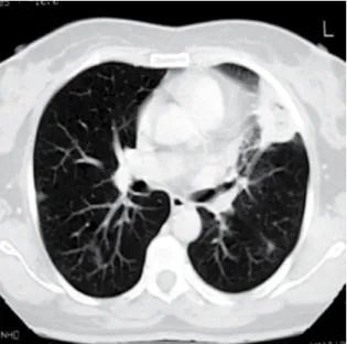
aggra-vated in the supine position, asthenia, anorexia, and weight loss (5 kg). One week after the onset of symp-toms, the patient presented progressively worsening chest pain (on the left side) with pleuritic character-istics, frequent fits of dry cough upon exertion, and an isolated peak of fever had appeared. The electro-cardiogram results were normal, and endoscopy of the upper digestive tract revealed a compliant cardia and antral gastritis. A chest X-ray showed a hetero-geneous hypotransparency, with loss of substance in its center, in the middle third of the left lung field. A computed tomography scan of the chest revealed a 4-cm mass with heterogeneous content and pleural extension to the level of the lingula, as well as two micronodules in the posterior region of the right lung field (Figure 1). An etiological study was carried out based on the working diagnosis of pulmonary neoplasia. From the analytical study, we highlight the following results: an erythrocyte sedimentation rate of 98 mm in the first hour; a cancer antigen 125 level of 63.3 U.mL–1 (<35.0);
a carcinoembryonic antigen level of 3.55 ng.mL–1
(<2.5); and a slight increase in the C-reactive protein level. The serum immunological study, the viral marker analysis, and the final analytical study
Figure 1 - Pretreatment computed tomography scan
of the chest showing a 4-cm heterogeneous mass with pleural extension at the level of the lingula and two micronodules in the posterior region of the right lung field.
Figure 2 - Cytological examination showing an
Treatment of pulmonary actinomycosis with levofloxacin
J Bras Pneumol. 2008;34(4):245-248 247
oral commensal type, typically Gram-negative, and can contribute to the pathogenesis of the disease.(3)
All thoracic tissue is susceptible to infection, and the symptoms often suggest malignancy. Pulmonary actinomycosis is typically caused by aspiration of infected oropharyngeal secretions. However, it can also result from the direct extension of cervicofacial infections. A typical lesion consists of a lung abscess filled with neutrophils and surrounded by dense fibrotic tissue. More rarely, consolidation without abscess formation can occur.
The onset of the pulmonary disease is insidious, with cough, expectoration, fever, and weight loss. In a small number of cases, hemoptysis or pleuritic pain can occur. Expectoration is usually yellow or white, with no characteristic odor. When it is foul smelling, it suggests the presence of other anaerobic microorganisms in combination with Actinomyces.(3)
Within the bronchus, there may be formation of inflammatory tissue, causing its partial obstruction. Often, the symptoms are surprisingly mild, given the extent of the disease. Leukocytosis, neutrophilia, and moderate anemia are common.(2-5) The
pulmo-nary lesion can present fistulation through the pleura into the chest wall, invading the ribs and the sternum. Patients can present a diffuse alveolar process with a bronchocutaneous or bronchopleural fistula, which can evolve to subcutaneous abscess or empyema, respectively.
Chronic infection can then manifest as a mass, similar to a bronchogenic carcinoma. A fistula forms between the mass and the pleural space or the chest wall, producing one or multiple abscesses, or causing bone loss. This form of dissemination of the pulmonary infection differs from that of other microorganisms, and can be initially confused with a malignant process.(1)
Pulmonary actinomycosis can mimic a variety of diseases, such as nocardiosis, tuberculosis, and bronchial carcinoma. On X-rays, the infiltrates are often dense and well circumscribed. Typically, the initial lesion presents as well-located consolidation that can evolve to cavitation in half of all cases, such cavitation generally being of small dimen-sions. Pleural effusion is common.(2) The infection
can affect multiple lobes, disseminating through the interlobular fissures and creating alveolar or nodular infiltrates. Occasionally, the erosion of a blood vessel leads to the hematogenous dissemina-tion of the disease, which takes the aspect of miliary there was complete radiological normalization.
Analytically, we observed a decrease in the erythro-cyte sedimentation rate and in the initially altered levels of both tumor markers, as well as normaliza-tion of the C-reactive protein level. The patient was also submitted to dental treatment.
Discussion
Actinomycosis is a non-contagious, chronic bacterial infection that occasionally occurs in humans and other animals. The causal agent is a commensal bacterium of the mouth, as well as of the respiratory, gastrointestinal, and genital tract of healthy indi-viduals. This commensal is a facultatively anaerobic Gram-positive bacterium, of the genus Actinomyces within the family Actinomycetaceae, which is fila-mentous and slow-growing.(1,2,5) Actinomycosis is a
rare disease, with a worldwide distribution, and its major risk factor is poor oral hygiene. The reduction in the incidence of this pathology might be related to improved oral hygiene and to the adequate use of antibiotics in infections with other agents.(1)
In tissue, Actinomyces develops in the form of dense granules or microcolonies that can reach 4 mm and are known as ‘sulfur granules’, due to their yellowish color.(5) Actinomycosis disseminates
without respecting anatomical planes.(3) It often
appears as a co-infection with other bacteria known as ‘associated bacteria’, which are usually also of the
Figure 3 - Computed tomography scan of the chest
248 Ferreira DFG, Amado J, Neves S, Taveira N, Carvalho A, Nogueira R
J Bras Pneumol. 2008;34(4):245-248
cycline, and clindamycin) can be used orally with good treatment results.(2) As in any bacterial disease
of the chest, surgery should be considered, since it is often necessary to relieve the obstruction of the mediastinal structures. Empyemas should be drained. Actinomycosis has a strong tendency toward recurrence, which is why the treatment should be extended to 6-12 months. The exact duration depends on the severity of the disease and its extent within and outside the chest, as well as on the response to the treatment. The prognosis is excellent, with a cure rate of 90% when the disease is diagnosed early and an efficient antibiotic therapy is administered.(1)
The therapeutic option used in the present case was unquestionably controversial, but, having been initiated with clinical efficacy before diag-nostic confirmation, it was maintained, with the informed consent of the patient, until the regres-sion of the symptoms and complete normalization of the radiological alterations. The evolution of this case suggests that levofloxacin represents an alter-native to the conventional treatment of pulmonary actinomycosis.
Referências
1. Gomes, MJ, Sotto-Mayor R. Infecções respiratórias não tuberculosas. Tratado de Pneumologia 2003;v. I;41:488-90. 2. Cheon JE, Im J, Lee J, Choi G, Yeon K. Thoracic actinomycosis:
CT findings. Radiology 1998;209(1):229-33.
3. Fishman AP. Infectious diseases of the lungs. Fishman’s Manual of Pulmonary diseases and disorders 2002;59:709-10. 4. Cendan I, Talavera W. Pulmonary actinomycosis. A cause of
endobronchial disease in a patient with AIDS. Chest 1993; 103(6):1886-7.
5. Gibson G, Duncan M, Costabel U, Sterk J, Corrin B. Respiratory Infections. Respiratory Medicine 2003;v. II;35:890-1.
6. Das DK. Actinomycosis in fine needle aspiration cytology. Cytopathology 1994;5(3):243-50.
disease (disseminated). Computed tomography scans can reveal the presence of cavities and their extension through the fissures, as well as pleural thickening or chest wall involvement, which are not always apparent on chest X-rays.(2)
The diagnosis is difficult, and less than 10% of the cases are diagnosed at the initial presenta-tion.(5) The cultures should be performed in strictly
anaerobic media and rapidly inoculated. A positive microbiological culture analysis of expectoration has little clinical significance, since Actinomyces is part of the normal oral flora. The presence of sulfur granules is suggestive of the diagnosis. However, such granules are rarely seen. Collection of secretion through fiberoptic bronchoscopy can also be contaminated by the normal flora of the oropharynx, unless protected catheters are used. Likewise, bacteriological examination of material obtained through transbronchial biopsy can be contaminated by upper airway secretions, whereas a histological diagnosis or a TNAB of such material are reliable. Obtaining an uncontaminated sample for the identification of the microorganism usually requires a needle TNAB or a surgical lung biopsy. In case of fistula, the presence of granules in the secretions is the key to diagnosis.(6) As in the case
presented here, TNAB can be important to allow collection in media that are appropriate for culture examination.