Sagittal and vertical aspects of Class II
division 1 subjects according to the
respiratory pattern
Laura de Castro Cabrera1, Luciana Borges Retamoso2, Raul Magnoler Sampaio Mei3, Orlando Tanaka4
Introduction: The teeth position, specially maxillary and mandibular incisors, in relation to basal bone and sur-rounding sot tissues must be considered in the elaboration of diagnosis, treatment planning and execution to obtain alignment, leveling, intercuspation, facial balance and harmony with stability of results. Objectives: To evaluate the modiications in the positioning of incisors in individuals with Angle Class II, division 1 malocclusion in two distinct moments of dentocraniofacial development, with mean interval of 2 years and 5 months. Methods: The measures were obtained by means of lateral cephalograms of 40 individuals, being 23 nasal breathers (NB) and 17 mouth breathers (MB). The analyzed measures were overjet, overbite, UCI-NA, LCI-NB, UCI.NA, LCI.NB, UCI.SN, LCI.GoGn, UCI.LCI, ANB, GoGn.SN, and OccPl.SN. Statistical analysis (2-way repeated-measures ANOVA) was applied to verify intergroups diferences. Results: Overjet, UCI-NA, LCI-NB, ANB, GoGn.SN, and OccPl.SN demonstrated statistically signiicant diference (p < 0.05) when observed the moment or the respiratory method. Conclusion: There is alteration in the positioning of incisors during growth with interference of the respiratory pattern.
Keywords: Angle Class II malocclusion. Mouth breathing. Breathing. Vertical dimension.
How to cite this article: Cabrera LC, Retamoso LB, Mei RMS, Tanaka O. Sagittal and vertical aspects of Class II division 1 subjects according to the respira-tory pattern. Dental Press J Orthod. 2013 Mar-Apr;18(2):30-5.
Submitted: December 22, 2008 - Revised and accepted: June 08, 2009
Contact address:Orlando Tanaka Rua Imaculada Conceição, 1155 CEP: 80.215-901 - Curitiba / PR, Brazil Email: [email protected]
1 MSc Student in Dentistry.
2 PhD Student in Dental Materials, PUCPR.
3 MSc in Orthodontics, PUCPR.
4 Responsible for the area of concentration in Orthodontics of PPGO-PUCPR.
» The authors report no commercial, proprietary or financial interest in the products or companies described in this article.
Introdução: a posição dos dentes, principalmente incisivos superiores e inferiores, em relação às bases ósseas e tecidos moles circundantes deve ser considerada na elaboração do diagnóstico, planejamento e execução do tratamento, para se obter alinhamento, nivelamento, intercuspidação, equilíbrio e harmonia facial com estabilidade dos resultados.
Objetivos: avaliar as modiicações no posicionamento dos incisivos em indivíduos com má oclusão de Classe II, divisão 1, de Angle, em dois momentos distintos do desenvolvimento dentocraniofacial, num intervalo médio de 2 anos e 5 meses.
Métodos: as medidas foram obtidas por meio de telerradiograias em norma lateral de 40 indivíduos, sendo 23 respi-radores predominantes nasais (RN) e 17 predominantemente bucais (RB). As medidas avaliadas foram sobressaliência, sobremordida, ICS-NA, ICI-NB, ICS.NA, ICI.NB, ICS.SN, ICI.GoGn, ICS.ICI, ANB, GoGn.SN, Plo.SN. Para veriicar a diferença intergrupos, utilizou-se a ANOVA a dois critérios com medidas repetidas. Resultados: sobressa-liência, ICS-NA, ICI-NB, ANB, GoGn.SN, Plo.SN apresentaram diferença estatisticamente signiicativa (p < 0,05) quando observado o momento ou o modo respiratório. Conclusão: existe alteração no posicionamento dos incisivos no decorrer do crescimento, com interferência do modo respiratório.
INTRODUCTION
Overbite is the vertical trespass and overjet is the horizontal trespass, which suffer significant altera-tions during development of dentition, from initial
mixed dentition to permanent occlusion.16 Overbite
is correlated to other measures that indicate facial dimensions, such as mandibular plane and occlusal plane angles. Overjet usually is a reflex of the
antero-posterior skeletal relation,8 and it is sensible to the
atypical function of lips and tongue. On the devel-opment of Class II and III malocclusions, these den-tal measurements tend to adapt to abnormal skeleden-tal relations. The position of maxillary and mandibular incisors, the relation between both and to the sur-rounding tissues, are important characteristics in the diagnosis, execution and stability of the treatment. The measures related to positioning of incisors affect the balance and harmony of facial profile. Due to its importance, since the introduction of craniometry, the position of mandibular incisor on sagittal plane
became a precious tool to assess a malocclusion.3,7 The
determination of positioning of maxillary and
man-dibular incisor is part of most cephalometric analysis.4
Downs6 and Riedel19 advocated specific values for the
position of mandibular incisor, however, other val-ues were suggested and used to predict the stability
of the treatment results.22,25,26 The maxillary incisors
perform an important role because they provide the
inclination for protrusive mandibular movement.20
Also, the position and specially the axial inclination of maxillary and mandibular incisors, are determina-tive on facial esthetics, as incisors with increased axial inclinations, create protruded lips and, many times, absence of passive lip seal. The orthodontic treatment is frequently performed during adolescence, between
10 and 16 years of age.17 Consequently, the evaluation
of incisors positioning, its relation to adjacent struc-tures, overjet and overbite during this period may provide information and contribute to the elabora-tion of diagnosis, planning, treatment and assessment of the post treatment stability. Thus, this work aims to assess the alterations on the position of maxillary and mandibular incisors in individuals with Angle Class II malocclusion,division 1, in two distinct mo-ments of the dentocraniofacial development, with mean interval of 2 years and 5 months according to respiratory pattern.
MATERIAL AND METHODS
To perform this research it was used lateral cephalo-grams of 40 individuals with Angle Class II division 1 malocclusion, where 23 were nasal breathers (NB) and 17 mouth breathers (MB), aged between 10 years and
9 months and 14 years (T1), and between 13 years and
4 months and 16 years and 6 months (T2). The
classii-cation of respiratory pattern was done according to
pro-tocol described by Wieler et al,27 which includes clinical
evaluation of lip seal performed by dental surgeon, sur-vey answered by the parents regarding respiratory habits, otorhinolaryngological assessment and speech evalua-tion. From these evaluations it was assigned scores and weighting for each evaluation, creating an index to clas-sify the individual’s predominant respiratory pattern. On each cephalogram it was ixed a sheet of acetate paper, 50 µm thick and 18 cm high x 17 cm wide. The cepha-lograms were traced with mechanical pencil Pentel P203 and graphite 2B, 0,3 mm of diameter, considering the interesting anatomic structures, and only on the let side, by a single operator, in a darkened room, being the only source of light the one from the negatoscope. The linear measures were performed with a single ruler with preci-sion of 0.5 mm, and the angular measures with a protrac-tor, precision of 0.5 degrees. The used angular and linear measures were the following, showed in Figure 1:
1. Overjet: Distance from vestibular surface of mandibular central incisor to palatine surface of maxillary central incisor, in millimeters.
2. Overbite: Distance, in millimeters, that the maxillary central incisor trespass the mandibular central incisor.
3. UCI-NA: Distance, in millimeters, from the vestibular surface of maxillary central incisor to the NA line.
4. LCI-NB: Distance, in millimeters, from the vestibular surface of mandibular central incisor to the NB line.
5. UCI.NA: Angle, measured in degrees, formed by the intersection of the long axis of maxillary central incisor and the NA line.
6. LCI.NB: Angle, measured in degrees, formed by the intersection of the long axis of mandibu-lar central incisor and the NB line.
and it was observed that, for all the studied measures, this did not exceed 3%, thus obtaining reliability for all obtained data.
RESULTS
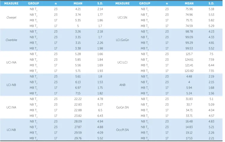
The statistical analysis was performed using the Sta-tistical Package for the Social Science 15.0 for Windows (SPSS, Inc., Chicago, IL, USA). The veriication of nor-mality was performed through Kolmogorov-Smirnov test, at signiicance level of 0.05. Once it was found the normal distribution, the veriication of existence or ab-sence of diference between the means (Table 1) of the two types of breathers for each one of the two moments, was performed with the aid of ANOVA with two crite-ria for classiication, with repeated measures.
When ANOVA demonstrated that there was a sta-tistically signiicant diference (p < 0.05) between the mean values of overjet, UCI-NA, LCI-NB, ANB, GoGn.SN and OccPl.SN according to moment or re-spiratory pattern, it was used Tukey’s HSD test of mul-tiple comparison to identify which groups were difer-ent from one another (Table 2).
DISCUSSION
The results of the present work showed that there were alterations on the measures related to positioning of incisors according to moments and respiratory
pat-tern, agreeing with Ceylan et al.4 It was observed
re-duction of the overjet from T1 to T2 in both analyzed
groups of individuals (nasal and mouth breathers). This behavior occurs because of the mandibular growth that tends to reduce the facial convexity and the overjet,
ac-cording to proved studies by Ceylan et al.4 Besides, the
modiication in the positioning of incisors may also have caused the reduction of overjet, for the measure LCI-NB increased in larger proportion than the UCI-NA. An-other factor to be considered, is the reduction of the oc-clusal plane, probably caused by the counterclockwise rotation of the mandible, and consequently reduction of overjet. It is emphasized that the respiratory pattern afected this measure, since mouth breathers present greater overjet than nasal breathers, according to
Mocel-lin15 and Ricketts,18 who point the respiratory pattern as
an etiologic factor for malocclusions. Generally, mouth breathing patients tend to present a protruded maxilla and maxillary atresia, consequence of the alteration on the tongue position, which becomes lower, breaking the Figure 1 - Linear and angular measurements used.
8. LCI.GoGn: Angle, measured in degrees, formed by the intersection of the long axis of mandibu-lar central incisor and the mandibumandibu-lar plane. 9. UCI.LCI: Angle, measured in degrees, formed
by the intersection of the long axis of maxillary central incisor and the long axis of mandibular central incisor.
10. ANB: Diference, measured in degrees, be-tween the angles SNA and SNB, determines the position of the maxilla and mandible in the an-teroposterior direction.
11. GoGn.SN: Angle, measured in degrees, that de-termines the facial pattern in vertical direction. 12. OccPl.SN: Angle, measured in degrees, that
de-termines the inclination of occlusal plane in rela-tion to SN line.
STUDY’S REPRODUCIBILITY ERROR
To evaluate the reproducibility error, it was ran-domly selected 30 teleradiographs and a single opera-tor performed the cephalometric evaluation for the second time, with interval of 30 days. It was
calculat-ed the systematic error5 between the two evaluations
2
1
4 8
6 9
5
3 12
11
Table 1 - Mean and standard deviation of linear and angular measures.
Table 2 - Mean, standard deviation and p value for the variables.
NOTE: Signiicance level for Tukey HSD multiple comparisons *p < 0.05; **p < 0.03; ***p < 0.01.
MEASURE GROUP n MEAn S.D. MEASURE GROUP n MEAn S.D.
Overjet
NB T1 23 4.21 2.14
UCI.SN
NB T1 23 75.96 5.18
NB T2 23 3.74 1.77 NB T2 23 74.96 5.53
MB T1 17 5.35 1.86 MB T1 17 75.71 5.82
MB T2 17 5 1.7 MB T2 17 74.59 5.29
Overbite
NB T1 23 3.26 2.18
LCI.GoGn
NB T1 23 98.78 4.23
NB T2 23 3.35 1.7 NB T2 23 99.09 4.33
MB T1 17 3.15 2.26 MB T1 17 99.29 4.81
MB T2 17 3.38 1.98 MB T2 17 99.53 5.52
UCI-NA
NB T1 23 5.28 1.66
UCI.LCI
NB T1 23 125.7 7.5
NB T2 23 5.85 1.84 NB T2 23 124.61 7.59
MB T1 17 5.56 1.69 MB T1 17 121.41 6.44
MB T2 17 5.71 1.93 MB T2 17 120.82 7.35
LCI-NB
NB T1 23 5.61 1.8
ANB
NB T1 23 4.48 2.19
NB T2 23 6.13 1.53 NB T2 23 4 2.15
MB T1 17 6.97 1.75 MB T1 17 5.94 1.68
MB T2 17 7.15 1.82 MB T2 17 5.24 1.56
UCI.NA
NB T1 23 22.22 4.78
GoGn.SN
NB T1 23 31.83 5.1
NB T2 23 22.83 5.27 NB T2 23 30.7 5.09
MB T1 17 22.88 6.5 MB T1 17 34.71 4.04
MB T2 17 23.82 6.43 MB T2 17 33.71 4.57
LCI.NB
NB T1 23 28.09 4.94
OccPl.SN
NB T1 23 16.48 4.83
NB T2 23 27.87 4.88 NB T2 23 14.83 5.21
MB T1 17 29.59 4.09 MB T1 17 19.12 2.26
MB T2 17 29.76 5.52 MB T2 17 17.53 2.21
MEASURE GROUP n MEAn S.D. intra and Intergroups diference
Overjet
NB T1 23 4.21 2.14 NS
NB T2 23 3.74 1.77 *NB T2 X MB T1
MB T1 17 5.35 1.86 *NB T2 X MB T1
MB T2 17 5 1.7 NS
UCI-NA
NB T1 23 5.28 1.66 *NB T1 X NB T2
NB T2 23 5.85 1.84 *NB T1 X NB T2
MB T1 17 5.56 1.69 NS
MB T2 17 5.71 1.93 NS
LCI-NB
NB T1 23 5.61 1.8 *NB T1 X NB T2, *NB T1 X MB T2
NB T2 23 6.13 1.53 *NB T1 X NB T2
MB T1 17 6.97 1.75 NS
MB T2 17 7.15 1.82 *NB T1 X MB T2
ANB
NB T1 23 4.48 2.19 NS
NB T2 23 4 2.15 **NB T2 X MB T1
MB T1 17 5.94 1.68 **NB T2 X MB T1
MB T2 17 5.24 1.56 NS
GoGn.SN
NB T1 23 31.83 5.1 **NB T1 X NB T2
NB T2 23 30.7 5.09 **NB T1 X NB T2
MB T1 17 34.71 4.04 NS
MB T2 17 33.71 4.57 NS
OccPl.SN
NB T1 23 16.48 4.83 ***NB T1 X NB T2
NB T2 23 14.83 5.21 ***NB T1 X NB T2, ***NB T2 X MB T1
MB T1 17 19.12 2.26 ***NB T2 X MB T1
stage, the diference can be explained by a genetic hori-zontal growth pattern disagreeing with results of Lessa
et al,14 which did not observe signiicant diferences on
these measures. Likewise, the measure OccPl.SN also reduced, due to reduction on the inclination of occlu-sal plane, consequence of the change on positioning of maxillary and mandibular incisors and the mandibular rotation. This measurement was afected by the
respira-tory pattern, according to Lessa et al,14 mouth
breath-ers present greater mandibular inclination and vertical growth pattern. This work obtained statistically signii-cant diferences in the positioning of incisors in indi-viduals with distinct respiratory patterns, agreeing with
results by Spinelli.21 Thus, the mouth breathing afected
growth of some facial structures, causing a mandibular rotation down and backwards, in relation to the palate; and reduction of angle formed by intersection of
man-dibular plane with nasal plane.13 According to
litera-ture, there is correlation between alterations caused by
mouth breathing and the occlusion,13 fact also veriied
in this work. It is assumed that alterations occurred on measures were consequence of genetic growth pattern, for with aging there is a tendency of the facial proile to
become relatively more straight28 associated to
environ-mental factors, especially the respiratory method. How-ever, this result disagree with Gwynne-Evans and
Bal-lard;11 Tomer and Harvold,24 which indicated that
mus-cle patterns and skeletal growth are genetically deined and, therefore, the individual characteristics, favorable or not, are inherited and little inluenced by the altera-tions on respiratory pattern. Therefore, the alteraaltera-tions in positioning of incisors and the individual’s respiratory pattern must be considered on the diagnosis, elaboration of treatment plan and execution of treatment in indi-viduals in growth stage.
CONCLUSION
It is concluded that there was alteration in the posi-tioning of incisors and overjet, during growth and with interference on the respiratory pattern.
balance with the buccinator muscle. It was veriied an increase of the linear measures UCI-NA and LCI-NB from 10 to 16 years of age, which may be related to pro-jection of maxillary and mandibular incisors, agreeing
with Bishara.2 On the other hand, Forsberg9 observed
verticalization of the incisors with facial growth. In this research, the measurement LCI-NB presented an in-crease proportionally larger than UCI-NA, explained as a way to camoulage the Class II skeletal relation. It is suggested that the alteration observed in the positioning of incisors can also be explained by the action of tongue
muscles. However, Baydas et al1 did not observe
statis-tically signiicant diference in the positioning of these teeth during growth. From analysis of the ANB angle it was veriied that there was a reduction on the difer-ence between maxillary and mandibular bone bases in the sagittal plane, which seems to be directly related to mandibular growth and overjet alterations. The fact that the linear measurements related to the positioning of the mandibular incisor increases in larger proportion than the maxillary incisor may have caused the reduction on ANB. The respiratory pattern afected this result, where mouth breathers presented a larger ANB, disagreeing
with the results of Frasson et al.10 It is suggested that the
decline on the tongue rest position, afected the
maxil-lary growth, according to Subtelny23 nasal breathing is
essential for a correct growth and development of the
craniofacial complex. However, Jakobsone et al12
1. Baydas B, Yavuz I, Atasaral N, Ceylan I, Dagsuyu I. Investigation of the changes in the positions of upper and lower incisors, overjet, overbite, and irregularity index in subjects with diferent depths of curve of Spee. Angle Orthod. 2004;74(3):349-55.
2. Bishara SE. Longitudinal cephalometric standards from 5 years of age to adulthood. Am J Orthod. 1981;79(1):35-44.
3. Broadbent BH. A new X-ray technique and its application in Orthodontics. Angle Orthod. 1931;1(2):45-6.
4. Ceylan I, Baydas B, Bolukbasi B. Longitudinal cephalometric changes in incisor position, overjet, and overbite between 10 and 14 years of age. Angle Orthod. 2002;72(3):246-50.
5. Dahlberg G. Statistical methods for medical and biological students. New York: Interscience; 1940.
6. Downs WB. Variation of facial relationships: their signiicance in treatment and prognosis. Am J Orthod. 1948;34(10):812-40. 7. Ellis EE, McNamara JA. Cephalometric evaluation of incisor position.
Angle Orthod. 1986;56(4):324-44.
8. Fleming H. An investigation of the vertical overbite during the eruption of the permanent dentition. Am J Orthod. 1961;31(1):53-62.
9. Forsberg CM. Facial morphology and ageing: a longitudinal cephalometric investigation of young adults. Eur J Orthod. 1979;1(1):15-23.
10. Frasson JMD, Magnani MBBA, Nouer DF, Siqueira VCV, Lunardi NC. Cephalometric study between nasal and predominantly mouth breathers. Braz J Otorhinolar. 2006;72(1):72-81.
11. Gwynne-Evans E, Ballard CF. The mouth-breather. Am J Orthod. 1958;44(7):559.
12. Jakobsone G, Urtane I, Terauds I. Soft tissue proile of children with impaired nasal breathing. Stomatol. 2006;8(2):39-43.
13. Kerr WJS, McWilliam JS, Linder-Aronson S. Mandibular form and position related to changed mode of breathing — a ive-year longitudinal study. Angle Orthod. 1989;59(2):91-6.
14. Lessa FCR, Enoki C, Feres MFN, Valera FCP, Lima WTA, Matsumoto MAN. Inluência do padrão respiratório na morfologia craniofacial. Rev Bras Otorrinolaringol. 2005;71(2):156-60.
REFERENCES
15. Mocellin M. Respirador bucal. In: Petrelli, E. Ortodontia para Fonoaudiologia. São Paulo: Lovise; 1992. p. 131-43.
16. Moyers RE. Ortodontia. 4ª ed. Rio de Janeiro: Guanabara Koogan; 1991. 786 p.
17. Prahl-Andersen B, Ligthelm-Bakker AS, Wattel E, Nanda R. Adolescent growth changes in soft tissue proile. Am J Orthod Dentofacial Orthop. 1995;107(5):476-83.
18. Ricketts RM. Perspectives in the clinical application of cephalometrics. Angle Orthod. 1981;51(2):115-50.
19. Riedel RA. The relation of maxillary structures to cranium in malocclusion and in normal occlusion. Angle Orthod. 1952;22(3):142-5.
20. Russouw PE, Preston CB, Lombard CJ, Truter JW. A longitudinal evaluation of the anterior border of the dentition. Am J Orthod Dentofacial Orthop. 1993;104(2):146-152.
21. Spinelli MLM, Casanova PC. Respiração bucal. [acesso em 2002 Fev 2] Disponível em: www.odontologia.com.br/artigos.
22. Steiner CC. Cephalometrics for you and me. Am J Orthod. 1953;39(10):729-55.
23. Subtelny JD. Oral respiration: facial mal development and corrective dentofacial orthopedics. Angle Orthod. 1980;50(3):147-64. 24. Tomer BS, Harvold EP. Primate experiments on mandibular growth
direction. Am J Orthod. 1982;82(2):114-9.
25. Tweed CH. The Frankfurt-mandibular incisor angle (FMIA) in
orthodontic diagnosis, treatment planning and prognosis. Angle Orthod. 1954;24(3):121-69.
26. Tweed CH. Clinical orthodontics. St. Louis: CV Mosby; 1966. v. 1. 27. Wieler WJ, Barros AM, Barros LA, Camargo EL, Ignácio SA, Maruo H, et
al. Combined protocol to aid diagnosis of breathing mode. Rev Clín Pesq Odontol. 2007;3(2):101-11.