SONOGRAPHIC EVALUATION OF THE SHOULDER JOINT
IN COMPETITIVE SWIMMERS*
Guilherme Moura da Cunha1, Edson Marchiori2, Elísio José Ribeiro3
OBJECTIVE: To evaluate the shoulders of symptomatic or asymptomatic competitive swimmers, quantify-ing the incidence of rotator cuff lesions in these athletes. MATERIALS AND METHODS: Eleven (eight male and three female) symptomatic and asymptomatic, competitive swimmers (master category) in the age range between 28 and 52 years, had both shoulders prospectively evaluated by ultrasonography for determining the prevalence of injuries in correlation with clinical findings. The studies included at least two orthogonal planes of rotator cuff tendons, as well as non-tendinous components of the shoulder joint. Tendinous find-ings were classified into tendinosis, probable full-thickness tears and partial-thickness tears. Non-tendinous findings were classified into present or absent. Additionally to the sonographic studies results, the analysis has taken the presence or absence of symptoms and the patients’ age into consideration. RESULTS: Over-all, the prevalence of symptomatic patients was higher (63.6%; seven athletes), with 75% of patients with bursitis, and 100% of those with partial-thickness tendon tears. Among isolated tendinous findings, the su-praspinatus tendon was the most frequently involved, showing echographic pattern alteration in 36.36% of cases. Tendinosis was the most prevalent finding, present in at least one tendon in 50% of the studied shoulders. The incidence of partial-thickness tendon tears was similar to the incidence reported for non-athlete individuals (13%), always present in individuals above 40 years of age. CONCLUSION: Swimmers do not seem to present a higher incidence of tendon tears or degeneration when compared with the general population. Individuals’ age is the most relevant determining factor, as far as rotator cuff lesions are con-cerned, no matter these individuals practice or not activities involving an overload of the shoulder joint.
Keywords: Impingement syndrome; Rotator cuff; Swimmers; Ultrasonography.
Avaliação ultra-sonográfica da articulação do ombro em nadadores de nível competitivo.
OBJETIVO: Avaliar o ombro de nadadores em nível competitivo, sintomáticos ou não, e quantificar a inci-dência de lesões do manguito rotador nesses atletas. MATERIAIS E MÉTODOS: Foram estudados, por meio de exame ultra-sonográfico, ambos os ombros de 11 nadadores de nível competitivo (categoria master), sendo oito homens e três mulheres, com idade variando de 28 a 52 anos, tanto sintomáticos quanto assintomáti-cos, para determinação de alterações e correlação com quadro clínico. Os exames foram realizados avaliando-se os tendões que compõem o manguito rotador em pelo menos dois planos ortogonais distintos, assim como as estruturas não-tendíneas que fazem parte da articulação do ombro. As alterações tendíneas foram clas-sificadas como tendinose, prováveis roturas intra-substanciais e roturas parciais. As lesões não-tendíneas fo-ram classificadas como presentes ou ausentes. Além do exame, fofo-ram consideradas, na análise dos resulta-dos, a presença ou ausência de sintomas e a idade dos pacientes. RESULTADOS: A prevalência geral de indivíduos sintomáticos foi de 63,6% (sete atletas). Neste grupo encontravam-se 75% das bursites e 100% das roturas tendíneas parciais. Entre as alterações tendíneas isoladas, o tendão mais freqüentemente aco-metido foi o supra-espinal, tendo alteração de seu padrão ecográfico em 36,36%. A tendinose foi o achado mais prevalente, estando presente em pelo menos um dos tendões em 50% dos ombros estudados. A inci-dência de roturas parciais foi semelhante à observada na população geral, sendo de 13%, estando estas, sempre que presentes, na faixa etária acima de 40 anos. CONCLUSÃO: Nadadores não parecem ter maior incidência de roturas ou degenerações tendíneas quando comparados com indivíduos da população geral. A idade dos indivíduos, sendo estes praticantes ou não de atividades que exijam sobrecarga desta articulação, é o fator com maior determinância em relação às lesões do manguito rotador.
Unitermos: Síndrome do impacto; Manguito rotador; Nadadores; Ultra-sonografia.
Abstract
Resumo
* Study developed at Clínica de Diagnóstico por Imagem (CDPI) and Hospital Universitário Clementino Fraga Filho (HUCFF) da Uni-versidade Federal do Rio de Janeiro (UFRJ), Rio de Janeiro, RJ, Brazil.
1. MD, Resident, Department of Radiology, Hospital Universi-tário Clementino Fraga Filho (HUCFF) da Universidade Federal do Rio de Janeiro (UFRJ), Rio de Janeiro, RJ, Brazil.
2. Full Professor of Radiology, Universidade Federal Fluminense (UFF), Niterói, RJ, Adjunct Coordinator for Course of Post-gradu-ation in Radiology, Universidade Federal do Rio de Janeiro (UFRJ), Rio de Janeiro, RJ, Brazil.
INTRODUCTION
Shoulder pain is the most frequent com-plaint among competitive swimmers, in such extent that the designation “swim-mer’s shoulder” was utilized in 1974 to call a painful syndrome caused repetitive im-pact on the shoulders of these athletes(1). 3. MD, Unit of Ultrasonography and Magnetic Resonance
Imaging, CDPI and Clínica Multi-Imagem, Rio de Janeiro, RJ, Brazil.
Mailing address: Dr. Guilherme Moura da Cunha. Avenida Pre-feito Dulcidio Cardoso, 3080, bloco 4, ap. 103, Barra da Tijuca. Rio de Janeiro, RJ, Brazil, 22631-054. E-mail: mouracunha@ hotmail.com
Shoulder pain hindering swimmers training is reported by 9% to 35% of com-petitive athletes, whereas 38% to 75% present with previous history of this symp-tom along their careers(1–3).
A study developed with the Canadian Olympic swimming team has concluded that 37% of the athletes’ orthopedic com-plaints are related to shoulders(3).
The high number of complaints related to swimmer’s shoulders of competitive ath-letes is due to the fact that their yearly workout involves about 1.32 million strokes, predisposing these athletes to over-use injuries of the shoulder joint(3).
Despite the wealth of the literature on the clinical syndrome involving swimmers shoulder pain, there is a scarcity of studies about anatomo-structural assessment in correlation with clinical findings in this population. The present study is aimed at evaluating, by means of ultrasonography, the shoulders of symptomatic or asymp-tomatic competitive swimmers, quantify-ing the incidence of rotator cuff lesions found in these athletes by this method.
MATERIALS AND METHODS
Eleven competitive swimmers, with ages ranging between 28 and 52 years (mean 32.9 years), eight male and three female, with a minimum five-year training (with a daily mean of > 2500 m under-taken), were selected according to their technical training load, differentiating them from amateur or recreative swimmers found in the general population. The ath-letes underwent ultrasound examination of the musculoskeletal apparatus, a same pro-tocol being adopted independently from the presence or not of shoulder pain. Later, the sonographic findings were correlated with their ages and the presence or not of symptoms.
The examinations were performed in an ultrasound equipment with a linear, 7–11 mHz transducer (Logic 500 – GE Medical Systems) in the period between April and July, 2004 at Clínica de Diagnóstico por Imagem (CDPI), Rio de Janeiro, RJ.
The sonographic images included, but were not limited to, longitudinal and trans-verse views of supraspinatus, infraspinatus, subscapular tendons, and the long head of
the biceps tendon, besides the evaluation of the acromioclavicular joint, posterior joint recess, and bursae.
The tendons were assessed as regards their diffuse abnormal echogenicity, echo-texture and thickness. Possible focal alter-ations were evaluated as regards echoge-nicity and localization (bursal surface, ar-ticular surface or intrasubstance). Thick-ened tendons with echogenicity alterations, heterogeneous echotexture and loss of usual fibrillar pattern, were considered as tendinosis(4).
Partial-thickness tendon tears were characterized by anechoic or hypoechoic areas, with tendinous fibers discontinuity, evaluated according to their dimensions, site, and presence of associated, and di-vided into probable partial-thickness tear, or compatible with partial-thickness tear. Hypoechoic tendinous areas only were considered as compatible with tear in the presence of fiber discontinuity and/or as-sociated, secondary findings of tear; other-wise, they were considered as tendinosis.
Secondary findings usually associated with tendinous tears and presence of extratendinous findings, like bursitis, acro-mioclavicular joint alterations, articular effusions and others were evaluated. All of the findings should be confirmed by at least two opposite orthogonal planes.
The findings were rated as follows: zero – normal tendons; type I – peritendinous inflammatory lesions(5); type II – thickened
tendons with heterogeneous echotexture (tendinosis)(4,5); type III – small, ill-defined,
hypoechoic foci, predictive or probably suggestive of tendinosis with small full-thickness tendon tears; type IV – well de-fined lesions with associated findings com-patible with partial- or full-thickness ten-don tear(6–8).
According to the literature(5,9)
pain-re-lated alterations are rated as types I and IV. Tendinosis, included in types II and III, may or not be associated with pain, these ten-dinous degenerative process being poorly correlated with the clinical condition of the patient.
After undergoing ultrasound examina-tions, these individuals were classified ac-cording two criteria: 1) presence of shoul-der pain; 2) age-range, based on the litera-ture about prevalence of rotator cuff
le-sions: individuals with < 40 years of age, or individuals with ≥ 40 years of age(9–11).
RESULTS
Sonographic findings
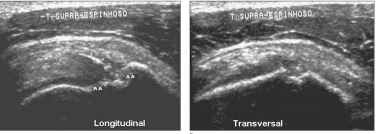
Twenty-two shoulders were evaluated with basis on the classification adopted, and the incidence of findings was distrib-uted as follows: type I alterations – four shoulders (18.18%); type II alterations (Figure 1) – 11 shoulders (50%); type III alterations (Figure 2) – three shoulders (13.63%); type IV alterations (Figures 3 and 4) – three shoulders (13.63%).
Amongst the sonographic findings, the most prevalent one was tendinosis.
Six of the examined athletes (54.5%) presented with at least one of theirs shoul-ders with types I and IV alterations directly related to shoulder pain. Three athletes (27.2%) presented with types II and III al-terations with a poor clinical significance. In two athletes (18.18%), no alteration was found on their shoulders ultrasound exami-nation.
Symptoms
In the present study, 63.6% (seven) of the swimmers complained of pain in at least one of their shoulders, frequently in association with the swimming practice. By correlating the data of these individuals with the pain-related findings, the authors could observe that 85.7% of these symp-tomatic patients presented type I and/or type IV alterations, and 57.1% presented non-rotator-cuff-related inflammatory al-terations (bursitis), with overlap between findings in four individuals.
Four individuals (36.3%) were asymp-tomatic, with no complaint of shoulder pain. In this group, three athletes presented only type II and/or type III alterations, and one, presented type IV alteration in one of the shoulders.
Ages
Figure 2. Longitudinal (A) and transverse (B) views. Thickened supraspinatus tendon, with heterogeneous echotexture, presenting hypoechoic focus, sugges-ting a probable partial-thickness tendon tear (arrows). Also in this patient, contralateral supraspinatus tendon heterogeneity and thickening were observed. Age-related sonographic findings are indicative of a degenerative process. Notwithstanding, the patients was completely asymptomatic.
A B
Figure 3. A: Supraspinatus tendinosis and small, partial-thickness tear (arrows). B: Observe the presence of fluid within the sheath of the long head of biceps, suggesting associated articular effusion.
A B
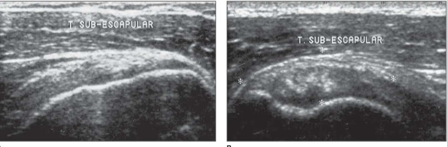
Figure 1. A: Asymptomatic, 31-year-old patient. Observe the subscapular tendon fibrillar pattern and homogeneous echotexture. B: Thirty-six-year-old patient with subscapular tendinosis. The tendon is thickened, with heterogeneous echotexture and fibrillar pattern (between asterisks).
alterations, with no sign of tendon tears or even probable tears in this age group. One individual in this age group presented type I alteration.
In the group with ≥ 40 years of age, in-cidence of tendinous alterations (II, III and IV types) was observed in 83.3% of indi-viduals. One hundred percent of cases with tears or even probable tears (III and IV types) were observed in this age range. Therefore, there was a clear correlation between higher ages and the presence of tendon lesions (Chart 1).
DISCUSSION
The shoulder pain mechanism in com-petitive swimmers is multifactorial, and, basically, results from the several forms of impingement of subacromial tissues. The repetitive compression of the supraspina-tus tendon and subdeltoid bursa under the coracoacromial arch leads to pain and in-dividual structural alterations(2,12).
The tendon affected by traction, im-pingement and abnormal vascular irriga-tion progresses with a degenerative process probably resulting in tear. In the bursa, the repetitive impingement may result in in-flammatory alterations. Both processes lead to pain as a final result(13,14).
Therefore, the prevalence of pain in these athletes is considerably high, ranging from 47% to 73% according reports in the literature(15,16). Similarly to the reviewed
lit-erature, the present study has found a
63.6% prevalence of shoulder pain among the participating individuals.
Inflammation of the bursa, a well inner-vated synovial structure, leads to a painful clinical picture. Tuite(12), in a study on
magnetic resonance imaging, has reported that, most frequently, lesion of tendinous structures of the rotator cuff are absent in young athletes with shoulder pain.
A B
Figure 4. Longitudinal (A) and transverse (B) views. Image compatible with partial-thickness supraspinatus tendon tear associated with secondary findings. Note the bone surface irregularity on the great tuberosity of the humerus (arrow heads) associated with an echogenic line permeating a hypoechogenic area (asterisks).
Chart 1 Findings according to the rating utilized.
Case
1
2
3
4
5
6
7
8
9
10
11
Age (years)
52
36
36
40
48
28
32
31
40
44
41
Shoulder
Right Left
Right Left
Right Left
Right Left
Right Left
Right Left
Right Left
Right Left
Right Left
Right Left
Right Left
Findings rating
I and II II and III
II 0
0 II
I and III II
II and IV I and II
0 0
I II
0 0
0 IV
II and IV II
III 0
Alteration found
Subscapular tendon
Infraspinatus and subscapular tendons
Subscapular tendon Normal results
Normal results Subscapular tendon
Supraspinatus tendon Supraspinatus tendon
Supraspinatus/infraspinatus and subecapular tendons Subscapular tendon
Normal results Normal results
Normal results Supraspinatus tendon
Normal results Normal results
Normal results Supraspinatus tendon
Supraspinatus tendon Supraspinatus tendon
Supraspinatus tendon Normal results
The prevalence of partial-thickness tears seen on ultrasound in the present ca-suistic (13.63%) was similar to the one re-ported by Matsen(11), who has found, in
studies with cadavers, a prevalence of 13% in the general population. Jacobson et al.(7)
au-thors such as Holsbeeck et al.(6) and Hodler
et al.(17). These studies have demonstrated
that, specifically for detecting these alter-ations, ultrasonography is an excellent method, with up to 100% sensitivity for full-thickness tears, and a 92% prospective accuracy for tendinous defects.
In the present study, the most prevalent finding was tendinosis (50% of cases), fol-lowed by bursitis (18.18%). Jacobson et al.(7), in their study about sonographic
find-ings in rotator cuff, have reported that the differentiation between tendinosis and small partial intrasubstance tear may be difficult. Both present a similar aspect at ultrasound and are part of a same progres-sive, degenerative process of the tendon. Therefore, due to the literature scarcity and differential diagnosis difficulty, the exact prevalence of tendinosis based on evalua-tion by ultrasonography in the present study and in the general population is still to be established. Furthermore, this abnormality cannot be arthroscopically identified, and studies in cadavers characterizing this per-centage are not available, so it is still more difficult to estimate its exact prevalence.
Taking all these aspects into consider-ation, type II and type III alterations were adopted as an initial phase of the degenera-tive process for including tendinosis as a finding. So, the results of the present study demonstrated that nine (81.8%) individu-als presented some kind of tendon degen-eration. These data correspond to the re-sults reported by Neumann et al.(18), who
have evaluated rotator cuff of asymptom-atic volunteers by means of magnetic reso-nance imaging, finding similar values. Eighty-nine percent of their patients pre-sented abnormalities compatible with de-generation, and only 2%, abnormalities compatible with partial-thickness tears. This result must be justified by the mean age (26 years) of the individuals included in their study.
As regards the presence of shoulder pain, 85.7% of athletes in the present study presented sonographic evidences justifying a painful clinical picture (bursitis or tears). Bursitis was present in 57.1% of these pa-tients, demonstrating that the lesion and pain mechanism observed in swimmers is part of a same physiological process of impingement, and, sometimes, there is an
overlap between bursitis and tendon tears. In the present study, tendinosis occurred in 85.7% of symptomatic, and in 50% of asymptomatic individuals. According to Maffulli et al.(5), the tendinosis process is
not necessarily symptomatic, and some-times the patient may progress to an ad-vanced degree of tendinosis in spite of never having experienced any discomfort in the tendon region. Up to the present moment, the reason for the disabling symp-toms resulting from tendinosis in some in-dividuals are still to be determined, whereas most of times the process is totally asymptomatic, even progressing to a more advanced degree resulting in tendon tear(5).
Notwithstanding, because of the scar-city, in the literature, of extensive studies about tendinosis in the general population, a linear relation between tendinosis and shoulder pain is still to be defined. There-fore the clinical meaning of tendinosis, up to the present moment, is poorly defined. Neumann et al.(18) have observed that 89% of the patients in their study presented ab-normalities potentially corresponding to tendinosis/tendon degeneration, and even so were asymptomatic(19).
Age, another factor considered in the present study, demonstrated a reasonable correlation with the literature reviewed. In the present casuistic, 100% of the patients with type IV alterations were in the age range above 40 year, in agreement with the literature. However, the analysis of the ath-letes included in the present casuistic, dem-onstrated a prevalence of tendon tears simi-lar to the prevalence in the general popu-lation, this prevalence being determined by the age of the individuals(20).
In the correlation between presence of symptoms and age range, a higher inci-dence of complaints of pain among the ath-letes included in the present study was observed as compared with studies evalu-ating the presence of pain in elite swimmers with a mean age of 19 years. However, if a previous history of shoulder pain during these athletes career is considered, the in-cidence is similar – about 64% in both groups(21).
The present study demonstrated that, despite the high incidence of shoulder pain in swimmers, this seems to be related to non-tendinous inflammatory processes or
partial-thickness tears found in the general population. Swimming seems not to in-crease the incidence of rotator cuff tendi-nous lesions, even in individuals practicing this activity for long periods of time(20).
In this context, the difficult character-ization and the low clinical significance of tendinosis, as well as the lack of studies evaluating its incidence in the general population, do not allow to evaluate if com-petitive swimmers present a more acceler-ated tendinous degenerative process than the general population.
A high prevalence of alterations in the echogenicity and echotexture of subscapu-lar tendons was observed in athletes in-cluded in the present study. These were the most frequently affected tendons after su-praspinatus tendons. No similar result was found in the literature, considering that, with the known impingement mechanisms supposedly affecting these individuals, the order of tears prevalence involves firstly the supraspinatus tendon, and secondly, the infraspinatus tendon. Even in studies evaluating the posterosuperior impinge-ment, which is the most frequent type of impingement among athletes involved in throwing and swimming, the infraspinatus was the second most frequently involved tendon.
The limitations of the present study were firstly related to the small size of the sample; secondly, to the absence of surgi-cal or histopathologisurgi-cal confirmation for the imaging findings.
Finally, competitive swimmers, despite the higher unnatural demands on their shoulder joints, do not seem to present a higher prevalence of rotator cuff tendon tears / early degeneration as compared with the general population. Most of times, the higher prevalence of shoulder pain in these athletes is a result of the inflammatory pro-cess involving the subacromiodeltoid bursa (bursitis) generated by the swimming mul-tifactorial impingement mechanism. The individuals´ age among swimmers or even among the general population plays the most significant role as a determining fac-tor of rotafac-tor cuff tendon tears.
REFERENCES
2. Kammer CS, Young CC, Niedfeldt MW. Swim-ming injuries and illnesses. Phys Sportsmed 1999;27:51–60.
3. McMaster WC, Troup J. A survey of interfering shoulder pain in United States competitive swim-mers. Am J Sports Med 1993;21:67–70. 4. Connell D, Burke F, Coombes P, et al. Sonographic
examination of lateral epicondylitis. AJR Am J Roentgenol 2001;176:777–782.
5. Maffulli N, Khan KM, Puddu G. Overuse tendon conditions: time to change a confusing terminol-ogy. Arthroscopy 1998;14:840–843.
6. van Holsbeeck MT, Kolowich P, Eyler WR, et al. US depiction of partial-thickness tear of the ro-tator cuff. Radiology 1995;197:443–446. 7. Jacobson JA, Lancaster S, Prasad A, van
Hols-beeck MT, Craig JG, Kolowich P. Full-thickness and partial-thickness supraspinatus tendon tears: value of US signs in diagnosis. Radiology 2004; 230:234–242.
8. Wohlwend JR, van Holsbeeck M, Craig J, et al. The association between irregular greater tuber-osities and rotator cuff tears: a sonographic study. AJR Am J Roentgenol 1998;171:229–233. 9. Uri DS. MR imaging of shoulder impingement
and rotator cuff disease. Radiol Clin North Am 1997;35:77–96.
10. Kjellin I, Ho CP, Cervilla V, et al. Alterations in the supraspinatus tendon at MR imaging: corre-lation with histopathologic findings in cadavers. Radiology 1991;181:837–841.
11. Matsen FA III. Rotator cuff tears. UW Medicine – Orthopaedics and Sports Medicine [On-line]. [Acessado em: 26/7/2004]. Disponível em: http:// www.orthop.washington.edu/uw/tablD_3351/ Default.aspx
12. Tuite MJ. MRimaging of sports injuries to the rotator cuff. Magn Reson Imaging Clin N Am 2003;11:207–219.
13. Brossmann J, Preidler KW, Pedowitz RA, White LM, Trudell D, Resnick D. Shoulder impinge-ment syndrome: influence of shoulder position on rotator cuff impingement – an anatomic study. AJR Am J Roentgenol 1996;167:1511– 1515.
14. Khan KM, Cook JL, Taunton EJ, Bonar F. Over-use tendinosis, not tendinitis. Part 1: a new para-digm for a difficult clinical problem. Phys Sports-med 2000;28:38–48
15. Dandachli W. Swimming shoulder injuries: wa-ter workouts are pretty safe. But top-level per-formers are more vulnerable to certain injuries. Sports Injury Bulletin.com [On-line]. [Acessado em 26/7/2004]. Disponível em: http://www.
sportsinjurybulletin.com/archive/swimming-shoulder-injuries.html
16. Johnson JN, Gauvin J, Fredericson M. Swimming biomechanics and injury prevention. New stroke techniques and medical considerations. Phys Sportsmed 2003;31:41–46.
17. Hodler J, Fretz CJ, Terrier F, Gerber C. Rotator cuff tears: correlation of sonographic and surgi-cal findings. Radiology 1988;169:791–794. 18. Neumann CH, Holt RC, Steinbach LS, Jahnke AH
Jr, Petersen SA. MR imaging of the shoulder: appearance of the supraspinatus tendon in asymp-tomatic volunteers. AJR Am J Roentgenol 1992; 158:1281–1287.
19. Hollister MS, Mack LA, Patten RM, Winter TC 3rd, Matsen FA 3rd, Veith RR. Association of sonographically detected subacromial/subdeltoid bursal effusion and intraarticular fluid with rota-tor cuff tear. AJR Am J Roentgenol 1995;165: 605–608.
20. Bak K, Faunl P. Clinical findings in competitive swimmers with shoulder pain. Am J Sports Med 1997;25:254–260.