www.rbo.org.br/
issn/$–see front matter © 2013 Sociedade Brasileira de Ortopedia e Traumatologia. Published by Elsevier Editora Ltda. All rights reserved. doi: 10.1016/j.rboe.2012.06.003
*Corresponding author at: Rua Monte Alegre, 253/121, CEP 09531-110, São Caetano do Sul, SP, Brazil. E-mail: [email protected]
A RT I C L E I N F O
Article history:
Received on February 8, 2012 Approved on June 6, 2012
Keywords: Rotator cuff
Arthroscopy/methods Evaluation studies
A B S T R A C T
Objectives: To assess the outcomes of the arthroscopic margin convergence of the posterior cuff to the biceps tendon. Methods: From October 2003 to December 2007, 20 patients with massive rotator cuff tear which include the rotator interval were treated with arthroscopic margin convergence of the posterior cuff to biceps tendon. Sixteen patients were female and four were male. The mean age was 58.95 years old. The dominant side was affected in 16 cases (80%). The outcomes were analysed according to the UCLA Score with a minimum follow-up period of two years. Results: The UCLA score improved, on average, 14 points (p < 0.001). Six patients had excellent results; nine good; three fair and two poor results. The mean improvement of forward flexion was 33o (p < 0.001), 3o of external rotation (p < 0.396) and two vertebral levels for internal rotation (p < 0.025). Conclusion: The arthroscopic margin convergence of the posterior cuff to the biceps tendon leads to satisfactory results.
© 2013 Sociedade Brasileira de Ortopedia e Traumatologia. Published by Elsevier Editora Ltda. All rights reserved.
Original Article
Evaluation of the clinical-functional results from repairing
extensive rotator cuff injury with inclusion of the tendon of
the long head of the biceps
Roberto Yukio Ikemoto,
1,*Joel Murachovsky,
2Luis Gustavo Prata Nascimento,
3Rogério Serpone Bueno,
4Luiz Henrique Oliveira Almeida,
5Eric Strose,
4Alberto Pires de Almeida
61PhD in Orthopedics and Traumatology from Santa Casa de Misericórdia de São Paulo (SCMSP); Head of the Orthopedics Service, Hospital
Ipiranga, and of the Shoulder and Elbow Group, Faculdade do ABC, São Paulo, SP, Brazil.
2PhD in Orthopedics and Traumatology from SCMSP; Attending Physician in the Shoulder and Elbow Group, Faculdade do ABC, and at
Hospital Ipiranga, São Paulo, SP, Brazil.
3MSc in Orthopedics and Traumatology from SCMSP; Attending Physician in the Shoulder and Elbow Group, Faculdade do ABC, and at
Hospital Ipiranga, São Paulo, SP, Brazil.
4MSc in Orthopedics and Traumatology from Faculdade de Medicina do ABC; Attending Physician in the Shoulder and Elbow Group,
Faculdade do ABC, and at Hospital Ipiranga, São Paulo, SP, Brazil.
5Attending Physician in the Shoulder and Elbow Group, Faculdade do ABC, and at Hospital Ipiranga, São Paulo, SP, Brazil. 6Trainee Physician in the Shoulder and Elbow Group, Faculdade do ABC, and at Hospital Ipiranga, São Paulo, SP, Brazil.
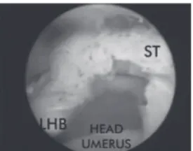
Fig. 1 - Lateral view of the left shoulder: extensive lesion of the supraspinatus with deficient rotator interval.
LHB: long head of the biceps; ST: supraspinatus tendon.
Introduction
The term “extensive rotator cuff injury” has been widely used to identify lesions that are particularly difficult to repair and are thus correlated with an uncertain prognosis.1,2 Cofield3
defined these injuries as complete tendon ruptures greater than or equal to 5 cm of its diameter, while Zumstein et al.4
defined extensive lesions as those that were complete and compromised two or more tendons that made up the rotator cuff.
Repairing chronic extensive rotator cuff tears is a challenge even for the most experienced shoulder surgeons. These tears are generally associated with atrophy of the musculature of the rotator cuff, with retraction and loss of mobility of the tendon, which greatly increases the difficulty in repairing it.2,5
Better knowledge of the injury patterns and advances in the quality and design of materials, along with improvements in surgical techniques, have made it possible to repair extensive tears by means of arthroscopy.6-10 In the case of extensive
tears of U-shaped pattern, with retraction of the supraspinatus tendon, without mobility and with deficiency of the rotator interval, a procedure to converge the margins of the posterior portion of the rotator cuff using the tendon of the long head of the biceps brachii muscle may be a good repair option.8
This study, conducted by the Shoulder and Elbow Group of the ABC Medical School (Faculdade de Medicina do ABC), had the aim of presenting the results obtained through this surgical technique, in treating extensive rotator cuff injuries.
Materials and methods
Between October 2003 and December 2007, 53 patients with extensive rotator cuff tears underwent arthroscopic surgical treatment performed by our group.
For inclusion in this study, patients in whom the supraspinatus tendon had retracted and lacked mobility, and in whom the rotator interval was deficient, were selected. The repair was performed using the margin convergence technique, with suturing of the posterior portion of the rotator cuff using the tendon of the long head of the biceps brachii muscle. Patients were excluded if they failed to respond to the request to come for a reassessment or if they had not yet completed 24 months of postoperative follow-up. Thus, 20 patients were evaluated: 16 women (80%) and four men (20%), of mean age 58.95 years (range: 42 to 75 years); 18 patients were right-handed and two were left-right-handed, and the lesion had occurred in the dominant limb in 16 patients (80%).
The preoperative joint mobility was assessed by means of the parameters described by Hawkins and Bokos.11 The
maximum elevation ranged from 60° to 160°, with a mean of 117.62°; the lateral rotation was from 20 to 80°, with a mean of 47.38°; and the medial rotation, which was evaluated according to the vertebral level that the patient was able to reach with the thumb, ranged from the ipsilateral greater trochanter to T7, with a mean of L2.
All the patients were evaluated using the UCLA score12
before the surgery, and the mean score was found to be
15.05 (range: 10-24). Magnetic resonance imaging was also performed preoperatively, and this showed fatty muscle degeneration of the supra and infraspinatus muscles, as assessed using Goutalier’s classification,5 with means of 2.9
and 2.4, respectively.
After the operation, the patients were reassessed clinically using the UCLA score12 and their joint mobility was reassessed
by means of the parameters described by Hawkins and Bokos.11
The statistical analysis was performed using the SPSS software (Statistical Package for the Social Sciences), version 17.0. The Wilcoxon signed rank test was used to investigate possible differences between the pre and postoperative UCLA scores and in relation to joint mobility. Spearman’s correlation analysis was used to ascertain the degree of correlation between the variables of interest (postoperative UCLA score, fatty degeneration of the supra and infraspinatus muscles before the operation, and re-rupture). We used the significance level of 5% (0.05) for applying the statistical tests.
Surgical technique
The patients underwent the surgical procedure in the “deckchair” position, under general anesthesia associated with brachial plexus block. The arthroscopic procedure began with an inspection of the joint using an optical device introduced through the posterior portal. The stability of the tendon of the long head of the biceps brachii muscle was tested by evaluating the competence of the medial pulley. In cases in which the upper portion of the subscapular tendon was torn, the lesser tubercle was repaired using an anchor and suture stitches, done by means of articular viewing. Going into the subacromial space, the rotator cuff was repaired by advancing the posterior portion of the lesion to the tendon of the long head of the biceps brachii. The repair was done using tendon-to-tendon stitches. Subsequently, this combination was reattached to the greater tubercle using anchors. In this manner, tenodesis of the long head of the biceps was performed without tenotomy at its origin. At the end of the procedure, the repair was also verified by means of articular viewing (Figs. 1-4).
UCLA n Mean Min Max Significance (p)
Pre 20 15.05 10 24
< 0.001
Post 20 28.95 14 35
Max: maximum; Min: minimum; Post: postoperative; Pre: preoperative.
Table 1 - Evaluation using the UCLA score before and after the operation.
Fig. 2 - Advancing of the posterior margin of the lesion to the long head of the biceps.
LHB: long head of the biceps; ST: supraspinatus tendon.
Fig. 3 - Repair of the supraspinatus at the long head of the biceps.
LHB: long head of the biceps; ST: supraspinatus tendon.
Fig. 4 - Final appearance of the repair, inserted into the greater tubercle.
LHB: long head of the biceps; ST: supraspinatus tendon.
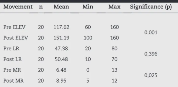
The mean postoperative length of follow-up among the patients in this study was 34 months, with a range from 24 to 60 months. The range of motion found in the reassessment was 151.19° (range: 100° to 160°) for elevation, 50.48° (range: 10° to 70°) for lateral rotation and T12 (range: L3 to T8) for medial rotation. For the statistical analysis on the medial rotation, we converted the vertebral level into a numerical scale from the trochanter to T7, represented as 0 to 13. The improvement in elevation, of 34 degrees (117° to 151.19°) was statistically significant (p = 0.001), as was the improvement in medial rotation of two vertebral levels (L2 to T12), with p = 0.025 (Table 2).
We evaluated the correlation between the results obtained using the postoperative UCLA score and the results from Goutalier’s classification on the supra and infraspinatus muscles before the operation, but we did not find any statistically significant difference (p = 0.829/0.410) (Table 3).
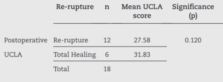
Out of the 20 cases included in this study, 18 underwent postoperative magnetic resonance imaging on the shoulder, 12 months after the surgery, which showed that six cases had healed completely (33.33%) and 12 had become torn again (66.66%). Two of the patients did not undergo the examination, which had been requested. Out of the 12 patients with renewed tears, only four cases presented any clinical signs of this. We evaluated the relationship between renewed tears and the postoperative UCLA score, but found that it did not have statistical significance (p = 0.120), probably because the sample was not representative (Table 4).
Six patients (30%) reported having acromioclavicular pain and underwent the Munford procedure during the arthroscopic repair.
Results
The postoperative evaluation using the UCLA score was 28.95 points, with a range from 14 to 35 points. There was an elevation of 13.9 points in relation to the preoperative levels (p < 0.001) (Table 1). Among the 20 patients, six had excellent results (30%), nine good (45%), three fair (15%) and two poor (10%).
Movement n Mean Min Max Significance (p)
Pre ELEV 20 117.62 60 160
0.001 Post ELEV 20 151.19 100 160
Pre LR 20 47.38 20 80
0.396
Post LR 20 50.48 10 70
Pre MR 20 6.48 0 13
0,025
Post MR 20 8.95 5 12
ELEV: elevation; LR: lateral rotation; Max: maximum; Min: minimum; MR: medial rotation; Post: postoperative; Pre: preoperative.
Table 2 - Relationship between preoperative and postoperative mobility.
Goutalier Significance (p) Correlation
coefficient for postoperative UCLA
Supra 0.829 0.052
Infra 0.41 -0.195
partial repairs in the literature. Burkhart et al.18 reported that
57% of their results were satisfactory, from partial repairs in 14 patients with extensive lesions, while Duralde and Bair19
found that 67% of their results were satisfactory, from partial repairs in 24 patients.
Partial repair has been shown to be a good treatment option for extensive rotator cuff injuries when complete anatomical repair of the lesion is not possible. Comparing the results from partial repair with those from debridement, for treating extensive lesions, partial repair has been shown to be superior. Berth et al.13 recently randomized 42 patients with extensive
lesions for treatment either with debridement or with partial repair and found that shoulder function was significantly better with partial repair.
Although surgical repair of extensive rotator cuff lesions provides good clinical results, a high rate of renewed rupture has been described in the literature.2,4,10,20-22
Gerber et al. showed improvements in shoulder function after open repair of extensive rotator cuff injuries, even though there was a 34% re-rupture rate.2 Galatz et al.20 reported that
17 cases out of 18 arthroscopic repairs on the rotator cuff presented renewed rupture but, despite this, they obtained excellent improvements in pain and shoulder function in their patients after 12 months of follow-up. They noted deterioration of the results after 24 months, but after this period 66.6% of the results were still satisfactory.
Like in the literature on repaired to extensive rotator cuff lesions,1,2,4,20-22 our rate of renewed rupture was high, affecting
66.6% (12 out of 18 cases), but even so, we obtained satisfactory clinical results (UCLA > 27 points) in eight of these 12 cases (66.6%). Although our sample was not representative, we did not find any statistically significant differences between the clinical results assessed using the postoperative UCLA scale and the re-rupture rate, or between UCLA and Goutalier’s classification.
Conclusion
In this study on cases of extensive rotator cuff injury, with a retracted supraspinatus tendon and a deficient rotator interval, repair of the posterior portion of the rotator cuff, with inclusion of the long head of the biceps, was shown to be a good option.
Conflicts of interest
The authors declare that there was no conflict of interests in conducting this study.
R E F E R E N C E S
1. Miyazaki NA, Fregoneze M, Santos PD, Silva LA, Pinto ECMM, Ortiz RT, et al. Lesões extensas do manguito rotador: avaliação dos resultados do reparo artroscópico. Rev Bras Ortop. 2009;44(2):148-52.
2. Gerber C, Fuchs B, Hodler J. The results of repair of massive tears of rotator cuff. J Bone Joint Surg Am. 2000;82(4):505-15.
Table 4 - Relação entre UCLA no período pós-operatório com re-ruptura.
Discussion
Primary anatomical repair of the rotator cuff is not always possible in cases of extensive lesions. Factors such as lesion size, fatty muscle degeneration, large retractions and tendon adherence have a great influence on the repair.1,10 In such
cases, the options for surgical treatment include debridement, partial repair, tenotomy of the biceps, tendon transfers and use of grafts (fascia lata, long head of the biceps or synthetic materials).10,13
Use of the tendon of the long head of the biceps has also emerged as a repair option for rotator cuff tears. Neviaser described use of the articular portion of the biceps, cut like an open book to aid in repairing rotator cuff lesions, in cases in which the edges of the lesion cannot be repaired primarily.14
Wolfgang described use of the proximal portion of the tendon the long head of the biceps as a pedicled graft for repairing cuff tears. This author believed that by suturing the proximal portion into the lesion and keeping it inserted in the glenoid, a blood supply would be taken to the repair and its distal part would undergo autotenodesis.15 Bigliani described use of this
tendon to facilitate repairs on extensive lesions.16 In 1997,
Snyder described inclusion of the tendon of the long head of the biceps in suturing the rotator cuff, without tenotomizing it, for cases of large retraction of the supraspinatus with a deficient rotator cuff interval.9
Tendon mobility is a decisive factor in choosing the treatment method, and this is essential in order to be able to completely or partially repair rotator cuff tears.8 Mobilization
of the anterior part of the retracted rotator cuff can be achieved by means of the technique of sliding the rotator interval, which was described by Tauro17, and by means of releasing
the adherences that surround the supraspinatus tendon. In cases in which the supraspinatus does not have mobility and the rotator interval is deficient, closure of the lesion seems to be impossible. In these cases, the technique of repairing the posterior portion of the rotator cuff tear together with the tendon of the long head of the biceps brachii, as described by Snyder, seems to be a solution.9 In 2004, Richards and Burkhart8
published this technique as a repair option in such cases, but without showing any sample.
We used this technique on 20 patients and achieved six excellent and nine good results, with significant improvement in shoulder function. Thus, 75% of the results were satisfactory. This was a good result in comparison with the results from
Re-rupture n Mean UCLA score
Significance (p)
Postoperative Re-rupture 12 27.58 0.120
UCLA Total Healing 6 31.83
3. Cofield RH. Current concepts review. Rotator cuff disease of the shoulder. J Bone and Joint Surg. 1985;67(7):974-9.
4. Zumstein MA, Jost B, Hempel J, Hodler J, Gerber C. The clinical and structural long-term results of open repair of massive tears of the rotator cuff. J Bone Joint Surg Am. 2008;(90):2423-31.
5. Goutallier D, Postel JM, Bernageau J, Lavau L, Voisin MC. Fatty muscle degeneration in cuff ruptures. Pre and postoperative evaluation by CT scan. Clin Orthop Relat Res. 1994;(304):78-83. 6. Burkhart SS, Athanasiou KA, Wirth MA. Margin convergence:
a method of reducing strain in massive rotator cuff tears. Arthroscopy. 1996;(12):335-8.
7. Burkhart SS. Arthroscopic treatment of massive rotator cuff tears. Clin Orthop Relat Res. 2001;(390):107-18.
8. Richards DP, Burkhart SS. Margin convergence of the posterior rotator cuff to the biceps tendon. Arthroscopy. 2004;20(7):771-5.
9. Snyder SJ. Shoulder arthroscopy. 2nd ed. Philadelphia: Lippincott Williams e Wilkins; 2006.
10. Bedi A, Dines J, Warren RF, Dines DM. Massive tears of the rotator cuff: current concepts review. J Bone Joint Surg Am. 2010;92(9):1894-908.
11. Hawkins RJ, Bokos DJ. Clinical evaluation of shoulder problems. In: Rockwood CA Jr, Matsen FA 3rd. The shoulder. 2nd ed. Philadelphia: Saunders; 1998. p.175-80.
12. Oku EC, Andrade AP, Stadiniky SP, Carrera EF, Tellini GG. Tradução e adaptação cultural do Modified – University of California at Los Angeles Shoulder Rating Scale para a língua portuguesa. Rev Bras Reumatol. 2006;46(4):246-52.
13. Berth A, Neumann W, Awiszus F, Pap G. Massive rotator cuff tears: functional outcome after debridement or arthroscopic partial repair. J Orthopaed Traumatol. 2010;11(1):13-20. 14. Neviaser JS. Ruptures of the rotator cuff. Arch Surg.
1971;102(5):483-5.
15. Wolfgang GL. Surgical repair of tears of the rotator cuff of the shoulder. J Bone Joint Surg Am. 1974;56(1):14-25.
16. Bigliani LU, Cordasco FA, Mcilveen SJ. Operative repair of massive rotator cuff tears: long-term results. J Shoulder Elbow Surg. 1992;1(3):120-30.
17. Tauro JC. Arthroscopic “interval slide” in the repair of large rotator cuff tears. Arthroscopy. 1999;15(5):527-30.
18. Burhart SA, Nottage WM, Ogilvie-Harris DJ, Kohn HS, Pachelli A. Partial repair of irreparable rotator cuff. Arthroscopy. 1994;10(4):363-70.
19. Duralde XA, Bair B. Massive rotator cuff tears: the result of partial rotator cuff repair. J Shoulder Elbow Surg. 2005;14(2):121-7.
20. Galatz LM, Ball CM, Teefey SA, Middleton WD, Yamaguchi K. The outcome and repair integrity of completely arthroscopically repaires large and massive rotador cuff tears. J Bone Joint Surg Am. 2004;86(2):219-24.
21. Miller BS, Downie BK, Kohen RB, Kijek T, Lesniak B, Jacobson JA, et al. When do rotator cuff repairs fail? Serial ultrasound examination after arthroscopic repair of large and massive rotator cuff tears. Am J Sports Med. 2011;39(10):2064-70. 22. Yoo JC, Ahn JH, Koh KH, Lim KS. Rotator cuff integrity after