Original article
Evaluation of the lumbar
multiidus
in rowers
during spinal stabilization exercise
Joseani Ceccato
Federal University of Health Sciences of Porto Alegre, Brazil
Jeam Marcel Geremia Alexandre Mayer Raquel de Oliveira Lupion
Marco Aurélio Vaz
Federal University of Rio Grande do Sul, Porto Alegre, Brazil
Abstract—Lumbar stabilization is important in high performance rowing due to the high incidence of low back pain. The purpose of this study was to evaluate the lumbar stabilizers muscles performance during an exercise of spinal
seg-mental stabilization and in lumbar multiidus muscle thickness in rowing athletes trained and untrained for this exercise.
Nine rowers trained with lumbar stabilization (TLS) and eight rowers without training (CON) participated in the study.
Lumbar stabilization performance and multiidus muscle thickness were measured during a maximal voluntary isometric
contraction. Lumbar stabilization performance was higher (p=0.015) in the TLS (mean 18.38 ± 8.00 mmHg) compared to the CON (9.31 ± 4.91 mmHg) group. Muscle thickness variation was higher (p=0.023) in the TLS (6.92% ± 3.98) compared to the CON (2.81% ± 1.40) group. Lumbar stabilization training is an eficient clinical tool to strengthen lumbar muscles and may help to prevent low back pain in rowers.
Keywords: spinal stabilization, multiidus, rowing, prevention
Resumo—“Avaliação dos multíidos lombares em atletas do remo durante exercício de estabilização vertebral.” A esta -bilização lombar é fundamental em esportes de alto rendimento como o remo, devido a elevada incidência de lombalgia. O objetivo do presente estudo foi avaliaro desempenho dos músculos estabilizadores lombares e a espessura do músculo
multíido lombar durante um exercício de estabilização segmentar vertebral em remadores. Nove remadores treinados
com exercícios de estabilização lombar (TLS) e oito remadores destreinados (CON) foram avaliados. O desempenho
dos músculos estabilizadores lombares e a espessura dos músculos multíidos foram mensurados durante uma contração
voluntária isométrica máxima. O desempenho foi maior (p=0,015) no grupo TLS (18,38±8,00 mmHg) quando comparado ao grupo CON (9,31±4,91 mmHg). A variação da espessura muscular foi maior (p=0,023) no grupo TLS (6,92%±3.98) quando comparado com o grupo CON (2,81%±1,40). Treinamento de estabilização lombar é uma ferramenta clínica
eiciente para fortalecimento da musculatura lombar e pode auxiliar na prevenção de lombalgia em remadores. Palavras-chave: estabilização vertebral, multíidos, remo, prevenção
Resumen—“Evaluación del multiidus lumbar en los atletas de remo durante el ejercicio en la estabilización de la columna
vertebral.” La estabilización lumbar es fundamental en la práctica de deportes como el remo de alto rendimiento, donde la
lumbalgia aparece con frecuencia. El objetivo de este estudio fue evaluar el rendimiento de los músculos estabilizadores
lumbares, así como el espesor del músculo multiido lumbar, durante un ejercicio de estabilización espinal segmentaria
lumbar (una contracción isométrica voluntaria máxima) en atletas de remo. Fueron evaluados nueve remadores sometidos a entrenamiento con ejercicios de estabilización lumbar (TLS) y ocho sin este adiestramiento (CON). El rendimiento fue mayor (p=0,015) en el grupo TLS (18,38±8,00 mmHg) e comparación con el grupo CON (9,31±4,91 mmHg). Va-riación del espesor muscular fue mayor (p=0,023) en el grupo TLS (6,92%±3,98) en comparación con el grupo CON (2,81%±1,40). El entrenamiento lumbar es una herramienta clínica eiciente para el fortalecimiento de la musculatura lumbar e puede auxiliar en la prevención de lumbalgias en remadores.
Lumbar multiidus evaluation
Introduction
High performance rowing is a sport with a high level of overload to the trunk, and the cause of lower back pain that frequently appears as the second most frequent injury complaint by rowers (Comerford & Mottram, 2001). During rowing, the force mag-nitude at the lumbar spine is substantial. In 70% of the training
cycle, rowers remain with the trunk lexed. The combination of this lexed position with compressive forces at the vertebrae has been identiied as one of the likely mechanisms for lumbar spine
injury (Rumball, Lebrun, Di Ciacca, & Orlando, 2005). Several factors can contribute to overload and low back pain in competitive rower athletes (Caldwell, Mcnair, & William’s, 2003; Reid & Mcnair, 2000; Rumball et al., 2005; Teitz, O’Ka-ne, & Lind, 2002). Spine extensors muscle fatigue, usually associated with large volume and training intensity, reduces the
contractility of muscle ibers, which can lead to an excessive lumbar lexion and increased stress on spinal structures.
Thus, spinal stability is necessary to reduce stress and de-pends on integrating two muscle systems: local and global. The global muscular system (consisting of the rectus abdominal, external obliques and the thoracic part of iliocostalis lumbar) when activated, leads to an overall stability of the trunk, but
is not able to directly inluence speciic spinal segments. The local muscle system (comprised of the multiidus, psoas major,
quadratus lumborum, lumbar portion of the lumbar iliocostalis,
longissimus, transversus abdominis, posterior ibers of the dia -phragm and internal oblique) acts directly on the lumbar spine and is responsible for providing segmental stability and directly control the lumbar segments (Bergmark, 1989; Panjabi, 1989).
These stabilizing muscles should be very it, especially in
athletes with overload at the spine. In clinical practice of sports physiotherapy, co-contraction of synergistic muscle groups
(trans-versus abdominis and multiidus) can be evaluated and followed
via functional tests such as, abdominal pressure or pressure biofe-edback unit (PBU) (Garnier et al., 2009; Mills, Taunton, & Mills, 2005). The PBU test is a reliable and valid clinical instrument to assess deep abdominal muscle function and/or performance during an abdominal wall contraction (Cynn, Oh, Kwon, & Yi, 2006; Garnier et al., 2009; Mills et al., 2005; Richardson, Jull, Toppenberg, & Comerford, 1992; Storheim, Pederstad, & Jahn-sen, 2002). Mills et al. (2005) used the PBU test to evaluate the effect of a 10-week training program on lumbo-pelvic stability in volleyball and basketball female athletes. The athletes presen-ted improvements on the lumbo-pelvic stability after training, showing evidence that the PBU test is an important and effective tool in the evaluation of lumbo-pelvic stability.
Studies (Costa, Maher, Latimer, Hodges, & Shirley, 2009; Dickx, Cagnie, Parlevliet, Lavens, & Danneels, 2010; Hall, Tsao, Macdonald, Coppieters, & Hodges, 2009; McEvoy, Cowling, Fulton, & Williams, 2008) showed evidence of
hypo-trophy or ahypo-trophy of the transversus abdominis and multiidus
muscles on the side of low back pain. These studies report a decrease in contractile performance of these muscles, emphasi-zing the importance of strengthening of deep abdominal muscles (Koppenhaver, Hebert, Parent, & Fritz, 2010; Mcgregor, Bull & Byng-Maddick, 2004; Mills et al., 2005).
Assuming that low back pain arises from a poor spinal seg-mental stabilization (Dickx et al. 2010; Hides, Stanton, Mendis, & Sexton, 2011), the aim of this study was to evaluate the changes
in PBU and the lumbar multiidus muscle thickness during a
segmental spinal stabilization exercise in trained and untrained
rowing athletes. The multiidus muscle group was chosen due to
the following reasons: (1) the possibility of simultaneous measu-rements of ultrasound and abdominal pressure with subjects in the prone position; (2) the ease of access to these muscles (posterior region) (Hides, Richardson, & Jull, 1998); (3) the synergism found
between strengthening of transversus abdominis and multiidus
muscles (Lehman, Story, & Mabee, 2005).
Therefore, our expectation was that subjects trained in
lum-bar stabilization (TLS) would have a larger change in multiidus
muscle thickness during the maximal voluntary contraction in the PBU test as an adaptation to the strength training of these synergistic muscles responsible for spinal stabilization compa-red to the untrained (CON) group.
Methods
Study design
The present study is an ex post facto and correlational study. All procedures were approved by the ethics committee and par-ticipants signed an informed consent to participate in the study.
Participants
The population of the present studied included 80% of the population of rowing athletes in junior and youth categories of Rio Grande do Sul State. Within this population, sixteen healthy male rower athletes (with no history of injury, lumbar spondylolisthesis, spondylolysis, lumbar disc herniations or protrusions, or any previous surgical treatment) were evaluated. The study included only rowing athletes with a minimum of two years of training.
The athletes were divided into two groups: the group trained in lumbar stabilization (TLS) consisted of eight athletes undergoing weekly training sessions to strengthen deep abdominal and back muscles (transversus abdominis and
multiidus) for more than two years, whereas the non-trained
or control group (CON) consisted of eight untrained athletes in lumbar stabilization. The study included only athletes who were enrolled on daily training (3 to 4 hours of rowing a day) and participating in competitions.
Segmental spinal stabilization training
standing positions. Muscle contractions were performed with emphasis on the expiratory phase of the respiratory cycle. The
athlete was asked to inspire by inlating the abdomen and during
the exhale, to tighten the abdomen, pulling the navel towards the back, aiming at the formation of a pelvic belt (Richardson et al., 1992; Richardson & Jull, 1995). Athletes performed four different exercises per session, with 20 repetitions from each
exercise, one-minute intervals between trials and ive-minute
intervals between exercises. Muscle contractions were empha-sized to be performed with emphasis on the expiratory phase of the respiratory cycle. The sessions began with stabilization exercises in the prone position, progressing to sitting and four
contact support positions (see below). In the irst exercise sub -jects laid prone in the same position depicted in Figure 1B. In
the second exercise subjects laid on their back, with knees lexed
and upper limbs resting besides the trunk. In the third exercise
subjects sat on the loor with hips abducted, knees lexed and feet plants facing each other. For the irst three exercises each
trial consisted of a maximal voluntary isometric contraction of the abdominal wall muscles lasting about ten seconds with four seconds in the inhalation phase and six seconds for the
expiratory phase. In the fourth exercise subjects lexed their
shoulders and hips, while maintaining the elbows extended and
wrists hyper-extended and knees lexed in the prone position,
supported in their hand palms and knees (four contacts prone position). The exercise consisted to contract the abdominal wall
muscles while simultaneously lexing the shoulder (with elbow
extended) and extending the contralateral hip and the knee. Subjects performed these movements alternating sides, with 10 repetitions for each side. All sessions were supervised by the same physiotherapist, and the unwanted movements during the exercises were observed and corrected.
Lumbar stabilizer muscles performance
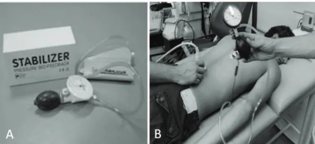
The lumbar stabilizer muscles performance was obtained by means of the PBU test (Stabilizer, Chattanooga Group, Australia; Figure 1A). The athletes were positioned prone
with the Stabilizer under the abdomen, in the region located between the anterior superior iliac spines and centered
rela-tive to the navel (Figure 1B). The Stabilizer was inlated to a
pressure of 70mmHg and the athletes were instructed to inhale and, during expiration, were asked to contract the abdomen as much as possible as if they were pulling the navel towards the back. During the exercise, the reduction in abdominal pressure over the Stabilizer was registered during four seconds within 10-second period (Garnier et al., 2009). The larger the reduction in pressure on the Stabilizer was considered the capacity of the athlete to produce spinal segmental stabilization (Garnier et al., 2009; Richardson & Jull, 1995).
In order to avoid any interference on the results during the voluntary contraction of the abdominal wall muscles, athletes were asked to: (1) empty their bladder before they started the test; (2) not consuming food at least two hours before testing; (3) not perform any abdominal strengthening exercise on the day of the test. This methodology was similar to that of a previous study (Storheim et al., 2002).
Muscle thickness variation
Simultaneously with the variation in abdominalpressure
measurement, the multiidus muscle thickness was assessed.
The linear array probe (sampling frequency = 7.5 MHz) was positioned longitudinally and unilaterally (Figure 1B) following the parameters of the study by Van, Hides and Richardson (2006). The thickness was measured on the dominant side of each athlete, at the L4-L5 level, with an ultrasound machine (Aloka SSD 4000, Tokyo, Japan, sampling frequency = 45 Hz).
After palpation, the spinous processes of L4 and L5 were marked on the skin. The athlete was positioned prone with the head relaxed to one side (left or right). Next, the probe was
positioned in the approximate direction of the muscle ibers
and laterally to the spinous processes. The measures were made at rest and during maximal voluntary contraction of the abdominal wall muscles during the stabilization exercise described above.
Lumbar multiidus evaluation
Data analysis
The abdominal pressure was obtained directly by reading the values on the Stabilizer display. To obtain the values of muscle thickness, the evaluation followed the same
methodo-logy described elsewhere (Van et al., 2006). The multiidus
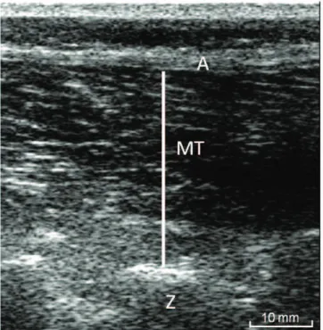
thickness (Figure 2) was measured from the tip of the L4-5
zigapophyseal joint to the superior border of the supericial aponeurosis (superior border of the multiidus muscle). The
equation used to calculate the variation in muscle thickness was as follows: [(thickness in contraction - thickness at rest) / thickness at rest x 100%].
The Shapiro-Wilk test was used to test the normality of the data. Age, height, body mass, abdominal pressure and changes in
multiidus muscle thickness were compared between the groups
(trained and untrained) using an independent Student t-test. Data were analyzed using the Sigma Plot Program (version 11) with
a signiicance level set to α=0.05.
Results
Athletes of the CON group had a mean age of 18.38 ± 5.10 years, height of 1.78 ± 0.6 m, body mass of 75.89 ± 7.32 kg and body mass index (BMI) of 23.79 ± 1.35 kg/m2. The TLS group had a mean age of 16.50 ± 2.67 years, height of 1.83 ± 0.62 m, body mass of 71.88 ± 8.00 kg and BMI of 21.44 ± 1.76 kg/m2. No difference was found in age (p=0.372), height (p=0.139) and body mass (p=0.313). The BMI was higher (p=0.010) in the CON group compared to TLS group.
Table 1 shows the PBU and the variation in lumbar mul-tifidus muscle thickness values of both groups. The PBU
values were higher in the TLS group compared to CON group (p=0.015). The TLS group showed a mean variation exceeding 50% of that of the CON group’s average. The variation in lumbar
multiidus muscle thickness was higher (p=0.023) in the TLS compared to the CON group.
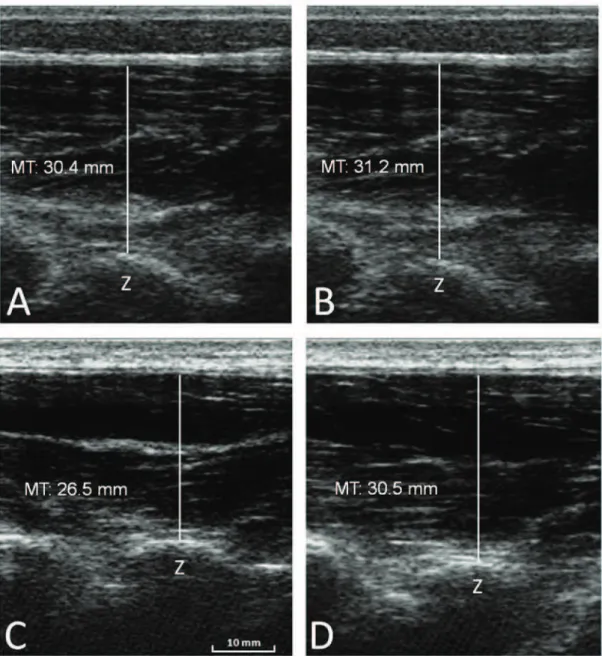
Figure 3 illustrates the multiidus thickness obtained at rest
(Figures 3A and 3C) and during the maximal abdominal volun-tary isometric contraction during the PBU test of an individual from the CON group (with small variations in thickness) and from an athlete of TLS group (with high thickness variation; Figures 3B and 3D), respectively.
Discussion
Spinal functional stability is dependent of both the local and global muscle integration systems (Bergmark, 1989). When spinal dysfunctions are present, there is a combination of normal movement restriction and compensations to maintain spinal stability. Training of the local stabilizing muscles is necessary to maintain proper alignment of the spine and to establish a stable base of support for body movement (Comerford & Mottram, 2001).
In sports such as rowing, there is a high demand of the trunk, and training the spinal stabilizing muscles might help in decreasing the risk for spinal injury. Although global mus-cle strengthening is important, a strong pelvic belt should be emphasized in rowing athletes to protect their spine during high performance training, as it may help to avoid spinal pain and/or injury.
A decrease in contractile performance of the multiidus mus -cle has been reported in patients with low back pain (Barker et al., 1989; Costa et al., 2009; Lee et al., 2006). If so, subjects with
low back pain should have more dificulty in contracting this
muscle, and therefore should present a smaller muscle thickness and also a smaller capacity to produce structural changes (i.e. changes in muscle thickness) when going from the resting state to a maximal effort condition.
A similar reasoning could be used when comparing trained and untrained subjects in spinal functional stability. Untrained subjects should present a smaller capacity to change muscle thickness from rest to full effort. The higher variation (6.31%)
of lumbar multiidus muscle thickness in TLS agrees with
this idea, and is similar to the results from a previous study (Wilke, Wolf, Claes, Arand, & Wiesend, 1995) which reported
that the lumbar multiidus promoted more than two thirds of
the rigidity in the L4-L5 segment. The greatest variation in Figure 2. Ultrasound image of the multiidus muscle thickness. Z= L4-5
zygapophyseal joint; A= aponeurosis; MT: multiidus muscle thickness.
CON (n=8)
TLS (n=8)
p
value
PBU (mmHg) 9.31 ± 4.91 18.38 ± 8.00 0.015
ΔMT (%) 2.81 ± 1.40 6.92 ± 3.98 0.023
from injury. However, due to the few studies that we were
able to ind in this area with a population of rowing athletes,
further longitudinal studies are necessary in order to better evaluate if strengthening training programs of abdominal muscles may really act effectively in the prevention of back injury in rowing athletes.
Conclusion
Rowing athletes trained on segmental spinal stabilization exercises showed great variation in abdominal pressure and in
multiidus thickness during maximal effort. These results suggest
that stabilization exercises for low back pain in young rowing athletes may help to decrease the injury risk due to sports training thereby reducing the incidence of low back pain in rowers. abdominal pressure, observed in TLS group (more than 50%
higher compared to the CON group), is also evidence that trained athletes in lumbar stabilization have a greater capacity
of lumbar spine stabilization. The above results conirmed
our initial hypothesis that rowers participating on a low back pain prevention program would show a greater variation
in abdominal pressure and a greater variation in multiidus
thickness during spinal stabilization exercise compared to untrained athletes.
These results may help in the understanding of spinal functional stabilization. In other words, the strengthening of
the transversus abdominis and multiidus muscles increases the
muscular thickness, improving the lumbar stabilization (Van et al., 2006). In addition, the exercises used in our strengthening program may help to avoid the incidence of excessive loads on skeletal tissues in this region, protecting the lumbar region
Lumbar multiidus evaluation
References
Barker, K. L., Shamley, D. R., & Jackson, D. (2004). Changes in the cross-sectional area of multiidus and psoas in patients with uni -lateral back pain. Spine, 29(22), 515-519. Retrieved from http:// journals.lww.com/spinejournal/toc/2004/11150
Bergmark, A. (1989). Stability of the lumbar spine. A study in me-chanical engineering. Acta Orthopedic Scandinavic Supplement, 60(230), 5-54. Retrieved from http://www.ncbi.nlm.nih.gov/ pubmed/2658468
Caldwell, J. S., Mcnair, J. P., & William’s, M. (2003). The effects of repetitive motion on lumbar lexion and rector spinae muscle activity in rowers. Clinical Biomechanics, 18(8), 704-711. Re-trieved from: http://www.sciencedirect.com/science/article/pii/ S0268003303001177
Comerford, M.J. & Mottram, S.L. (2001). Functional stability re-trai-ning: principles and strategies for managing mechanical dysfunc-tion. Manual Therapy, 6(1), 3-14. Retrieved from: http://www. ncbi.nlm.nih.gov/pubmed/11243904
Costa, L. O. P., Maher, C. G., Latimer, J., Hodges, P. W., & Shirley, D. (2009). An investigation of the reproducibility of ultrasound measures of abdominal muscle activation in patients with chronic non-speciic low back pain. European Spine Journal, 18(7), 1059-1065. Retrieved from: http://www.ncbi.nlm.nih.gov/pmc/articles/PMC2899591 Cynn, H. S., Oh, J. S., Kwon, O. Y., Yi, & C. H. (2006). Effects of
lumbar stabilization using a pressure biofeedback unit on muscle activity and lateral pelvic tilt during hip abduction in sidelying. Ar-chives of Physical Medicine and Rehabilitation, 87(11), 1454-1458. Retrieved from: http://www.ncbi.nlm.nih.gov/pubmed/17084119 Dickx, N., Cagnie, B., Parlevliet, T., Lavens, A., & Danneels, L (2010).
The effect of unilateral muscle pain on recruitment of the lumbar multiidus during automatic contraction. An experimental pain study. Manual Therapy, 15(4), 364-369. Retrieved from http:// www.ncbi.nlm.nih.gov/pubmed/20207187
Garnier, K., Köveker K., Rackwitz, B., Kober, U., Wilke, S., Ewert, T., & Stucki, G. (2009). Reliability of a test measuring transverses abdominis muscle recruitment with a pressure biofeedback unit. Physiotherapy, 95(1), 8-14. Retrieved from http://www.ncbi.nlm. nih.gov/pubmed/19627680
Hall, L., Tsao, H., Macdonald, D., Coppieters, M., & Hodges, P. W. (2009). Immediate effects of co-contraction training on motor control of the trunk muscles in people with recurrent low back pain. Journal of Electromyography and Kinesiology, 19(5), 763-773. Retrieved from http://www.sciencedirect.com/science/article/ pii/S1050641107001708
Hides, J. A., Richardson, C. A., Jull, G. A. (1998). Use of real-time ultrasound imagining for feedback in rehabilitation. Manual The-rapy, 3(3), 125-131. Retrieved from http://www.sciencedirect.com/ science/article/pii/S1356689X98800027
Hides, J., Stanton, W., Mendis, M., & Sexton, M. (2011). The rela-tionship of transversus abdominis and lumbar multiidus clinical muscle tests in patients with chronic low back pain. Manual Therapy, 16, 573-7
Koppenhaver, S. L., Hebert, J. J., Parent, E. C., & Fritz, J.M. (2009). Rehabilitative ultrasound imagining is a valid measure of trunk muscle size and activation during most isometric sub–maximal contractions: a systematic review. Australian Journal of Physio-therapy, 55(3), 153-169. Retrieved from http://www.ncbi.nlm.nih. gov/pubmed/19681737
Lee, S., Chan, C.K., Lam, T., Lam, C., Lau, N., Lau, R. W., & Chan, S. (2006). Relationship between low back pain and lumbar multiidus size at different postures. Spine, 31(19), 2258-2262. Retrieved from http://journals.lww.com/spinejournal/toc/2006/09010
Lehman, G. J., Story, S., & Mabee, R. (2005). Inluence of static lumbar lexion on the trunk muscles response to sudden arm movements. Chiropractic and Osteopathy, 13(23),1-5. Retrieved from http:// www.ncbi.nlm.nih.gov/pmc/articles/PMC1315330
Mcevoy, P. M., Cowling, A. J., Fulton, I. J., & Williams, M. T. (2008) Transversus abdominis: changes in thickness during an incremen-tal upper limb exercise test. Physiotherapy Theory and Practice, 24(4), 265-273. Retrieved from http://informahealthcare.com/doi/ abs/10.1080/09593980701773340
Mcgregor, A. H., Bull, A. M., & Byng-Maddick, R. (2004). A compa-rison of rowing technique at different stroke rates: a description of sequencing, force production and kinematics. International Journal of Sports Medicine, 25(6), 465-470. Retrieved from https://www. thieme-connect.com/DOI/DOI?10.1055/s-2004-820936
Mills, J. D., Taunton, J. E., & Mills, W. A. (2005). The effect of a 10-week training regimen on lumbo-pelvic stability and athletic performance in female athletes: a randomized-controlled trial. Physical Therapy in Sport, 6(2), 60-66. Retrieved from http:// www.sciencedirect.com/science/article/pii/S1466853X05000386 Panjabi, M., Abumi, K., Duranceau, J., & Oxland, T. (1989). Spinal
stability and intersegmental muscle forces. A biomechanical model. Spine, 14(2), 194-200. Retrieved from http://www.ncbi.nlm.nih. gov/pubmed/2922640
Reid, D. & Mcnair, P. (2000). Factors contributing to low back pain in rowers. British Journal of Sports Medicine, 34(5), 321-322. Retrieved from http://www.ncbi.nlm.nih.gov/pubmed/11049136 Richardson, C. A., Jull, G. A., Toppenberg, R., & Comerford, M.
(1992). Techniques for active lumbar stabilization for spinal pro-tection: a pilot study. Australian Journal of Physiotherapy, 38(2), 105-112. Retrieved from http://ajp.physiotherapy.asn.au/AJP/ vol_38/2/AustJPhysiotherv38i2Richardson.pdf
Richardson, C. A. & Jull, G. A. (1995). Muscle Control – Pain Control. What exercises would you prescribe? Manual Therapy, 1(1), 2-10. Retrieved from http://www.sciencedirect.com/science/article/pii/ S1356689X85702433
Rumball, J. S., Lebrun, C. M., Di Ciacca, S. R., & Orlando, K. (2005). Rowing injuries. Sports Medicine, 35(6); 537-555. Retrieved from http://www.ncbi.nlm.nih.gov/pubmed/15974636
Storheim, K., Bo, K., Pederstad, O., & Jahnsen, R. (2002). Intra-tester reproducibility of pressure biofeedback in measurement of trans-verses abdominis function. Physiotherapy Research International, 7(4), 239-249. Retrieved from http://www.ncbi.nlm.nih.gov/ pubmed/12528579
Teitz, C., O’kane, J., Lind, K., Bonnie, K. L., & Hannain, J. A. (2002). Back pain in intercollegiate rowers. American Journal of Sports Medicine, 30(5), 674-679. Retrieved from http://ajs.sagepub.com/ content/30/5/674.abstract
Van, K., Hides, J. A., & Richardson, C.A. (2006). The use of real-time ultrasound imaging for biofeedback of lumbar multiidus muscle contraction in healthy subjects. Journal of Orthopaedic Sports Physical Therapy, 36(12), 920-925. Retrieved from http://www. ncbi.nlm.nih.gov/pubmed/17193869
Wilke, H. J., Wolf, S., Claes, L. E., Arand, M., & Wiesend, A. (1995). Stability increase of the lumbar spine with different muscle groups. A biomechanical in vitro study. Spine, 20(2), 192-198. Retrieved from http://www.ncbi.nlm.nih.gov/pubmed/7716624
Authors’ note
Jeam Marcel Geremia, M.Sc. is a Ph.D. student, Exercise Research La-boratory, Federal University of Rio Grande do Sul, Porto Alegre, Brazil.
Alexandre Mayer, M.Sc. is researcher at the Exercise Research Labo-ratory, Federal University of Rio Grande do Sul, Porto Alegre, Brazil.
Raquel de Oliveira Lupion, B.Sc., is a master’s student, Exercise Research Laboratory, Federal University of Rio Grande do Sul, Porto Alegre, Brazil.
Marco Aurélio Vaz, Ph.D., professor, is afiliated with the Exercise Research Laboratory, Federal University of Rio Grande do Sul, Porto Alegre, Brazil.
Corresponding author
Joseani Ceccato
Clube Grêmio Náutico União, Rua Quintino Bocaiúva, n°500, Porto Alegre 90440-050 RS, Brazil.
Phone: 55-51-3025 3929 E-mail: [email protected]
Acknowledgements
This study was supported by CNPq grant awarded to Marco A Vaz. The authors would like to acknowledge the Clube Grêmio Náutico União and the athletes for their cooperation.