Concurrent validity of the pressure biofeedback
unit and surface electromyography in measuring
transversus abdominis muscle activity in patients
with chronic nonspecific low back pain
Validade concorrente da unidade de biofeedback pressórico e eletromiografia
de superfície na mensuração da atividade muscular do transverso abdominal em
pacientes com dor lombar crônica inespecífica
Pedro O. P. Lima1, Rodrigo R. Oliveira1, Alberto G. Moura Filho2, Maria C. F. Raposo3, Leonardo O. P. Costa4, Glória E. C. Laurentino2
Abstract
Background: The Pressure biofeedback unit (PBU) is an assessment tool used in clinical practice and research aimed to indirectly analyze the transversus abdominis (TrA) muscle activity. The concurrent validity of the PBU in a clinically relevant sample is still unclear. Objective:
The purpose of this study was to evaluate the concurrent validity and diagnostic accuracy of the PBU in measuring TrA muscle activity in patients with chronic nonspecific low back pain. Method: This study was performed using a validation, cross-sectional design. Fifty patients with chronic nonspecific low back pain were recruited for this study. To test the concurrent validity both PBU measures (index test) and superficial electromyographic measures (reference-standard test) were compared and collected by a physical therapist in a series of voluntary contraction maneuvers of TrA muscle. Results: Participants were on average 22 years old, weighed 63.7 kilos, 1.70 meters height and mean low back pain duration was 1.9 years. It was observed a weak and non-significant Phi coefficient (r=0.2, p<0.20). With regards to diagnostic accuracy tests, our results suggest a low sensitivity (60%) and specificity (60%) of the PBU. The positive predictive value was high (0.8) and negative predictive value was low (0.2). Conclusions: Concurrent validity of the PBU in measuring TrA muscle activity in patients with chronic nonspecific low back pain is poor given the low correlation and diagnostic accuracy with superficial EMG.
Keywords: pressure biofeedback unit; low back pain; physical therapy.
Resumo
Contextualização: A Unidade de Biofeedback Pressórico (UBP) é uma ferramenta de avaliação usada na prática clínica e pesquisa
científica para analisar indiretamente a atividade muscular do transverso abdominal (TrA). A validade concorrente da UBP em uma amostra clinicamente relevante ainda não está esclarecida. Objetivo: Avaliar a validade concorrente e acurácia diagnóstica da UBP em mensurar a atividade muscular do TrA em pacientes com dor lombar crônica inespecífica. Método: Este estudo foi realizado usando um delineamento de validação. Cinquenta pacientes com dor lombar crônica inespecífica foram recrutados. Para testar a validade concorrente, ambas as medidas pressóricas (teste índice) e eletromiográficas superficiais (teste padrão de referência) foram comparadas e coletadas por um fisioterapeuta a partir de uma manobra de contração voluntária do músculo TrA. Resultados: Os participantes tinham em média 22 anos, 63,7 kg, 1,70 m de altura, e a duração média de dor lombar era de 1,9 ano. Observou-se um coeficiente Phi fraco e não significativo (r=0,2; p<0,20). Com relação aos testes de acurácia diagnóstica, os resultados sugerem uma baixa sensibilidade (60%) e especificidade (60%) da UBP. O valor preditivo positivo foi elevado (0,8), e o valor preditivo negativo foi baixo (0,2). Conclusões: A validade concorrente da UBP em mensurar a atividade muscular do TrA em pacientes com dor lombar crônica inespecífica é pobre, considerando a baixa correlação e acurácia diagnóstica com a EMG de superfície.
Palavras-chave: biofeedback pressórico; dor lombar; fisioterapia.
Received: 11/06/2011 – Revised: 03/26/2012 – Accepted: 05/18/2012
1 Physical Therapy Department, Universidade Federal do Ceará (UFC), Fortaleza, CE, Brazil 2 Physical Therapy Department, Universidade Federal do Pernambuco (UFPE), Recife (PE), Brazil 3 Department of Statistics, UFPE, Brazil
4 Master’s Program in Physical Therapy, Universidade da Cidade de São Paulo (UNICID), São Paulo, SP, Brazil
Introduction
Low back pain is considered as a major public health prob-lem and is associated with high socioeconomic costs, work
ab-senteeism and disability1,2. Usually, acute low back pain presents
a favorable prognosis with most patients returning to their usual
activities between 4 to 8 weeks, but recurrence is common3.
Ap-proximately 10–40% of acute low back pain patients become chronic and most of them (85%) are classiied as nonspeciic.
Patients who develop chronic nonspeciic low back pain (i.e.,
persistent pain and disability for more than three months) are responsible for more than 80% of health care that are spent for
spinal problems4,5. he European Guidelines for the
Manage-ment of Low Back Pain recommends motor control exercises and spinal manipulative therapy as possible choices for the
clini-cal management of chronic nonspeciic low back pain6-8.
A delay in the onset of activation of the transversus ab-dominis (TrA) muscle has been observed in patients with
re-current low back pain compared to asymptomatic controls9,10.
In addition, longitudinal studies have shown that speciic exercise for TrA muscle is efective in reducing low back pain
recurrences11,12. In general, the gold-standard method used for
measuring the activity of the deep abdominal wall muscles is ine-wire electromyography. However, some factors such as high cost, pain, discomfort and risk of infection make the use of this method less likely to be used in clinical practice. Ultrasound imaging and palpation tests are also used for measuring activity these muscles, however recent evidence have suggested that reproducibility studies are not
accept-able for these evaluation tools13,14. Several studies have used
surface electromyography in measuring of TrA muscle activ-ity15-18. Surface EMG has reasonable measurement properties
such as validity (cross-correlation of 0.69) and reproducibility (Intraclass Correlation Coeicient of 0.90) during the drawing-in maneuver, but high cost hdrawing-inders the widespread its use drawing-in
clinical practice19.
Other valid and reliable instruments that are noninvasive and low cost could be useful to measure the TrA muscle activ-ity favoring the clinical management of patients with chronic
nonspeciic low back pain20,21. Accordingly, the pressure
bio-feedback unit (PBU) could be considered as an alternate
ap-proach to indirectly measure TrA muscle activity22-24. he PBU
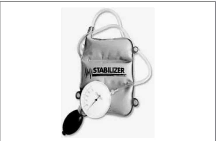
is a simple pressure transducer consisting of a three-chamber air-illed pressure bag, a catheter and a sphygmomanometer
gauge24,25. he pressure bag has 16.7×24 cm in size and made
from non-elastic material. he sphygmomanometer scale ranges from 0 mmHg to 200 mmHg, with 2 mmHg intervals on the scale. Movement or change in position causes volume changes in the pressure bag, which is registered by this device.
It is necessary that pressure measures of the abdominal muscle activity have a minimum level of validity to be use-ful. Validity is the degree to which the measure represents the phenomenon of interest, and refers to how much an
in-strument measures what it would be supposed to measure26.
hus, when an instrument undergoes a validation process, in fact is not the instrument itself that is being validated, but
the purpose for which the instrument is being used27. here
are ive types of validity described in the literature: face, con-tent, criterion, construct and concurrent validity. Concurrent validity refers to the extent to which scores on a particular instrument relate to a reference standard (i.e., not a gold stan-dard) and is consistent with theoretically derived hypotheses
concerning the concepts that are being measured27-30.
Clinimetric properties of the PBU in measuring TrA
muscle activity have been discussed in many studies22,25,31-34,
but most of them sampled solely healthy individuals22,25,32,33,
and two31,34 recruited subjects with history of low back pain.
Of the six studies, only one31 investigated the validity of this
instrument and, despite the authors have concluded that the PBU provides valid measures, their indings are not conclu-sive due to the suboptimal methodological quality, such as small sample size (n=15) and use of diferent motor tasks
to assess TrA muscle activity31. A recent systematic review
from our research group has summarized main methodologi-cal limitations of studies on measurement properties of the
PBU35. Furthermore, a recently low risk of bias study on the
reproducibility of the PBU was conducted and observed ICCs of 0.74 and 0.76 for intra and inter-examiner reproducibility,
respectively36. Scientiic indings are more accepted when
diferent studies investigate the efects of an intervention or
test, obtain similar conclusions37. hus, clinimetric properties
of the measures should be studied and presented for that the quality of the results can be evaluated.
herefore, considering the importance of the use of vali-dated measures that can support clinical practice and consid-ering the scarcity of studies concerning the validation of the PBU in a clinically relevant sample, the purpose of this study was to evaluate the concurrent validity and diagnostic accu-racy of the PBU in measuring TrA muscle activity in patients with chronic nonspeciic low back pain.
Method
This study used a validation, cross-sectional design38, and
of the Universidade Federal do Pernambuco (UFPE), Recife, PE, Brazil in 2009.
The sample was selected from students of the UFPE with chronic nonspecific low back pain. This study was approved by the Institutional Research Ethics Commit-tee (Agamenon Magalhães Hospital, Recife, PE, Brazil) (#00490236000-09). All participants signed an informed consent form. The required sample size was estimated based upon the recommendations of specific guidelines on clinimetric properties that suggest a sample of at least 50
individuals for validation studies28.
We included participants with chronic nonspeciic low back pain (i.e., low back pain of at least three months duration
and without a speciic cause)39. We excluded participants who
had acute low back pain (i.e., an episode of low back pain of
less than 6 weeks), those who were pregnant, who had previ-ous abdominal wall or spinal surgery, with a body mass index (BMI) of 25 or over, were menstruating during the tests and
those with suspected or conirmed serious pathologies (i.e,
nerve root compromise; fractures; cancer; infectious diseases of the spine; cauda equina syndrome and widespread neuro-logical disorders).
All participants received basic information about the anatomy, biomechanics and functions of the TrA muscle, as well as they received training of the TrA muscle contraction,
which has been described in detail in the literature22,25,33,34,40. All
subjects were previously instructed to fast for 2 hours prior to testing (including water), empty the bladder immediately be-fore the tests and to not perform abdominal exercises two days
prior to the tests22. For both tests, participants and examiners
have adopted the same positioning, environmental and tem-poral conditions to avoid external inluences or internal errors during data collection. Participants were positioned in a prone position on a hard surface, with the lower limbs positioned with the feet of the plinth and with the arms beside the trunk.
To test the concurrent validity of PBU, surface electromy-ography (EMG) was considered as the reference-standard test,
based on the indings of previous studies on the topic19,41,42 and
considering the questions of the ethics committees in Brazil (our ethics committee did not allowed us to use ine-wire EMG). For all patients, we irstly tested the TrA muscles using
the surface EMG (Miotec®Miotool 400) under the conditions
described below. Before placing the electrodes, detection sur-faces were properly cleaned, exfoliated and, when necessary shaving was performed following the recommendations of ISEK (International Society of Electrophysiology and Kinesiol-ogy) and SENIAM (Surface Electromyography for Non Invasive
Assessment of Muscles)43 to allow a low impedance between
the skin and the electrodes. he bipolar surface electrodes
(Ag/AgCl - Meditrace®) were placed with a distance of 20 mm
between their centers, and placed at right side in the area cor-responding to the TrA/OI (transversus abdominis/internal oblique) muscles, located 2 cm from the anterior superior iliac spine, following the instructions established by Hodges,
Gandevia and Richardson41 and adapted by Chanthapetch
et al.42. he reference electrode was placed in the right lateral
epicondyle. Participants were positioned on supine position with knees lexed at 90°.
We irstly recorded 30 seconds at rest to detect the param-eters of electromyographic silence (basal tonus). hen, it was performed the protocol of maximal voluntary isometric
con-traction (MVIC) in agreement with the standards of SENIAM43
three contractions of 5 seconds with intervals of 3 seconds between each contraction, with three repetitions. Each par-ticipant had one minute of rest between repetitions. To inish the electromyographic collection was requested a TrA muscle contraction maintained for 10 seconds. Overall, there were ive electromyographic records from each subject. he signs of elec-trical activity of TrA muscle were collected and transmitted to a monitor that was connected to the EMG equipment. EMG data were sampled at 1000 Hz over a bandwidth of 20-450 Hz (band-pass ilter). he raw EMG signals generated were analyzed using the root mean square (RMS). EMG data were normalized by the percentage of MVIC and signiicant electromyographic activity (positive result - success) was deined as an activity at least 10%
of the MVIC44,45.
After obtaining EMG recordings and respecting an in-terval of 15 minutes, we measured the TrA muscle activity from the PBU. Participants were positioned on prone posi-tion and the inlatable bag was placed between the anterior superior iliac spine and navel. Before starting the contrac-tion, the bag was inlated to a pressure of 70 mmHg with the valve closed and participants were instructed to breathe deeply using mainly abdominal wall, then the inlatable bag was adjusted to 70 mmHg again. Patients were requested to perform three TrA muscle contractions with following verbal commands standardized by examiner: “Draw in your abdo-men without moving the spine or pelvis” and maintain these
contractions for ten seconds22.he examiner have checked by
palpation if the participants were moving (in contrast to the instructions) the spine or pelvis. According to the
manufac-turer of PBU (Stabilizer®, Chattanooga Group Inc., Hixson,
TN, USA), the ability to contract the TrA muscle results in a pressure reduction from 4 to 10 mmHg, which is recorded by
pressure gauge of PBU24. hus, based on this information and
previous studies on the topic24,35,36, a pressure reduction of at
Variables Mean (SD)
Pressure Measures
PBU* (mmHg) - 4.1 (2.4)
Electromyographic Measures
MVIC** (µV) 127.1 (95.5)
MVIC (%) 100
Rest (µV) 2.3 (1.9)
Rest (%) 2.9
Contraction maintained for 10 seconds (µV) 29.6 (21.8)
Contraction maintained for 10 seconds (%) 24.4
Table 2. Description of the pressure measures and electromyographic measures during the TrA muscle activity.
*PBU: pressure biofeedback unit; **MVIC: maximal voluntary isometric contraction.
sEMG** (reference-standard test)
Positive Negative Total
PBU* (index test)
Positive 25 (50%) 4 (8%) 29 (58%)
Negative 15 (30%) 6 (12%) 21 (42%)
Total 40 (80%) 10 (20%) 50 (100%)***
Table 3. 2x2 Table between the PBU ratings and the surface EMG ratings.
*PBU: pressure biofeedback unit; **sEMG: surface electromyography; ***Phi (f) Coefficient (Phi=0.2, p<0.20).
that changes in pressure were not due to a leak in the pressure biofeedback unit cushion, the PBU was calibrated according to
the recommendations established by von Garnier34.
A trained and experienced examiner performed both measurements of surface EMG and PBU. All information was recorded using a digital evaluation form provided by speciic
software (Miograph®). Pain intensity was measured by the
Nu-merical Pain Rating Scale and disability was measured by the Roland Morris Disability Questionnaire, both cross-culturally adapted and clinimetrically tested for Brazilian-Portuguese
speakers46-48.
he sample characteristics are presented using
descrip-tive statistics49. To describe the validity, we used the Phi ( f )
Coeicient, whose values range from -1 to 1 and if it lower than 0.5 can be classiied as weak, values between 0.5 and 0.7 are classiied as moderate and over 0.7 are classiied as strong
correlation50,51. he relationship between the TrA muscle
con-traction maintained for 10 seconds measured by the surface EMG (%MVIC) and the scores of pressure reduction measured by the PBU (mmHg) was the speciic hypothesis formulated to analyze the concurrent validity. We hypothesized that the PBU (index test) would have a moderate and positive
cor-relation with the surface EMG (reference-standard test)28,52.
Furthermore, we calculated the values of diagnostic accu-racy tests, such as sensitivity, speciicity, positive predictive value (PPV) and negative predictive value (NPV). Data were analyzed with SPSS (Statistical Package for Social Sciences) version 15.0, considering the signiicance level of p<0.05 in all calculations.
Results
he sample characteristics are presented in Table 1. Of the 50 participants, 38 (76%) were women and were on average 22 years old, weighed 63.7 kilos and had an average height of 1.70 meters. he mean low back pain duration was 1.9 years. he mean low back pain intensity measured by the Pain Intensity Numerical Rating Scale was 5.1 points and the mean disabil-ity measured by the Roland Morris Disabildisabil-ity Questionnaire was 9.1 points.
he measures performed from the surface EMG obtained mean MVIC of 127.1 (95.5) µV, representing 100% MVIC. he records at rest had a mean of 2.3 (1.9) µV, representing 2.9% MVIC. he contraction maintained for 10 seconds was 29.6 (21.8) µV on average, representing 24.4%MVIC. he measures performed from the PBU obtained mean pressure values of -4.1 (2.4) mmHg (Table 2).
Figure 1. Pressure Biofeedback Unit (Stabilizer®).
According to predetermined criteria to characterize the results as positive (success), 31 (62%) patients received similar ratings by the two assessment instruments, but 19 (38%) were classiied as false-positive (8%) and false-negative (30%), which shows a weak and non-signiicant correlation of Phi=0.2 (p<0.20) between the ratings of the PBU and the surface EMG (Table 3).
Variables Mean (SD)
Gender (n, %) Female 38 (76%)
Male 12 (24%)
Age (years) 22.0 (2.3)
Low back pain duration (years) 1.9 (3.4)
Weight (kg) 62.7 (12)
Height (m) 1.7 (0.1)
Pain Intensity Numerical Rating Scale (0–10) 5.1 (1.8)
Roland Morris Disability Questionnaire (0–24) 9.1 (5.9)
Regarding to the diagnostic tests, the results showed low sensitivity (60%), low speciicity (60%), a high PPV (0.8) and a low NPV (0.2) of the PBU.
Discussion
Our study showed a poor concurrent validity of the PBU in measuring TrA muscle activity during voluntary contraction maneuver in patients with nonspeciic chronic low back pain. he usefulness of a measure in research or in clinical practice depends on how much we can rely on the accuracy of the data as indicators of the behavior or the phenomenon assessed. Ideally, any measurement should be practical, easy to obtain and able to produce reliable and valid data, since no consis-tent measures produce unreliable results, leading to erroneous
conclusions53.
With regards to the correlation between the ratings of the PBU and the surface EMG, the high disagreement of 38% showed that the pressure data are not statistically correlated
to the EMG data (Phi=0.2, p<0.20)50. Similar results were found
by Hodges, Richardson and Jull54, who preliminarily found a
non-signiicant correlation between the absolute estimates of the PBU and ine-wire EMG (r=0.48, p<0.07), but subsequently showed that the same data, when converted into an ordinal scale to classify the two measures had high probability of being correlated (Fisher’s exact test, p<0.006). Methodological difer-ences can be used as a possible explanation for these
conlict-ing results, since in the study of Hodges, Richardson and Jull54,
the PBU was used to evaluate the presence of TrA muscle activ-ity and the EMG was used to evaluate the latency period be-tween the onset of contraction of the TrA and anterior deltoid
muscles (i.e., the instruments evaluated distinct phenomena).
In this study, the two instruments (PBU and surface EMG) were used to evaluate the same phenomenon (i.e., presence of TrA muscle activity).
he scarcity of studies investigating the validity of the PBU makes it harder to perform a detailed analysis of our results.
Besides the study of Hodges, Richardson and Jull31 other
study conducted by Costa et al.22 tried to estimate the validity
of the PBU with a palpation test, and showed a positive and signiicant correlation between the two tests (r=0.99, p<0.01). However, it is observed that their study compared two clinical tests and did not take into account any laboratory inding, and its methodological approach are questioned by Richardson et
al.55 and Falla et al.56 who found a correlation of 0.56 (p<0.001)
between PBU and EMG data (contact electrodes attached to a suction catheter), but this study was conducted in individuals
with chronic nonspeciic neck pain, unlike our sample of chronic low back pain participants.
he possibility of existence of the phenomenon of cross-talk would be a likely explanation for the weak correlation observed
between the instruments, however the SENIAM43 assumes
that the placement of the surface electrodes could collect the electromyographic signals from the TrA and OI muscles. Furthermore, the validity of an assessment instrument is
re-stricted by the reliability of test27. his point was not clear in
the literature35, but a recently published study36 about the PBU
shows strong evidences of the excellent reproducibility of test. his study was conducted with an adequate sample size, clini-cally relevant participants and procedures for data collection
rigorously executed. Results indicated an ICC2,1 of 0.74 and 0.76
for the intra and inter-examiner reproducibility, respectively. he intra and inter-examiner agreement were within the limits of agreement (LOA) on 95% of occasions. herefore, the repro-ducibility of PBU in measuring TrA muscle activity in patients with chronic nonspeciic low back pain can be considered as excellent.
Among many factors that can afect the accuracy of an outcome measure, the lack of standardization is often referred to as an important source of error. In this sense, the deinition of certain criteria is essential for a critical assessment as the best evidence available when diferent studies investigating
the efects of a test27,37,38. One inding during the development
of this study was the benchmark for deining what would be considered “positive result” and “negative result” of the PBU. he criterion of pressure reduction cannot be not considered as biased, because the criterion adopted (at least 4 mmHg) is
the most cited in the literature22,32,33,35,36,57,58 and recommended
by the equipment manufacturer24. he point is that there are
no available studies with a ROC (Receiver Operating Char-acteristics) curve analysis to determine the score-by-score results of the PBU compared to the gold-standard tool. he
study conducted by Hodges, Richardson and Jull54 is the one
that could have clariied this issue, but this statistical method was not performed. herefore, despite to be a consolidated criterion and repeated by most researchers on the topic and by the manufacturer, this can only be conirmed with a ROC curve
analysis27,38,59,60. We suggest a future study with primary aim to
and movement is not direct. Although these tests have evalu-ated the same task (voluntary contraction of TrA muscle), the surface EMG is able to assess amount of muscle activation (and not amount of force or movement) and the PBU is able to evaluate the depression of abdominal wall (which is the ability to generate movement by TrA muscle) indirectly by measur-ing the pressure reduction recorded by the device. herefore, a muscle that is more active in the electromyography signal will not necessarily generate more movement. his may happened
due to muscle weakness61-63.
Regarding to the diagnostic tests, the results showed a low sensitivity (60%) with small ability of the PBU detect TrA muscle activity when it is present, also showed a low speciicity (60%) with small ability of the PBU to rule outTrA muscle activity when it is absent. Based on these results we can infer that the pressure measures were not sensitive to discriminate the presence of TrA muscle activity and were not speciic to identify the absence of the same muscle activ-ity, making questionable the usefulness of the PBU to assess TrA muscle activity in patients with chronic nonspeciic low
back pain60,64. With regards to diagnostic tests, the high PPV
(0.8) found, ensures that if result of the PBU is positive, the chance to be a real TrA muscle activity is 80%. While the low NPV (0.2) does not assume that the negative tests of the PBU represent absence of TrA muscle activity, since if results of the PBU are negative, the chance that there is no TrA muscle activity is only 20%.
herefore, based on the results observed and the literature, it is emphasized that there was no evidence suicient to sup-port the concurrent validity of the PBU in measuring TrA mus-cle activity in patients with chronic nonspeciic low back pain.
Acknowledgements
he authors would like to thank CAPES (Coordenação de Aperfeiçoamento de Pessoal de Nível Superior), Brasilia, DF, Brazil - Ministério da Educação and FAPESP (Fundação de Am-paro a Pesquisa do Estado de São Paulo), São Paulo, SP, BRazil.
References
1. Chou R, Qaseem A, Snow V, Casey D, Cross JT Jr, Shekelle P, et al. Diagnosis and treatment of low back pain: a joint clinical practice guideline from the American College of Physicians and the American Pain Society. Ann Intern Med. 2007;147(7):478-91.
2. Chou R, Huffman LH; American Pain Society; American College of Physicians. Nonpharmacologic therapies for acute and chronic low back pain: a review of the evidence for an American Pain Society/American College of Physicians clinical practice guideline. Ann Intern Med. 2007;147(7):492-504.
3. Swinkels-Meewisse EJ, Swinkels RA, Verbeek AL, Vlaeyen JW, Oostendorp RA. Psychometric properties of the Tampa Scale for kinesiophobia and the fear-avoidance beliefs questionnaire in acute low back pain. Man Ther. 2003;8(1):29-36.
4. Ferreira ML, Ferreira PH, Latimer J, Herbert RD, Hodges PW, Jennings MD, et al. Comparison of general exercise, motor control exercise and spinal manipulative therapy for chronic low back pain: A randomized trial. Pain. 2007;131(1-2):31-7.
5. Macedo LG, Maher CG, Latimer J, McAuley JH. Motor control exercise for persistent, nonspecific low back pain: a systematic review. Phys Ther. 2009;89(1):9-25.
6. Ferreira ML, Ferreira PH, Latimer J, Herbert R, Maher CG. Does spinal manipulative therapy help people with chronic low back pain? Aust J Physiother. 2002;48(4):277-84.
7. Macedo LG, Latimer J, Maher CG, Hodges PW, Nicholas M, Tonkin L, et al. Motor control or graded activity exercises for chronic low back pain? A randomised controlled trial. BMC Musculoskelet Disord. 2008;9:65.
8. Airaksinen O, Brox JI, Cedraschi C, Hildebrandt J, Klaber-Moffett J, Kovacs F, et al. Chapter 4. European guidelines for the management of chronic nonspecific low back pain. Eur Spine J. 2006;15 Suppl 2:S192-300.
9. Hodges PW, Richardson CA. Altered trunk muscle recruitment in people with low back pain with upper limb movement at different speeds. Arch Phys Med Rehabil. 1999;80(9):1005-12. 10. Hodges PW, Richardson CA. Delayed postural contraction of transversus abdominis in low back
pain associated with movement of the lower limb. J Spinal Disord. 1998;11(1):46-56. 11. Tsao H, Galea MP, Hodges PW. Reorganization of the motor cortex is associated with postural
control deficits in recurrent low back pain. Brain. 2008;131(Pt 8):2161-71.
12. Hides JA, Jull GA, Richardson CA. Long-term effects of specific stabilizing exercises for
first-13. Sedaghat N, Latimer J, Maher C, Wisbey-Roth T. The reproducibility of a clinical grading system of motor control in patients with low back pain. J Manipulative Physiol Ther. 2007;30(7):501-8. 14. Costa LO, Maher CG, Latimer J, Smeets RJ. Reproducibility of rehabilitative ultrasound
imaging for the measurement of abdominal muscle activity: a systematic review. Phys Ther. 2009;89(8):756-69.
15. Kane K, Barden J. Contributions of trunk muscles to anticipatory postural control in children with and without developmental coordination disorder. Hum Mov Sci. 2011. [Epub ahead of print]. 16. Sapsford RR, Hodges PW, Richardson CA, Cooper DH, Markwell SJ, Jull GA. Co-activation of the
abdominal and pelvic floor muscles during voluntary exercises. Neurourol Urodyn. 2001;20(1):31-42. 17. Sjödahl J, Kvist J, Gutke A, Oberg B. The postural response of the pelvic floor muscles
during limb movements: a methodological electromyography study in parous women without lumbopelvic pain. Clin Biomech (Bristol, Avon). 2009;24(2):183-9.
18. Stüpp L, Resende AP, Petricelli CD, Nakamura MU, Alexandre SM, Zanetti MR. Pelvic floor muscle and transversus abdominis activation in abdominal hypopressive technique through surface electromyography. Neurourol Urodyn. 2011;30(8):1518-21.
19. Marshall P, Murphy B. The validity and reliability of surface EMG to assess the neuromuscular response of the abdominal muscles to rapid limb movement. J Electromyogr Kinesiol. 2003;13(5):477-89. 20. Critchley DJ, Coutts FJ. Abdominal muscle function in chronic low-back pain patients:
measurements with real-time ultrasound scanning. Physiotherapy. 2002;88(6):322-32. 21. McMeeken JM, Beith ID, Newham DJ, Milligan P, Critchley DJ. The relationship between EMG and
change in thickness of transversus abdominis. Clin Biomech (Bristol, Avon). 2004;19(4):337-42. 22. Costa LOP, Costa LCM, Cançado RL, Oliveira WM, Ferreira PH. Confiabilidade do teste
palpatório e da unidade de biofeedback pressórico na ativação do músculo transverso abdominal em indivíduos normais. Acta Fisiátrica. 2004;11(3):101-5.
23. Hodges PW, Pengel LH, Herbert RD, Gandevia SC. Measurement of muscle contraction with ultrasound imaging. Muscle Nerve. 2003;27(6):682-92.
24. Chattanooga G. Stabilizer pressure bio-feedback. Operating instructions. Hixson: Chattanooga Group Inc.; 2005.
26. Fletcher RH, Fletcher SW. Epidemiologia Clínica: elementos essenciais. 4th ed. Porto Alegre:
Artmed; 2006.
27. Portney LG, Watkins MP. Foundations of clinical research: applications to practice. 3rd ed. Upper
Saddle River: Prentice Hall; 2008.
28. Terwee CB, Bot SD, de Boer MR, van der Windt DA, Knol DL, Dekker J, et al. Quality criteria were proposed for measurement properties of health status questionnaires. J Clin Epidemiol. 2007;60(1):34-42.
29. Johnston MVK, Keith RA, Hinderer SR. Measurement standards for interdisciplinary medical rehabilitation. Arch Phys Med Rehabil. 1992;73(12-S):S3-23.
30. Armitage P, Colton T. Encyclopedia of biostatistics. 1st ed. Boston: John Wiley & Sons Ltd; 1998.
31. Hodges PW, Richardson CA. Inefficient muscular stabilization of the lumbar spine associated with low back pain. A motor control evaluation of transversus abdominis. Spine (Phila Pa 1976). 1996;21(22):2640-50.
32. Costa LOP, Costa Lda C, Cancado RL, Oliveira WdeM, Ferreira PH. Short report: intra-tester reliability of two clinical tests of transversus abdominis muscle recruitment. Physiother Res Int. 2006;11(1):48-50.
33. Figueiredo MK, Chaves Júnior IP, Figueiredo VGC, Costa LOP, Menezes LC. Estudo da confiabilidade intra e entre-examinadores da unidade de biofeedback pressórico na medida da contração do músculo transverso abdominal. Rev Bras Ciênc Mov. 2005;13(4):93-100. 34. von Garnier K, Köveker K, Rackwitz B, Kober U, Wilke S, Ewert T, et al. Reliability of a test
measuring transversus abdominis muscle recruitment with a pressure biofeedback unit. Physiotherapy. 2009;95(1):8-14.
35. Lima POP, de Oliveira RR, Costa LO, Laurentino GE. Measurement properties of the pressure biofeedback unit in the evaluation of transversus abdominis muscle activity: a systematic review. Physiotherapy. 2011;97(2):100-6.
36. Lima POP, Oliveira RR, Moura Filho AG, Raposo MCF, Costa LOP, Laurentino GEC. Reproducibility of the pressure biofeedback unit in measuring transversus abdominis muscle activity in patients with chronic nonspecific low back pain. J Bodyw Mov Ther. 2012;16(2):251-7. 37. Sampaio RF, Mancini MC. Estudos de revisão sistemática: um guia para síntese criteriosa da
evidência científica. Rev Bras Fisioter. 2007;11(1):83-98.
38. Hulley SB, Cumming SR, Browner WS, Grady D, Hearst N, Newman TB. Delineando a pesquisa clínica - Uma abordagem epidemiológica. 3rd ed. Porto Alegre: Artmed; 2008.
39. van Middelkoop M, Rubinstein SM, Verhagen AP, Ostelo RW, Koes BW, van Tulder MW. Exercise therapy for chronic nonspecific low-back pain. Best Pract Res Clin Rheumatol. 2010;24(2):193-204.
40. Richardson CA, Jull GA. Muscle control-pain control. What exercises would you prescribe? Man Ther. 1995;1(1):2-10.
41. Hodges PWG, Gandevia SC, Richardson CA. Contractions of specific abdominal muscles in postural tasks are affected by respiratory maneuvers. J Appl Physiol. 1997;83(3):753-60. 42. Chanthapetch P, Kanlayanaphotporn R, Gaogasigam C, Chiradejnant A. Abdominal muscle
activity during abdominal hollowing in four starting positions. Man Ther. 2009;14(6):642-6. 43. Hermens HJ, Freiks B, Disselhorst-Klug C, Rau G. Development of recommendations for SEMG
sensors and sensors placement procedures. J Electromyogr Kinesiol. 2000;10(5):361-74. 44. Vasseljen O, Fladmark AM, Westad C, Torp HG. Onset in abdominal muscles recorded
simultaneously by ultrasound imaging and intramuscular electromyography. J Electromyogr Kinesiol. 2009;19(2):e23-31.
45. Yang YS, Koontz AM, Triolo RJ, Mercer JL, Boninger ML. Surface electromyography activity of trunk muscles during wheelchair propulsion. Clin Biomech (Bristol, Avon). 2006;21(10):1032-41.
46. Costa LOP, Maher CG, Latimer J. Self-report outcome measures for low back pain: searching for international cross-cultural adaptations. Spine (Phila Pa 1976). 2007;32(9):1028-37. 47. Maher CG, Latimer J, Costa LOP. The relevance of cross-cultural adaptation and clinimetrics for
physical therapy instruments. Rev Bras Fisioter. 2007;11(4):245-52.
48. Costa LOP, Maher CG, Latimer J, Ferreira PH, Ferreira ML, Pozzi GC, et al. Clinimetric testing of three self-report outcome measures for low back pain patients in Brazil: which one is the best? Spine (Phila Pa 1976). 2008;33(22):2459-63.
49. Soares JF, Siqueira AL. Introdução à estatística médica. 2ª ed. Belo Horizonte: Coopmed; 2002. 50. Santos C. Estatística descritiva: manual de auto-aprendizagem. 2ª ed. Lisboa: Sílabo; 2007. 51. Conover WJ. Practical nonparametric statistics. 3rd ed. New York: Wiley; 1999.
52. Munro H. Statistical Methods for Health Care Research. 5th ed. Philadelphia, PA: Lippincott
Williams & Wilkins; 2005.
53. Gadotti IC, Vieira ER, Magee DJ. Importance and clarification of measurement properties in rehabilitation. Rev Bras Fisioter. 2006;10(2):137-46.
54. Hodges P, Richardson C, Jull G. Evaluation of the relationship between laboratory and clinical tests of transversus abdominis function. Physiother Res Int. 1996;1(1):30-40.
55. Richardson CA, Snijders CJ, Hides JA, Damen L, Pas MS, Storm J. The relation between the transversus abdominis muscles, sacroiliac joint mechanics, and low back pain. Spine (Phila Pa 1976). 2002;27(4):399-405.
56. Falla D, Jull G, Dall’Alba P, Rainoldi A, Merletti R. An electromyographic analysis of the deep cervical flexor muscles in performance of craniocervical flexion. Phys Ther. 2003;83(10):899-906. 57. Hides JA, Richardson CA, Jull GA. Multifidus muscle recovery is not automatic after resolution
of acute, first-episode low back pain. Spine (Phila Pa 1976). 1996;21(23):2763-9.
58. Jull G, Richardson C, Toppenberg R, Comerford M, Bui B. Towards a measurement of active muscle control for lumbar stabilisation. Aust J Physiother. 1993;39(3):187-93.
59. Rothstein JM. Measurement in physical therapy. New York: Churchil Livingstone; 1985. 60. Finch E, Brooks D, Stratford PW, Mayo NE. Physical rehabilitation outcome measures. 2nd ed.
Hamilton, Ontario: Lippincott Williams & Wilkins; 2002.
61. Rice DA, McNair PJ, Lewis GN. Mechanisms of quadriceps muscle weakness in knee joint osteoarthritis: the effects of prolonged vibration on torque and muscle activation in osteoarthritic and healthy control subjects. Arthritis Res Ther. 2011;1(5)3:R151.
62. Philippon MJ, Decker MJ, Giphart JE, Torry MR, Wahoff MS, LaPrade RF. Rehabilitation exercise progression for the gluteus medius muscle with consideration for iliopsoas tendinitis: an in vivo electromyography study. Am J Sports Med. 2011;39(8):1777-85.
63. Distefano LJ, Blackburn JT, Marshall SW, Padua DA. Gluteal muscle activation during common therapeutic exercises. J Orthop Sports Phys Ther. 2009;39(7):532-40.
64. Fleiss J, Levin B, Paik M. Statistical methods for rates and proportions. 3rd ed. New York: John