Imaging evaluation of vocal cord paralysis*
Avaliação por imagem da paralisia de pregas vocaisMarcelo de Mattos Garcia1, Fabiana Pizanni Magalhães2, Gabriela Bijos Dadalto1, Marina Vimieiro Timponi de Moura3
Vocal cord paralysis is a common cause of hoarseness. It may be secondary to many types of lesions along the cranial nerve X pathway and its branches, particularly the laryngeal recurrent nerves. Despite the idiopathic nature of a great number of cases, imaging methods play a very significant role in the investigation of etiologic factors, such as thyroid and esophagus neoplasias with secondary invasion of the laryngeal recurrent nerves. Other conditions such as aortic and right subclavian artery aneurysms also may be found. The knowledge of local anatomy and related diseases is of great importance for the radiologist, so that he can tailor the examination properly to allow an appropriate diagnosis and therapy planning. Additionally, considering that up to 35% of patients with vocal cord paralysis are asymptomatic, the recognition of radiological findings indicative of this condition is essential for the radiologist who must warn the referring physician on the imaging findings. In the present study, the authors review the anatomy and main diseases related to vocal cord paralysis, demonstrating them through typical cases evaluated by computed tomography and magnetic resonance imaging, besides describing radiological findings of laryngeal abnormalities indicative of this condition.
Keywords: Vocal cord; Paralysis; Imaging.
Paralisia das pregas vocais é causa frequente de rouquidão, podendo ser secundária a várias lesões ao longo do trajeto do X par craniano e seus ramos, particularmente os nervos laríngeos recorrentes. Apesar de grande parte dos casos ser idiopática, os métodos de imagem são muito importantes na pesquisa de fatores etioló-gicos, tais como lesões neoplásicas da tireoide e esôfago com invasão secundária dos nervos laríngeos re-correntes. Além destas, outras anormalidades como aneurismas do arco aórtico e da artéria subclávia direita podem ser encontradas. É fundamental que o radiologista conheça a anatomia pertinente a esta região e as principais afecções que podem ocorrer, para que o estudo seja corretamente planejado, auxiliando o diag-nóstico e o planejamento terapêutico. Além disso, como até 35% dos casos de paralisia da prega vocal são assintomáticos, o conhecimento dos sinais radiológicos que indicam esta condição é indispensável, cabendo ao radiologista alertar o médico assistente sobre os achados do exame. Neste trabalho realizamos uma revi-são da anatomia e das principais doenças responsáveis pela paralisia de cordas vocais, demonstrando-as por meio de estudos de tomografia computadorizada e ressonância magnética de casos típicos. Mostramos, tam-bém, as alterações radiológicas próprias da laringe que indicam a presença de paralisia das pregas vocais.
Unitermos: Cordas vocais; Paralisia; Imagem. Abstract
Resumo
* Study developed at Axial Centro de Imagem, Belo Horizonte, MG, Brazil.
1. Titular Members of Colégio Brasileiro de Radiologia e Diag-nóstico por Imagem (CBR), MDs, Radiologists, Axial Centro de Imagem, Belo Horizonte, MG, Brazil.
2. Titular Member of Colégio Brasileiro de Radiologia e Diag-nóstico por Imagem (CBR), MD, Radiologist, Axial Centro de Imagem and Hospital de Pronto Socorro João XXIII, Belo Hori-zonte, MG, Brazil.
3. MD, Radiologist, Axial Centro de Imagem and Biocor Insti-tuto, Resident in Radiology at Axial Centro de Imagem, Belo Horizonte, MG, Brazil.
Mailing address: Dr. Marcelo de Mattos Garcia. Avenida Ber-nardo Monteiro, 1472, Funcionários. Belo Horizonte, MG, Bra-zil, 30150-280. E-mail: [email protected] or [email protected]
nance imaging (MRI) studies were focused in the research of the main causes of the condition, illustrating them with typical cases.
ANATOMY
The vagus nerve is the longest of the cranial nerves, extending from the brain-stem to the abdomen(1). It originates from
four nuclei in the bulb, three of them con-verging in the basal cistern to form a single nerve that emerges from the skull through the jugular foramen, passing through the neck and chest to the abdomen. In this long course, the vagus nerve gives rise to a num-ber of branches to innervate the larynx and
Garcia MM, Magalhães FP, Dadalto GB, Moura MVT. Imaging evaluation of vocal cord paralysis. Radiol Bras. 2009;42(5):321– 326.
(paired cranial nerve X) pathway and its branches. The recognition of specific radio-logical findings, as well as the knowledge on the anatomy and diseases that may af-fect the paired cranial nerve X are ex-tremely important for the choice and pro-gramming of the best imaging method to be utilized, and for an appropriate interpreta-tion of images.
In the present study, the authors sought to demonstrate radiological findings in-dicative of vocal cord paralysis. Addition-ally, the strategies for evaluation of com-puted tomography (CT) and magnetic
reso-INTRODUCTION
Vocal cord paralysis is a frequent cause of hoarseness, and may be secondary to several conditions along the vagus nerve
the pharynx. In the cervical region, it passes between the carotid artery (medially) and the internal jugular vein (laterally). There are three major branches of the vagus nerve in this region: the pharyngeal, superior la-ryngeal and the recurrent lala-ryngeal nerves. The pharyngeal branch originates from the inferior ganglion of the vagus nerve and is part of the pharyngeal plexus, respon-sible for the motor innervation of the pha-ryngeal and levator veli palatine muscles. The superior laryngeal nerve also origi-nates from the inferior ganglion of the va-gus nerve, caudally to the pharyngeal branch, and is divided into external laryn-geal nerve (motor) and internal larynlaryn-geal nerve (sensory). It is responsible for the cricothyroid muscle innervation.
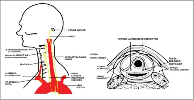
The recurrent laryngeal nerves (RLNs) (Figure 1) are responsible for the innerva-tion of the other intrinsic muscles of the larynx(2). These nerves carry motor, sensory and parasympathetic fibers, dividing itself into internal branch, responsible for the sensory function of the vocal cords and subglottic region, and external branch that bears the motor function of four intrinsic laryngeal muscles: thyroarytenoid, anterior and posterior cricoarytenoid, oblique and transverse arytenoid muscles(3). At right,
the RLN emerges anteriorly from the vagus
nerve at the intersection with the right sub-clavian artery to loop under and around the artery to reach its posterior surface. Then, it runs superiorly in the tracheoesophageal cleft towards the larynx. The left RLN emerges from the vagus nerve in the medi-astinum, after crossing it anterolaterally to the aortic arch to loop under the arch and running between the aorta and the left pul-monary artery, extending posteriorly to the tracheoesophageal cleft to reach the larynx. Because of its longer and partially intratho-racic course, the left RLN may be affected by mediastinal diseases(1). Both nerves
en-ter the larynx in the region of the cricothy-roid joint through the fibers of the inferior constrictor muscle of the pharynx.
In the RLN ascending course from the chest to the larynx, a number of anatomi-cal variations may be found. Rustad(4), in
a post-mortem series has observed that 43% of the RLNs split into one or more branches on both sides. The findings of nerve branching before reaching the larynx have also been surgically confirmed by Nemiroff & Katz(5) in 40% of cases. The
path of the RLNs may change because of congenital vascular abnormalities or ana-tomical distortions resulting from the pres-ence of goiter, neoplasias or inflammatory processes. In cases of aberrant subclavian
artery, the right RLN runs directly from the vagus nerve to the larynx, without looping around the respective artery. This variation is closely related to surgical injuries and is known as “non-recurrent” inferior laryn-geal nerve.
The RLN is localized in the tracheoe-sophageal cleft that is a relevant anatomi-cal landmark, in 65% of cases at right, and in 77%, at left. The nerve is found at the right side of the trachea in 33% of cases, and at left, in 22%. It rarely ascends antero-laterally to the trachea and, in this case, it is more susceptible to surgical injury.
CLINICAL FINDINGS
Usually, patients with vocal cord paraly-sis present with complaint of hoarseness. Other more severe symptoms include
fre-quent aspiration and pneumonia(6).
How-ever, up to 35%f patients may be asymptom-atic(5). In these cases, vocal cord paralysis
will be incidentally identified, and this find-ing should alert the clinician to the neces-sity of additional evaluation. The previous and direct evaluation by laryngoscopy must be initially performed to rule out the pres-ence of mucosal and submucosal lesions like those caused by squamous cell carcinoma, to justify a subsequent imaging study.
Vocal cord paralysis may be acute or chronic, and may occur in one or both vo-cal folds. The left vovo-cal cord is most fre-quently affected, because of the longer course of the left recurrent laryngeal nerve, with about 12 cm in length from the aorta to the cricothyroid joining. At clinical ex-amination, the identification of the affected side is relatively simple: the truly involved vocal fold will be with a complete or par-tially reduced mobility.
IMAGING DIAGNOSIS
The classic findings of vocal cord pa-ralysis can be observed on plain radio-graphs and, particularly, on sectional imag-ing methods such as CT and MRI. These studies should include a scan from the skull base thru the aortic-pulmonary window, covering the whole course of the RLN. CT should include pre- and post-contrast phases, sometimes with a phonation phase with vocalization of the vowel i. This ac-quisition is useful in the evaluation of the true vocal folds mobility, but should be cau-tiously used to minimize the exposure of the patient to ionizing radiation. The study in-cludes axial acquisitions with later 3D coro-nal and sagittal multiplanar reconstructions. The CT and MRI parameters utilized in the authors’ institution are shown on Table 1. The bulging of the oropharyngeal and hypopharyngeal contour in association with the thinning of the constrictor muscle
constitutes an evidence of a lesion in the ipsilateral pharyngeal plexus(7). Such
find-ings indicate that the abnormality is local-ized immediately below the skull base or more cranially in the posterior fossa.
Most of imaging findings related to RLN paralysis are secondary to the thy-roarytenoid muscle atrophy (Figure 2)(7). At
least ten findings associated with this nerve paralysis are described by Landman(8) in
laryngoscopic studies. Notwithstanding the limitations of the method, these findings were transferred for analysis by axial CT. Several findings correlated the CT images with vocal cord paralysis(7); among them,
the following can be mentioned:
– thickening and medial displacement of the ipsilateral aryepiglottic fold (the most frequent finding) (Figure 3);
– dilatation of the pyriform sinus (Fig-ure 4);
– dilatation of the ipsilateral laryngeal ventricle (Figure 5);
– antero-medial displacement of the ip-silateral arytenoid cartilage (Figure 6);
– dilatation of the ipsilateral vallecula; – flattening of the subglottic arch dem-onstrated on coronal images.
The first two findings above described are observed in more than 75% of cases, constituting the most reliable diagnostic criteria.
Figure 2. Retraction of left true vocal cord corresponding to thyroarytenoid muscle hypertrophy.
Figure 3. Thickening of right aryepiglottic fold.
Table 1 Technical parameters utilized in computed tomography and magnetic resonance imaging stud-ies.
Computed tomography (multislice, 64-channel equipment)
Pre-contrast Post-contrast Collimation 1.5 mm 0.75 mm mAs 150 150 kV 140 140 Acquisition time 8 s 15 s
Magnetic resonance imaging (1.5 T, 8-channel coil equipment)
Coronal STIR
Coronal T1
Axial T1
Axial T2 FAT
Figure 7. Pancoast tumor obliterating aortic-pulmonary region.
Figure 6. Anteromedial displacement of left arytenoid cartilage. With the advent of multidetector CT,
high-resolution reformatted coronal images have provided a higher capacity for analyz-ing the larynx. However, the authors em-phasize that axial images allow a correct di-agnosis in most of cases.
CAUSES
There are several causes of vocal cord paralysis, and approximately 50% of re-ported cases are toxic or idiopathic(7). Thus,
in at least half of patients, sectional images may fail in identifying lesions along the vagus and recurrent laryngeal nerves path.
Figure 5. Left ventricle dilatation demonstrated by 3D reconstruction of multislice CT image.
In the past, the most common cause of vocal cord paralysis was thyroid surgery with dissection of the Berry’s ligament, the site where the RLN penetrates the larynx. In these cases, the lesions result from an excessive traction of the thyroid gland, or occur at the moment of the hemostasis. The RLN may be connected with the inferior thyroid vessels, therefore its identification during the surgical procedure is essential. Recently, lung (Figure 7) and skull base
neoplasms, as well as surgical injuries have been the most frequent causes of vocal cord paralysis.
Several diseases may affect the vagus nerve tract (Figure 8), leading to vocal cord paralysis. Thus, it is necessary to evaluate this nerve from the brainstem to the carina. The causes of vocal cord paralysis may be divided into central origin (vagal neuropa-thy) and peripheral (RLN neuropaneuropa-thy), the latest being most frequently observed, achieving up to 90% of cases in some se-ries. However, in most (up to 85%), lesions have not been identified along the nerves pathway.
Among the central causes, lesions af-fecting the brainstem, the skull base and the carotid bifurcation can be mentioned, all of them also associated with vagal neuropa-thy. In this group, vagal paragangliomas, hypervascularized masses involving or compressing the whole cranial nerve X pathway, as well as their nuclei in the jugu-lar foramen can be clearly demonstrated on contrast-enhanced phases at CT and MRI(9). Vascular events involving, for
ex-ample, the posterior-inferior cerebellar ar-tery leading to occlusion, may compromise the ambiguous nucleus in the dorsolateral aspect of the bulb. Thus, infarcts in this area may justify ipsilateral vocal cord paraly-sis(10). Central vagal neuropathy may be
suspected in the setting of paralysis or
Figure 8. Axial CT image demonstrating left jugular foramen enlargement. Contrast-enhanced coronal MRI spin-echo, T1-weighted sequence with fat-satura-tion demonstrating a solid mass in the left jugular foramen, with intense and homogeneous contras uptake: vagal schwannoma.
B A
sis of the pharyngeal constrictor muscles in cases where the pharyngeal plexus is in-volved. The pharyngeal plexus is formed by branches of cranial nerves IX, X and XI, besides branches of the sympathetic trunk. The vagal branches emerge from the no-dose ganglion localized below the skull base, and penetrate the pharyngeal muscles, branching into the superior and inferior constrictor muscles. A lesion affecting the pharyngeal plexus will cause paresis or paralysis of the ipsilateral constrictor muscles, eventually leading to atrophy.
Peripheral causes include RLN invasion by thyroid and cervical esophageal tumors (respectively Figures 9 and 10). Four
per-Figure 10. Esophageal neoplasia with invasion of right tracheoesophageal cleft and ipsilateral vocal cord paralysis.
cent of patients with unilateral vocal cord paralysis present thyroid disease, but only 0.7% are benign. Locally invasive thyroid cancers must be correctly evaluated by imaging methods, considering that the laryngotracheal invasion justifies the RLN involvement with subsequent vocal cord paralysis(11). Advanced malignant
esoph-ageal tumors may extend towards the tra-cheoesophageal groove, the RLN path-way(12). On the left side, aortic aneurysms
(Figure 11), cardiomegaly, and upper lobe lung tumors are implicated as potential causes of vocal cord paralysis. On the right side, supraclavicular tumors and subcla-vian artery aneurysms are considered as causal factors. Pancoast tumors, also known as superior sulcus tumors, are typi-cally bronchogenic non-small-cell carci-noma, usually of squamous-cell type, af-fecting the superior sulcus that is the path-way of the right RLN(13).
Vocal cord paralysis may also be asso-ciated with peripheral neuritis triggered by alcoholism, viruses, acute bacterial infec-tions and drugs toxicity. Neurological dis-eases associated with multiple sclerosis, poliomyelitis, myasthenia gravis, amyo-trophic lateral sclerosis, cerebrovascular disease and acromegaly complications are also implicated.
CONCLUSION
Considering that up to 35% of patients with vocal cord paralysis are asymptom-atic, the recognition of radiological find-ings indicative of this condition is essen-tial for the radiologist who must warn the referring physician on the imaging find-ings. Additionally, imaging methods, par-ticularly the sectional ones, allow the study of the whole paired cranial nerve X and RLNs identifying the several diseases af-fecting this region. Thus, the knowledge of local anatomy and related diseases is of
great importance for the radiologist, so that he can tailor the examination properly to allow an appropriate diagnosis and therapy planning.
REFERENCES
1. Curtin HD. The larynx. In: Som PM, Curtin HD, editors. Head and neck imaging. 4th ed. St. Louis: Mosby; 2003. p. 1601–3.
2. O’Rahilly R. Cabeça e pescoço: faringe e laringe. In: Gardner E, Gray DJ, O’Rahilly R, editors. Anatomia: estudo regional do corpo humano. 4ª ed. Rio de Janeiro: Guanabara Koogan; 1988. p. 730–50.
3. Ardito G, Revelli L, D’Alatri L, et al. Revisited anatomy of the recurrent laryngeal nerves. Am J Surg. 2004;187:249–53.
4. Rustad VH. Revised anatomy of recurrent laryn-geal nerves: surgical importance based on dissec-tion of 100 cadavers. J Clin Endocrinol Metab. 1954;14:87–96.
5. Nemiroff PM, Katz AD. Extralaryngeal divisions of the recurrent laryngeal nerve. Surgical and clini-cal significance. Am J Surg. 1982;144:466–9. 6. Ramadan HH, Wax MK, Avery S. Outcome and
changing cause of unilateral vocal cord paralysis. Otolaryngol Head Neck Surg. 1998;118:199–202. 7. Chin SC, Edelstein S, Chen CY, et al. Using CT to localize side and level of vocal cord paralysis. AJR Am J Roentgenol. 2003;180:1165–70. 8. Landman GHM. Laryngography and
cinelaryn-gography. Baltimore: Williams & Wilkins; 1970. 9. Rao AB, Koeller KK, Adair CF. From the Archives of the AFIP. Paragangliomas of the head and neck: radiologic-pathologic correlation. Radiographics. 1999;19:1605–32.
10. Cornier PJ, Long ER, Russell EJ. MR imaging of posterior fossa infarctions: vascular territories and clinical correlates. Radiographics. 1992;12:1079– 96.
11. Dedivitis RA, Guimarães AV. Carcinoma papilí-fero da tireóide localmente invasivo. Rev Bras Otorrinolaringol. 2002;68:687–91.
12. Duarte BB, Mikinev RM, Costa KC, et al. Para-lisia bilateral em abdução de pregas vocais como manifestação de câncer de esôfago: relato de caso e revisão de literatura. Arq Int Otorrinolaringol. 2006;10:327–30.