Magnetic resonance imaging and computed tomography
of the temporomandibular joint: beyond dysfunction*
Ressonância magnética e tomografia computadorizada da articulação temporomandibular: além da disfunçãoMarcelo de Mattos Garcia1, Karina Freitas Soares Machado1, Marcelo Henrique Mascarenhas2
Several diseases should be considered in the differential diagnosis of disorders affecting the temporomandibular joints. Internal derangement is the main condition responsible for pain related to this joint. Clinical signs may, though, be quite non-specific, and many other conditions present with similar and not infrequently indistinguishable signs and symptoms. In the present study, the authors describe several non-dysfunctional conditions affecting the temporomandibular joints through computed tomography and magnetic resonance imaging, emphasizing the importance of these imaging methods in the diagnosis of inflammatory, neoplastic and traumatic diseases of this region. Considering that clinical presentations are frequently non-specific, radiologists play a critical role in the differential diagnosis.
Keywords: Temporomandibular joint; Computed tomography; Magnetic resonance imaging.
Várias doenças devem ser consideradas no diagnóstico diferencial dos distúrbios que comprometem as ar-ticulações temporomandibulares. A disfunção interna é a principal entidade responsável pelos quadros dolo-rosos desta articulação. Entretanto, os achados clínicos podem ser bastante inespecíficos e diversas outras condições se manifestam com sinais e sintomas semelhantes e, não raramente, indistinguíveis. Neste traba-lho demonstramos, por meio de imagens de tomografia computadorizada e ressonância magnética, várias doenças não-disfuncionais, enfatizando a importância dos métodos de imagem no diagnóstico de doenças inflamatórias, neoplásicas e traumáticas desta região. O papel do radiologista é fundamental no diagnóstico diferencial, uma vez que o quadro clínico é, com freqüência, inespecífico.
Unitermos: Articulação temporomandibular; Tomografia computadorizada; Imagem por ressonância mag-nética.
Abstract
Resumo
* Study developed at Clínica Axial Centro de Imagem, Belo Horizonte, MG, Brazil.
1. Titular Members of Colégio Brasileiro de Radiologia e Diagnóstico por Imagem (CBR), MDs, Radiologists at Clínica Axial Centro de Imagem, Belo Horizonte, MG, Brazil.
2. Specialist in Oromaxillofacial Surgery, Physiotherapist, Co-ordinator for the Course of Specialization in Temporomandibular Disorder and Orofacial Pain - Associação Brasileira de Odontologia de Minas Gerais, Belo Horizonte, MG, Brazil.
Mailing address: Dr. Marcelo de Mattos Garcia. Axial Centro de Imagem. Avenida Bernardo Monteiro, 1472, Funcionários. Belo Horizonte, MG, Brazil. 30150-280. E-mail: marcelomgarcia @superig.com.br; [email protected]
Received September 17, 2007. Accepted after revision Janu-ary 8, 2008.
of the isotropic acquisition properties of the method(3). The high-resolution of the
method is ideal for assessing bone struc-tures and their abnormalities. Currently, a 16-channel multislice CT equipment is uti-lized for 0.75 mm collimation volume ac-quisitions, with open and closed mouth views. Later, images are analyzed on the axial, coronal and sagittal planes, and the volume rendering technique is utilized for 3D images reconstruction.
MRI is currently the method of choice for evaluating this joint, because of its ex-cellent capability to demonstrate the TMJ anatomy, particularly when a specific sur-face coil is employed(1). High contrast
reso-lution images demonstrate soft tissues and can be obtained with the mouth closed or at different degrees of mouth opening, therefore providing functional data. In the author’s institution, the protocol described on Table 1 is utilized, and the utilization of Garcia MM, Machado KFS, Mascarenhas MH. Magnetic resonance imaging and computed tomography of the temporoman-dibular joint: beyond dysfunction. Radiol Bras. 2008;41(5):337–342.
dysfunction. However, this clinical picture is quite specific and several other non-dysfunctional diseases, conditions not di-rectly localized in these joints included, may be responsible for symptoms reported by the patients(2). Not rarely,
non-dysfunc-tional diseases such as arthritis and expansile lesions, among others, are ini-tially confused with internal derangement, delaying the definitive diagnosis and con-sequently the adoption of an appropriate management. Like in the evaluation of dysfunctional conditions, computed to-mography (CT) and magnetic resonance imaging (MRI) are quite useful in the as-sessment of these patients, with higher ac-curacy than conventional radiographic studies.
The advent of the multislice technology has significantly improved the images ac-quisition time, besides allowing high-qual-ity multiplanar images acquisition because INTRODUCTION
The most prevalent alteration involving temporomandibular joints (TMJs) is inter-nal derangement(1). This term refers to an
intravenous paramagnetic contrast agent is reserved for cases where an evaluation of a possible inflammatory process is re-quired.
The present study sought to demonstrate by means of CT and MRI, different non-dysfunctional lesions of the TMJ, high-lighting the significance of these pathologi-cal conditions in the differential diagnosis for patients with complaints regarding these joints.
ARTHRITIS
Different inflammatory processes may affect the TMJ, usually characterized by edema and synovial proliferation, such as retrodiskitis, rheumatoid arthritis, ankylos-ing spondilitis, psoriatic arthritis, systemic lupus erythematosus and juvenile rheuma-toid arthritis(4). Both the pathophysiology
of these conditions as well as the patients’ clinical complaints are similar(5).
Gener-ally, symptoms correspond to pain during the active phase of the disease, limited mouth opening, morning rigidity and crepitation as result of secondary osteoar-thritis. Clicking, however, is not a frequent symptom.
Imaging studies can demonstrate the presence of cortical erosion and signs of secondary osteoarthritis, ankylosis at termi-nal stages of the disease included. Bone al-terations can be better characterized by CT. In the active phase of the disease, articular effusion, bone marrow edema and synovial contrast-enhancement can be better visual-ized by MRI (Figure 1), corresponding to the presence of pannus. The contrast-en-hancement, however, is non-specific and does not allow the differentiation between primary inflammation and osteoarthritis(5).
Similarly, there is no significant difference in the imaging findings of the different in-flammatory processes that may involve this joint (Figure 2). Granulation and pannus formation, like in other joints, typically occur in naked areas of cartilage adjacent to capsular insertions.
A recent study utilizing technetium-99m-labeled leukocyte in an animal model has demonstrated that this scintigraphic method can early and accurately identify an inflammatory process before structural al-terations have developed(6).
INFECTION
Granulomatous or pyogenic infections are uncommon, and may occur as a result of hematogenic dissemination of a distant infection or, most frequently, as a result of a direct extension of an oral infection or following TMJ surgeries(4).
Radiological findings are similar to the ones described for arthritis in general(4),
with evident intra-articular effusion, thick-ening and synovial contrast-enhancement (Figure 3), which are better characterized at MRI. Subtle bone irregularities can be better detected by computed tomography (Figure 4). The clinical progression of the disease is rapid, and the clinical history and physical examination are quite characteris-tic, considering that local increase in vol-ume can be observed as a result of edema and intra-articular effusion. Usually, in-tense local pain and marked functional limitation are observed.
CORONOID PROCESS HYPERPLASIA
Coronoid process hyperplasia is a gen-erally unpainful disorder characterized by restricted jaw movement. The cause for this
Figure 1. Rheumatoid arthritis. Coronal, T1-weighted, fat-suppressed image after paramagnetic contrast injection. Note the left TMJ involvement, where reduction of the articular space, irregular contour of the condyle and synovial contrast-en-hancement (arrow) can be observed.
Figure 2. Ankylosing spondilitis. Sagittal, T1-weighted, fat-suppressed image after intravenous paramagnetic contrast injection. Note the irregu-larity of the mandibular condyle articular surface, with contrast-enhanced subchondral cystic images (arrow). Unaltered intra-articular disk morphology, signal intensity and position (arrowhead).
Table 1 Magnetic resonance imaging sequences utilized for TMJ evaluation at Clínica Axial Centro de Imagem.
Weighting DP (FSE) DP (FSE) T2 (FSE) DP (FSE) DP (FSE)
T1 (FSE with fat suppression) SPGR* (pseudodynamic study)
flip angle 30
Plane Sagittal Sagittal Coronal Coronal Coronal Sagittal Sagittal Mouth positioning Closed Open Closed Closed Open Closed Progressive opening TE (ms) 24 24 120 24 24 Min. Min. TR (ms) 1250 1150 3330 1200 1200 450 100 Bandwidth (Hz) 15.63 15.63 20.83 19.23 19.23 20.83 10.42 FOV (mm) 12 12 17 17 17 12 12 Slice thickness (mm) 2 2 3 3 3 2 4 Matrix
256 x 192 256 x 192 320 x 192 320 x 192 320 x 192 256 x 224 256 x 128
Nex 2 2 3 2 2 2 2
condition is still to be determined, and both unilateral(7) and bilateral(8) presentations are
reported in the literature. In these cases, the coronoid process elongates at least 1 cm above the lower border of the zygomatic arch with possible impingement on the zygomatic bone (Figure 5) of the maxilla, where frequently the presence of bone re-modeling and sclerosis is observed. These alterations can be detected by conventional radiography, but CT is more effective for characterizing morphological alterations as well as alterations secondary to the im-pingement of the hyperplasied coronoid process against the zygomatic bone and the proximal contiguous segment of the zygo-matic arch(9). Considering that this
condi-tion is poorly known by radiologists, and its occurrence may be underestimated(10).
Generally, the surgical management with coronoid process resection is successful.
SECONDARY NEOPLASTIC PROCESS
Metastases to the TMJ are not frequent, but this hypothesis should not be disre-garded in the differential diagnosis of pain-ful syndromes affecting this region. Most frequently, secondary lesions arising from adenocarcinomas (Figure 6) affect the man-dible. In order of frequency, metastases from primary lesions in the breasts, kid-neys, lungs, colon, prostate, thyroid, stom-ach, skin and testicles may affect this re-gion(11). Lesions are most frequently found
blastic bone lesions, depending on the na-ture of the primary lesion(12). MRI,
how-ever, presents a higher resolution for dem-onstrating soft tissue components and re-spective extent, particularly on T1-weighted, fat-suppressed sequences after intravenous paramagnetic contrast agent injection.
SYNOVIAL CHONDROMATOSIS
Synovial chondromatosis is character-ized by a cartilaginous metaplasia in the synovial membrane producing small carti-lage nodules that can break off from the synovial membrane. These fragments may be found freely within the joint cavity and eventually calcify. Rarely this condition affects the TMJ, but, generally, cases where this joint is involved affect patients in their fourth and fifth decade of life, and most frequently involve women(13). Symptoms
reported by patients are vague, and usually their complaints are related to pain and in the molar and premolar regions, with
involvement of the mandibular condyle being less frequent. Computed tomography can demonstrate the presence of lytic or Figure 3. Pyogenic arthritis. Coronal, T1-weighted,
fat-suppressed image after paramagnetic contrast injection. Note an extensive inflammatory process involving the masticatory space at right, with an evident and intense synovial contrast enhancement on the right TMJ (arrow).
Figure 4. Pyogenic arthritis (the same patients in Figure 3). Axial CT image with bone window dem-onstrating the cortical irregularity in the medial face of the mandibular condyle (arrow).
Figure 5. Coronoid process hyperplasia. Sagittal CT image reconstruction demonstrating coronoid pro-cess elongated against the zygomatic bone (arrowhead).
swelling in the region, with facial asymme-try and occlusal problems included. CT studies demonstrate the presence of gener-ally multiple intra-articular calcified bod-ies (Figure 7). The mandibular condyles may be preserved or with signs of osteoar-thritis, with irregularities on the joint sur-face being observed(14). In cases where a
higher aggressiveness is observed, destruc-tion of the condyle and articular fossa may be found, with intracranial extension in-cluded, as already reported(15,16).
MRI can demonstrate the presence of intra-articular effusion, signs of paramag-netic contrast-enhanced synovitis, and presence of intra-articular masses with het-erogeneous signal, generally, with hyperin-tensity on T2-weighted sequences(12), which
may be located within the upper as well as in the lower compartments of the joint, and also posteriorly to the mandibular condyle. Calcified lesions present with hypointense signal on T1- and T2-weighted images(14),
especially on gradient-echo sequences.
OSTEONECROSIS
Avascular necrosis of the mandibular condyle corresponds to an area with corti-cal and medullary infarction, resulting in structural weakening that predisposes to collapse and degenerative alterations(17).
This condition may be associated with he-matological disorders, bone dysplasia, che-motherapy, corticosteroid therapy, trauma, or occurring as a result of orthognatic sur-gery complications(18). However, the
major-ity of cases are related to advanced stages of internal derangement of the TMJ(18).
Pain is the predominant symptom, and may be constant, sometimes reported as pulsatile, exacerbated by the articular movement. Other frequent complaints in-clude headache, otalgia, masticatory muscle spasm and pain, limited mouth opening and crepitation.
Radiographic and tomographic findings include asymmetric condylar morphology, focal defects and depression on the articu-lar surface of the condyle, besides decrease in the condylar volume (Figure 8).
MRI is quite sensitive in the diagnosis of osteonecrosis(17) and, besides
morpho-logical alterations, can demonstrate the presence of altered signal intensity in the condylar bone marrow. This alteration is characterized by signal hypointensity on T1-weighted sequences, and variability in signal intensity on T2-weighted sequences (Figure 8), depending on the phase where the study is performed. In a earlier stage, signal hyperintensity can be observed on T2-weighted sequences, corresponding to the presence of edema resulting from vas-cular congestion. On the other hand, at the later phases of the process, signal hypoin-tensity can be observed on T2-weighted sequences, corresponding to medullary re-placement by fibrotic tissue and bone scle-rosis. However, it should be highlighted that these alterations are not pathogno-monic of avascular necrosis, and MRI does not allow rule out the hypothesis of med-ullary fibrosis not resulting from osteone-crosis(11). Additionally, histological
evi-dences may demonstrate the presence of edema, with no evidence of osteonecrosis, or as a possible precedent factor(19).
CALCIUM PYROPHOSPHATE DIHYDRATE DEPOSITION DISEASE (PSEUDOGOUT)
Metabolic arthritis rarely affects the TMJs. Most frequently this condition af-fects women with > 40 years of age. The pathophysiology of the disease is still to be completely known. Crystals of calcium py-rophosphate dehydrate must originate from articular chondrocytes and their release would occur because of a mechanism of reparation of cartilaginous surface lesions. Therefore, the level of these crystals may be high in the synovial fluid of the affected joint, and normal in the blood and urine of these patients(13).
The clinical picture is generally charac-terized by acute onset of pain, with local pain and edema. Imaging studies can dem-onstrate the presence of gross or subtle in-tra-articular calcifications, the subtle ones being more easily detected by CT (Figure 9). Erosion may be found both in the man-dibular condyle and in the articular fossa; however, late detection of the correspond-ing radiological alterations may occur in cases of few, sparse crisis episodes along an extensive period of time(20).
TRAUMA
The prevalence of condylar process fractures corresponds to 25%-50% of all mandibular fractures, the condylar/sub-condylar region being the most frequently fractured portion of the mandible.
These fractures can be classified as condylar neck fractures and condylar head
Figure 7. Synovial chondromatosis. Axial CT image showing free intra-articular bodies at right (arrow).
Figure 8. Osteonecrosis. Sagittal MRI T2-weighted image: note the marked irregularity of the condyle contour (arrowhead), with heterogeneous signal, and medullary sclerosis in the remaining condyle characterized by the signal hypointensity (arrow).
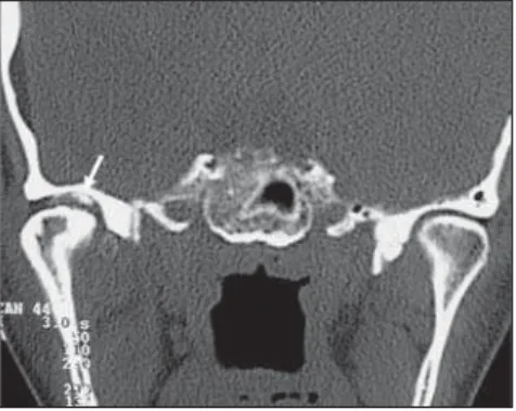
fractures. Condylar neck fractures can be high, medium or low (Figure 10), depend-ing on their positiondepend-ing. On the other hand, condylar head fractures can be subdivided into intracapsular (Figure 11) and extracap-sular(5).
Intracapsular fractures are less common and, most frequently affect children. Ext-racapsular fractures involve the subcon-dylar region and most frequently occur unilaterally, eventually in association with contralateral mandibular angle fracture(21).
The differentiation between high condylar neck fracture and extracapsular condylar head fracture is somewhat arbitrary. Condylar process fractures can further be subdivided into displaced and nondis-placed fractures. Typically, because of the traction exerted by the pterygoid muscle, fractures are medially displaced.
The evaluation of the type and degree of disk displacement is critical for the
therapeutic planning, considering the angle between the mandibular head and ramus, the contact between the fracture ends (par-ticularly the degree of contraction in the vertical plane that affects the occlusion), the transverse displacement, and the posi-tion of the fractured head as related to the articular eminence and articular fossa, these parameters being useful in the decision making about a surgical or non-surgical ap-proach(5).
Generally, the initial evaluation is per-formed by means of conventional radiog-raphy, particularly panoramic radiography of the mandible, because of the low expen-siveness, high availability and experience with the method. CT is considered as the imaging method of choice for evaluating facial traumas. The multislice technology has considerably reduced the CT images acquisition time, achieving an improved coverage of the sample volume, besides allowing imaging of different high-resolu-tion angles and planes, and excellent 3D images reconstruction, constituting a quite useful tool in the planning of reconstruc-tive surgeries(21).
MRI can be useful in the identification of post-traumatic disk displacement, as well as capsular, cartilaginous and ligamen-tous lesions, playing a significant role in the pretreatment evaluation of condylar process fractures. The treatment is aimed at an appropriate reduction and consolida-tion of the fracture, preservaconsolida-tion of the dental occlusion and articular function, besides a good aesthetic outcome. Func-tional disorders, dental maloccusion, pseudoarthrosis, pain, ankylosis and os-teoarthritis constitute complications of in-appropriate fracture consolidation(5)
(Fig-ure 12).
The selection of open or closed reduc-tion techniques depends on the injury type and severity, presence of unilateral or bilat-eral fracture, as well as the patient’s age and comorbidities.
Generally, the conservative treatment is preferred in cases of unilateral or bilateral nondisplaced fractures in children. The surgical management is reserved for cases of displaced fractures, even the unilateral ones, provided the articular movement amplitude is impaired or dental malocclu-sion is present.
CONCLUSIONS
Internal derangement corresponds to the main pathologic entity found in patients with TMJ-related complaints. Pain, click-ing and functional limitation predominate in the clinical picture. However these signs and symptoms are non-specific and may be found in several non-dysfunctional condi-tions, including inflammatory processes, expansile lesions and post-traumatic alter-ations, among others.
Imaging methods are quite useful in the diagnosis of these conditions which fre-quently are not considered as primary clini-cal suspicion.
MRI and CT present a higher diagnos-tic accuracy as compared with conventional radiology, considering their higher ana-tomical resolution. CT is ideal for evaluat-ing bone structures, while MRI allows the evaluation of soft tissues, the intra-articular disk included. Frequently, these methods are complementary in the study of the TMJ abnormalities, playing a relevant role in the differential diagnosis of several conditions affecting this region.
Additionally, other studies demonstrate that differentiated and symmetrical mor-phological alterations of the styloid process radiographically evaluated in patients with temporomandibular joints disorder occur independently from gender and age(22).
REFERENCES
1. Rao VM, Bacelar MT. MR imaging of the tem-poromandibular joint. Magn Reson Imaging Clin N Am. 2002;10:615–30.
Figure 10. Multislice CT. 3D reconstruction dem-onstrating low condylar neck fracture with displace-ment (arrow).
Figure 12. 3D reconstruction by the volume ren-dering technique (VRT) demonstrating the inad-equate positioning of the right mandibular condyle in relation to the articular fossa (arrow).
2. Heo MS, An BM, Lee SS, et al. Use of advanced imaging modalities for the differential diagnosis of pathoses mimicking temporomandibular dis-orders. Oral Surg Oral Med Oral Pathol Oral Radiol Endod. 2003;96:630–8.
3. Dalrymple NC, Prasad SR, El-Merhi FM, et al. Price of isotropy in multidetector CT. Radiographics. 2007;27:49–62.
4. Murphy WA, Kaplan PA. Temporomandibular joint. In: Resnick D, editor. Diagnosis of bone and joint disorders. Philadelphia: Saunders; 1995. p. 1699–754.
5. Sommer OJ, Aigner F, Rudisch A, et al. Cross-sec-tional and funcCross-sec-tional imaging of the temporoman-dibular joint: radiology, pathology and basic bio-mechanics of the jaw. Radiographics. 2003;23: e14. Epub 2003 Aug 14.
6. Brasileiro CB, Cardoso VN, Ruckert B, et al. Ava-liação de processos inflamatórios na articulação temporomandibular empregando leucócitos autó-logos marcados com tecnécio-99m em modelo animal. Radiol Bras. 2006;39:283–6. 7. Lucaya J, Herrera M, Vera J. Unilateral
hyperpla-sia of the coronoid process in a child: a cause of restricted opening of the mouth. Radiology. 1982; 144:528.
8. Kreutz RW, Sanders B. Bilateral coronoid hyper-plasia resulting in severe limitation of
mandibu-lar movement: report of a case. Oral Surg Oral Med Oral Pathol. 1985;60:482–4.
9. Munk PL, Helms CA. Coronoid process hyper-plasia: CT studies. Radiology. 1989;171:783–4.
10. Isberg A, Isacsson G, Nah KS. Mandibular coro-noid process locking: a prospective study of fre-quency and association with internal derange-ment of the temporomandibular joint. Oral Surg Oral Med Oral Pathol. 1987;63:275–9.
11. Westesson PL, Yamamoto M, Sano T, et al. Tem-poromandibular joint. In: Som P, Curtin HD, edi-tors. Head and neck imaging. St Louis: Mosby; 2003. p. 995–1053.
12. Larheim TA, Westesson PL. Temporomandibular joint. In: Larheim TA, Westesson PL, editors. Maxillofacial imaging. Berlin, Heidelberg, New York: Springer; 2006. p. 143–78.
13. Fernandes JL, Viana SL. Avaliação por imagem das artropatias por deposição de cristais. In: Fer-nandes JL, Viana SL. Diagnóstico por imagem em reumatologia. Rio de Janeiro: Guanabara Koogan; 2007. p. 178–204.
14. Heffez LB. Imaging of internal derangements and synovial chondromatosis of the temporomandibu-lar joint. Radiol Clin North Am. 1993;31:149–62. 15. Quinn PD, Stanton DC, Foote JW. Synovial chondromatosis with cranial extension. Oral Surg Oral Med Oral Pathol. 1992;73:398–402.
16. Sun S, Helmy E, Bays R. Synovial chondro-matosis with intracranial extension. A case report. Oral Surg Oral Med Oral Pathol. 1990;70:5–9.
17. Gillespy T 3rd, Genant HK, Helms CA. Magnetic resonance imaging of osteonecrosis. Radiol Clin North Am. 1986;24:193–208.
18. Schellhas KP, Wilkes CH, Fritts HM, et al. MR of osteochondritis dissecans and avascular ne-crosis of the mandibular condyle. AJR Am J Roentgenol. 1989;152:551–60.
19. Larheim TA, Westesson PL, Hicks DG, et al. Os-teonecrosis of the temporomandibular joint: cor-relation of magnetic resonance imaging and his-tology. J Oral Maxillofac Surg. 1999;57:888–98. 20. Laskin DM. Diagnosis of pathology of the tem-poromandibular joint. Clinical and imaging per-spectives. Radiol Clin N Am. 1993;31:135–47.
21. Cinnamon J, Shanmuganathan K, Robertson BC. Computed tomography of maxillofacial trauma: patterns of osseous injury and associated soft tis-sue involvement. In: Mirvis SE, Shanmuganathan K, editors. Imaging in trauma and critical care. Philadelphia: Saunders; 2003. p. 133–83. 22. Guimarães SMR, Carvalho ACP, Guimarães JP,