Sonographic determination of liver size in healthy
newborns, infants and children under 7 years of age*
Determinação do tamanho do fígado de crianças normais, entre 0 e 7 anos,por ultrassonografia
Silvia Maria Sucena da Rocha1, Ana Paula Scoleze Ferrer2, Ilka Regina Souza de Oliveira3, Azzo Widman4, Maria Cristina Chammas5, Luiz Antonio Nunes de Oliveira6, Giovanni Guido Cerri7
OBJECTIVE: The present study was aimed at sonographically determining the liver size in healthy newborns, infants and children under 7 years of age, correlating results with age, sex, height, body weight and body mass index. MATERIALS AND METHODS: A total of 584 healthy children subdivided into 11 age groups were evaluated with measurements of the left lobe craniocaudal diameter at the midsternal line, and the craniocaudal diameter of the right lobe posterior surface at the midclavicular line. The following tests were utilized for statistical analysis: a) Pearson’s correlation coefficient (correlation study); b) non-paired Student’s
t-test (comparison of measures between sexes); c) nonlinear regression models (nomograms). RESULTS:
The liver size presented a progressive growth from the birth up to the age of 7, proportionally lower than the body growth, in correlation with age, height and body weight (r > 0.70). Correlation with the body mass
index was not observed (r < 0.11). There was no significant difference in liver size between male and
female individuals. CONCLUSION: Liver size was sonographically determined in healthy children under the age of 7 by means of a standardized method, demonstrating a strong correlation with age and anthropometric indicators. Nomograms demonstrate the typical variations of the liver size in the population evaluated with a different growth pattern for each hepatic lobe.
Keywords: Liver; Liver size; Biometry; Child; Ultrasonography.
OBJETIVO: Determinar o tamanho do fígado de crianças normais, entre 0 e 7 anos de idade, por ultrassono-grafia, correlacionando os valores obtidos com: idade, sexo, estatura, peso corporal e índice de massa cor-pórea. MATERIAIS E MÉTODOS: Foram examinadas 584 crianças saudáveis, subdivididas em 11 grupos etários, sendo medidos o diâmetro crânio-caudal do lobo esquerdo, na linha médio-esternal, e o diâmetro crânio-caudal da superfície posterior do lobo direito, na linha hemiclavicular. Na análise estatística foram aplicados: a) coeficiente de correlação de Pearson (estudo de correlação); b) teste t de Student não-pareado
(comparação das medidas entre os sexos); c) modelos de regressão não linear (nomogramas). RESULTADOS: O tamanho hepático apresentou aumento progressivo, do nascimento aos 7 anos de idade, proporcional-mente menor que o crescimento corporal, correlacionado com idade, estatura e peso corporal (r > 0,70),
não havendo correlação com índice de massa corpórea (r < 0,11). Não se observou diferença consistente
das medidas hepáticas em relação ao sexo. CONCLUSÃO: Valores do tamanho do fígado de crianças nor-mais (entre 0 e 7 anos) foram determinados mediante aplicação de técnica padronizada, verificando-se forte correlação com a idade e indicadores antropométricos. Nomogramas demonstram as variações normais do tamanho hepático na população estudada, com crescimento diferenciado para cada lobo.
Unitermos: Fígado; Tamanho do fígado; Biometria; Criança; Ultrassonografia.
Abstract
Resumo
* Study developed at Serviço de Apoio Diagnóstico e Tera-pêutico (SADT) do Instituto da Criança “Prof. Pedro de Alcân-tara” do Hospital das Clínicas da Faculdade de Medicina da Universidade de São Paulo (Icr/HC-FMUSP) and Instituto de Ra-diologia do Hospital das Clínicas da Faculdade de Medicina da Universidade de São Paulo (InRad/HC-FMUSP), São Paulo, SP, Brazil.
1. MD, Radiologist, Assistant at Serviço de Apoio Diagnóstico e Terapêutico (SADT) do Instituto da Criança “Prof. Pedro de Alcântara” do Hospital das Clínicas da Faculdade de Medicina da Universidade de São Paulo (ICr/HC-FMUSP), São Paulo, SP, Brazil.
2. MD, Pediatrician, Assistant at General Pediatric Ambulatory of Hospital Universitário da Universidade de São Paulo (HU-USP) and Liga de Puericultura do Instituto da Criança “Prof. Pedro de Alcântara” do Hospital das Clínicas da Faculdade de Medicina
Rocha SMS, Ferrer APS, Oliveira IRS, Widman A, Chammas MC, Oliveira LAN, Cerri GG. Sonographic determination of liver size in healthy newborns, infants and children under 7 years of age. Radiol Bras. 2009;42(1):7–13.
da Universidade de São Paulo (ICr/HC-FMUSP), São Paulo, SP, Brazil.
3. PhD, Professor at Department of Radiology of Faculdade de Medicina da Universidade de São Paulo (FMUSP), Technical Director for Division of Imagenology – Hospital Universitário da Universidade de São Paulo (HU-USP), São Paulo, SP, Brazil.
4. PhD, Physician Assistant Supervisor at Divison of Surgery of Digestive System II – Hospital das Clínicas da Faculdade de Medicina da Universidade de São Paulo (HC-FMUSP), São Pau-lo, SP, Brazil.
5. PhD, Technical Director at Division of Ultrasonography of Instituto de Radiologia do Hospital das Clínicas da Faculdade de Medicina da Universidade de São Paulo (InRad/HC-FMUSP), São Paulo, SP, Brazil.
6. Head for Serviço de Apoio Diagnóstico e Terapêutico (SADT) at Instituto da Criança “Prof. Pedro de Alcântara” do Hospital das
Clínicas da Faculdade de Medicina da Universidade de São Paulo (ICr/HC-FMUSP), São Paulo, SP, Brazil.
7. Titular Professor at Department of Radiology – Faculdade de Medicina da Universidade de São Paulo (FMUSP), Head for Instituto de Radiologia do Hospital das Clínicas da Faculdade de Medicina da Universidade de São Paulo (InRad/HC-FMUSP), São Paulo, SP, Brazil.
Mailing address: Dra. Silvia Maria Sucena da Rocha. Serviço de Apoio ao Diagnóstico e Terapia do Instituto da Criança “Prof. Pedro de Alcântara”, Hospital das Clínicas da Faculdade de Medicina da Universidade de São Paulo. Avenida Doutor Enéas de Carvalho Aguiar, 647, 2° andar. São Paulo, SP, 05403-001, Brazil. E-mail: [email protected]
INTRODUCTION
Hepatomegaly is a frequent clinical finding in children, and may be caused by intrinsic liver diseases or by systemic alter-ations(1), and in case of clinical suspicion,
ultrasonography (US) is generally the method of choice for starting diagnostic investigation in pediatric patients. Biom-etry studies in children by means of US, however, propose different methods(1–15),
none of them with consensus acceptance. In a previous study(16) the authors have
standardized an easy and reproducible ultrasonographic biometry method for uti-lization in the pediatric age range(16,17)
based on the measurement of the hepatic length in two longitudinal planes. The sin-gular aspect of such technique lies in the proposition of intrahepatic anatomical re-pairs in association with external orienta-tion lines and the introducorienta-tion of a new parameter for measurement of the right hepatic lobe, resulting in higher accuracy in the definition of section planes and the measurements themselves.
The present study is aimed at determin-ing the liver size in healthy children in the age range between 0 and 7 years, by apply-ing the technique described by Rocha et al.(16), and correlating the results with the
following variables: age, height, body weight, body mass index and sex.
MATERIALS AND METHODS
Casuistic
Between April 2003 and April 2005, a cross-sectional study with measurements of children livers by B-mode ultrasonography was developed. The sample included 584 children whose parents or guardians signed a term of free and informed consent. Among these children, 301 were girls (51.54%) and 283 boys (48.46%) aged from 0.23 months (7 days) to 83.8 months (6 years, 11 months and 25 days) (mean = 42.2 months).
The project was approved by the Ethics Committee for Analysis of Research Projects (Comissão de Ética para Análise de Projetos de Pesquisa – CAPPesq) of Board of Clinical Directors of Hospital das Clínicas da Faculdade de Medicina da Uni-versidade de São Paulo (HC-FMUSP).
The children were referred by the fol-lowing institutions related with Univer-sidade de São Paulo:
a) Liga de Puericultura do Instituto da Criança (Childcare League of the Children’s Institute) (ICr) of HC-FMUSP (32 children); b) General Pediatrics Ambulatory (AGEP) of Hospital Universitário da Uni-versidade de São Paulo (HU-USP) (18 chil-dren);
c) Day care unit of HC-FMUSP (248 children);
d) Municipal School for Children (EMEI) “Prof. Antonio Branco Lefèvre” located in the HC-FMUSP Complex (286 children);
The selected children population deter-mined the age range covered by the study. The children were divided into groups ac-cording to age, as follows:
– group 1: 0 to 2 months and 29 days (n
= 32; 13 girls, 19 boys);
– group 2: 3 to 5 months and 29 days (n
= 34; 19 girls, 15 boys);
– group 3: 6 to 8 months and 29 days (n
= 42; 17 girls, 25 boys);
– group 4: 9 to 11months and 29 days (n
= 36; 21 girls, 15 boys);
– group 5: 12 to 17 months and 29 days (n = 32; 18 girls, 14 boys);
– group 6: 18 months to 23 months and 29 days (n = 36; 20 girls, 16 boys);
– group 7: 2 years to 2 years, 11 months and 29 days (n = 34; 19 girls, 15 boys);
– group 8: 3 years to 3 years, 11 months and 29 days (n = 48; 23 girls, 25 boys);
– group 9: 4 years to 4 years, 11 months and 29 days (n = 70; 47 girls, 23 boys);
– group 10: 5 years to 5 years, 11 months and 29 days (n = 111; 53 girls, 58 boys);
– group 11: 6 years to 6 years, 11 months and 29 days (n = 109; 51 girls, 58 boys);
All the children were weighted, mea-sured and evaluated by a pediatrician. Weight and height were recorded and com-pared with the weighted stature standard of the National Center of Health Statistics (NCHS) 2000 USA.
Subsequently to the clinical evaluation, the US scans were performed. All the chil-dren were examined by the same profes-sional, an experienced radiologist special-ized in US.
Exclusion criteria: Exclusion criteria were the following: a) children with altered
clinical or US examinations; b) children bearing any chronic or acute diseases.
Inclusion criteria: All the children with ages up to 6 years, 11 months and 29 days, whose parents authorized the partici-pation in the research, until the minimum number of individuals for each age range was achieved.
Method
Children assisted in the Childcare League of The Children’s Institute of ICr/ HC-FMUSP were submitted to US scan in the Division of Diagnostic and Therapeu-tic Support of the Institute, with a Logic 7 unit(General Electric Medical Systems; Milwaukee, USA) and an Apogee 800 Plus unit (ATL Inc.; Bothell, USA), utilizing convex, multifrequency 2–10 MHz and 2– 7 MHz transducers, respectively.
Children assisted in the AGEP of HU-USP underwent US scan in the Imagenol-ogy Division of HU-USP, with a conven-tional US equipment model SSA 270-A (Toshiba; Tokyo, Japan), utilizing a convex, multifrequency, 3–7 MHz transducer.
The children enrolled at EMEI under-went US scan with a portable Logiq Book unit (General Electric Medical Systems; Milwaukee, USA), utilizing a convex, mul-tifrequency 3-7 MHz transducer.
The equipment settings were standard-ized according to the protocol for pediat-ric abdomen.
The examinations were performed with the children in the supine position, with the upper limbs extended along the body, and extended lower limbs, without any support under the head, and without any prepara-tion or sedaprepara-tion. The transducer was posi-tioned below the costal cage, with longitu-dinal orientation, in orthogonal position relative to spine plane.
Measurements of the liver were based on external orientation lines in correlation with intra- and extrahepatic anatomic re-pairs (Figure 1).
through an oblique line traced between the upper extremity and the lower hepatic bor-der.
Statistical analysis
The measurements were correlated with age, height and weight of the children evaluated, with the Pearson’s correlation coefficient. The non-paired Student’s t test
was utilized for comparing measurements between the female and male groups. The significance level utilized was 0.05.
Normality curves for liver size, as a func-tion of age and the anthropometric variable with highest correlation coefficient, were elaborated by means of non-linear regres-sion models, utilizing the Curve Expert 1.3 software.
RESULTS
Of the 584 children included in the sample, 301 were girls (51.54%0 and 283 were boys (48.46%), aged between 0.23 months (7 days) and 83.8 months (6 years, 11 months and 25 days) (mean = 42.2 months, median = 47.2 months).
The sample profile in terms of anthro-pometric indicators is very close to the ex-pected distribution standard, according to the reference curve of the NCHS 2000 (Figure 2). The height/age curve practically matches the expected distribution, while the weight/age curve shows a small devia-tion to the right as compared with the ref-erence curve, indicating a trend towards overweight in the target population of this study.
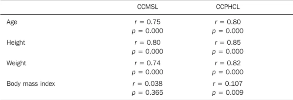
Analysis of correlation between liver measures, age, anthropometric indicators (height and weight) and body mass index
The correlation analysis showed a posi-tive and significant correlation between the liver measures, age, height and body weight, with high correlation coefficients (r > 0.70), while for the body mass index,
the correlation coefficients were close to zero (r < 0.11) for measures of both lobes
(Table 1).
Figure 1. Schematic representation and photographic documentation of a ultrasonography study, showing the section planes and reference points for liver measurements. Section plane A established by the midsternal line (LME) and Section plane B established by the hemiclavicular line (LHC). Intra-hepatic anatomic repairs: left hepatic vein (VHE) in section plane A and cross section of right portal branch (RPD) in section plane B, where one uses the diaphragm (DIAF) with intra-abdominal extra-hepatic repair. VCI, inferior vena cava; RD, right kidney; LC, caudate lobe (modified from Rocha et al.(16)).
Table 1 Correlation of liver measures and age, height, body weight and body mass index (n = 584).
Age
Height
Weight
Body mass index
CCMSL
r = 0.75 p = 0.000
r = 0.80 p = 0.000
r = 0.74 p = 0.000
r = 0.038 p = 0.365
CCPHCL
r = 0.80 p = 0.000
r = 0.85 p = 0.000
r = 0.82 p = 0.000
r = 0.107 p = 0.009
CCMSL, craniocaudal diameter in the midsternal line; CCPHCL, craniocaudal diameter of the posterior surface of the liver on the hemiclavicular line; r, Pearson’s correlation coefficient, p, probability of significance.
significant difference was observed be-tween sexes with respect to the CCPHCL measure (p = 0.006 and p = 0.009,
respec-tively).
In age group 8 (3 to 4 years) there was significant difference between sexes with respect to CCMSL measure (p = 0.004).
Comparison of liver measures in the different age ranges
Both the left hepatic lobe and the right hepatic lobe presented progressive increase in size with aging (Figure 4).
The average of CCMSL measures ranged from 3.41 cm in neonates to 6.91 cm in children between 6 and 7 years of age (Table 2).
The mean CCPHCL ranged from 6.61 cm in neonates to 10.94 cm in children between 6 and 7 years of age (Table 3).
Figure 4. Graphical representation of right and left hepatic lobes tabulated mean CCPHCL and CCMSL measures in the different age groups.
Figure 3. Graphical representation of the comparison between mean CCMSL (a) and CCPHCL (b) in the female and male groups by age.
Nomograms
Nomograms of size of left and right hepatic lobes according to age (Figures 5 and 6) and height (Figures 7 and 8) were established by means of non-linear regres-sion models. The curves selected were those that more closely matched the data, with higher correlation coefficients, smaller errors and with an evolution pattern compatible with the biologic phenomenon.
DISCUSSION
The present study relied on a large sample of healthy children. It is known that, in population studies, the high number of Comparison of liver measures between
the female and male groups by age The comparison of CCMSL and CCPHCL between the female and male groups in the different age ranges
demon-strated small but significant difference in three of them (p > 0.05), with higher
val-ues for the boys (Figure 3):
Figure 5. Percentile curves (3, 5, 10, 25, 50, 75, 90, 95 and 97) of left hepatic lobe size, represented by the CCMSL diameter, according to age.
Figure 6. Percentile curves (3, 5, 10, 25, 50, 75, 90, 95 and 97) of right hepatic lobe, represented by the CCPHCL diameter, according to age.
Table 2 Mean, standard deviation, median and minimum and maximum values for measurements of craniocaudal diameter in the midsternal line in the different age groups.
Age (months)
0 to 3
3 to 6
6 to 9
9 to 12
12 to 18
18 to 24
24 to 36
36 to 48
48 to 60
60 to 72
72 to 84 n 32 34 42 36 32 36 34 48 70 111 109 Mean 3.41 4.05 4.80 4.77 5.51 5.65 6.32 6.57 6.62 6.79 6.91 Standard deviation 0.55 0.77 0.65 0.70 0.62 0.77 0.83 0.71 0.69 0.87 0.85 Median 3.27 4.04 4.88 4.74 5.40 5.53 6.23 6.60 6.58 6.85 6.87 Minimum 2.48 2.83 3.20 3.40 4.49 3.77 4.91 5.15 5.20 4.61 3.90 Maximum 4.91 5.50 6.27 6.03 6.87 6.91 7.78 8.28 8.56 8.86 9.34
Table 3 Mean, standard deviation, median, minimum and maximum val-ues for measurements of craniocaudal diameter of the posterior surface of the liver on the hemiclavicular line, in the different age groups.
Age (months)
0 to 3
3 to 6
6 to 9
9 to 12
12 to 18
18 to 24
24 to 36
36 to 48
48 to 60
60 to 72
72 to 84 n 32 34 42 36 32 36 34 48 70 111 109 Mean 6.61 7.66 8.40 8.44 9.00 9.35 9.47 9.98 10.28 10.68 10.94 Standard deviation 0.57 0.72 0.91 0.69 0.78 0.74 0.74 0.67 0.79 0.85 0.87 Median 6.62 7.75 8.32 8.42 8.95 9.20 9.37 10.02 10.30 10.69 10.89 Minimum 4.76 5.80 6.35 6.56 7.38 7.83 8.14 7.87 8.67 8.75 9.19 Maximum 7.50 9.09 10.14 9.61 10.83 11.17 11.28 11.53 12.24 13.09 13.33
Figure 7. Regression curve for estimated size of left hepatic lobe (CCMSL) according to height.
sample elements decreases the standard error of the estimated value in the re-search(18). The US studies for determination
of the liver size variability in healthy chil-dren usually utilized as a reference in the ultrasonography practice were based on smaller samples(5–8,12–14).
In spite of being a sample of conve-nience, the present sample presents as fa-vorable characteristics, racial diversity, similar number of boys and girls, and the prevalence of average socioeconomic background, thus assuring a minimal risk for malnutrition in this group, allowing the inclusion of small but healthy children (be-low the 3rd percentile in NCHS weighted growth curves). One also opted for the in-clusion of children overweight (above the 97th percentile of the reference curve), since no relevant clinical or anatomic cor-relation was found between liver size and body mass index (r < 0.11), indicating that
overweight does not represent an influenc-ing factor on the hepatic dimensions.
The liver size in the selected children population presented a continuous and pro-gressive increase from birth to 7 years of age. The increase in dimensions was ob-served in both hepatic lobes, however in a distinctive manner. The left hepatic lobe, represented by the CCMSL parameter, pre-sented an accelerated growth in the first three years of life, and after that a practi-cally inexpressive growth, with decreasing yearly increments (≤ 0.25 cm). The right hepatic lobe, represented by the CCPHCL parameter, presented a gradual, progressive and significant growth, from birth to 7 years of age, with a faster growth in the first 9 months of life (increments around 1.0 cm per quarter), and progressive growth, how-ever with smaller increments in the subse-quent age groups.
The growth pattern observed in the present study corresponds to the expected standard, with a higher rate in the first years of life, following the somatic growth, which is more accelerated in this age group19). The differentiated growth of each
lobe was also expected, as embryology and anatomy studies describe a progressive dis-proportion of hepatic lobe dimensions, with a relative reduction of the left hepatic lobe, from the intrauterine period until af-ter birth(20). The body growth was
propor-tionally larger than the hepatic growth in the studied age groups, with an average increase in height of 125.5%, while the hepatic left lobe grew 103.5% and the right hepatic lobe grew 71%.
The measurements of the liver pre-sented a positive and significant correlation with age, height and body weight, in agree-ment with results found in literature(2,3,5– 8,12,13,15,21–25). The height was the variable
that presented the highest correlation coef-ficients for both parameters for liver size evaluation (r = 0.80 for CCMSL and r =
0.85 for CCPHCL). The analysis of corre-lation between the mean hepatic diameter measures and the variables age and body weight also evidenced high and very close coefficients: r = 0.75 for CCMSL and r =
0.80 for CCPHCL, in relation to age, and
r = 0.74 for CCMSL and r = 0.82 for
CCPHCL, in relation do weight.
The differences found between the he-patic measure averages in relation to sex, were not constant amongst the several age groups, therefore it was not possible to characterize a differentiated growth pattern for boys and girls, and not even a trend. In spite of being statistically significant, these measures variations in relation to sex were punctual and small in absolute values, therefore being inexpressive from a clini-cal point of view. Thus, in agreement with other authors(2,6,8,11,12,22,23,26), the authors
believe that such finding in not consistent enough to justify the construction of differ-ent reference tables for both sexes.
The normal liver size values in the sample evaluated presented great variation, in all age groups, and in both measured diameters. Similar results are found in the several hepatic biometry studies reviewed, particularly stressed by KonuÕ et al.(12). A
possible explanation for the wide spectrum of values for the normality range of liver size may lie in the complex and variable morphology of the organ.
The liver size variation values based on age for the children included in the present study, were presented in the form of: a) percentile curves, with high degree of ad-justment (r = 0.98 for CCMSL and r = 0.99
for CCPHCL); b) tables showing means, minimum and maximum values and the standard deviations by age group. The tables are practical for use in the routine of
the sonographist. On the other hand, the curves allow the precise determination of values in the class intervals of the tables. Ancillary normality curves as a function of height were also established, to contem-plate the evaluation of children with growth deficit, for whom the tables as a function of age were not appropriate.
The comparison between the liver size values in the population of healthy children of the present sample with those obtained in studies with children populations from other countries could only be made in an approximate manner, as each study applies different methods both in what refers to the measurements, as well as in data process-ing.
The variation in size of the left hepatic lobe was similar to that in other popula-tions(5,12). However, with respect to the right hepatic lobe, the mean measures of the present study were significantly larger, in all age groups, when compared to those determined in other studies(5,12). This can be
explained by the difference in size between the measured hepatic surfaces: the poste-rior surface in the present study and the anterior surface in those of other authors. The length of the posterior hepatic surface is clearly larger than that the anterior sur-face, its size being affected, to a certain extent, by the anteroposterior hepatic diam-eter. The authors believe that the utilization of CCPHCL as a parameter to estimate the dimensions of the right hepatic lobe has the potential of representing both alterations of the longitudinal axis size as well as, indi-rectly, alterations in the anteroposterior axis. Additionally, this measurement allows the simultaneous assessment of the mor-phological characteristics of the right lobe (surface and inferior border), which consti-tutes an additional factor in favor of the adoption of this parameter.
FINAL CONSIDERATIONS
The determination of the liver size in healthy children by means of US was yet to be made in our environment. The results obtained reflect a very small fraction of the children population in our region, and rig-orously they should not be extended as a reference standard. However, they do con-stitute a starting point for a future popula-tion study, ideally covering the entire pe-diatric age group.
REFERENCES
1. Walker WA, Mathis RK. Hepatomegaly. An ap-proach to differential diagnosis. Pediatr Clin North Am. 1975;22:929–42.
2. Holder LE, Strife J, Padikal TN, et al. Liver size determination in pediatrics using sonographic and scintigraphic techniques. Radiology. 1975; 117:349–53.
3. Wladimiroff JW, Sekeris A. Ultrasonic assess-ment of liver size in the newborn. J Clin Ultra-sound. 1977;5:316–20.
4. Rylance GW, Moreland TA, Cowan MD, et al. Liver volume estimation using ultrasound scan-ning. Arch Dis Child. 1982;57:283–6. 5. Dittrich M, Milde E, Dinkel E, et al. Sonographic
biometry of liver and spleen size in childhood. Pediatr Radiol. 1983;13:206–11.
6. Phuapradit P, Assadamongkol K, Udompanich O, et al. Liver size and serum alkaline phosphatase in normal preschool Thai children. J Med Assoc Thai. 1986;69 Suppl 2:69–76.
7. Assadamongkol K, Phuapradit P, Udompanich O, et al. Liver size and serum alkaline phosphatase in normal Thai school-aged children. J Med Assoc Thai. 1989;72 Suppl 1:88–93.
8. Friis H, Ndhlovu P, Mduluza T, et al. Ultrasono-graphic organometry: liver and spleen dimensions among children in Zimbabwe. Trop Med Int Health. 1996;1:183–90.
9. Hessel G. Hepatometria na infância – compara-ção entre o método clínico e ultra-sonográfico [dissertação de mestrado]. Campinas: Universi-dade Estadual de Campinas; 1991.
10. Chen CM, Wang JJ. Clinical and sonographic as-sessment of liver size in normal Chinese neonates. Acta Paediatr. 1993;82:345–7.
11. Jungthirapanich J, Kaewtubtim J, Poovorawan Y. A new reference line for measuring the liver size in healthy newborns. J Med Assoc Thai. 1998; 81:938–43.
12. KonuÕ ÖL, Özdemir A, Akkaya A, et al. Normal liver, spleen, and kidney dimensions in neonates, infants, and children: evaluation with sonography. AJR Am J Roentgenol. 1998;171:1693–8. 13. Haddad-Zebouni S, Hindy R, Slaba S, et al.
Éva-luation échographique de la taille des reins, du foie et de la rate chez l’enfant. Arch Pédiatr. 1999; 6:1266–70.
14. Sarac K, Kutlu R, Yakinci C, et al. Sonographic evaluation of liver and spleen size in school-age children. Turk J Med Sci. 2000;30:187–90. 15. Safak AA, Simsek E, Bahcebasi T. Sonographic
assessment of the normal limits and percentile curves of liver, spleen, and kidney dimensions in healthy school-aged children. J Ultrasound Med. 2005;24:1359–64.
16. Rocha SMS, Oliveira IRS, Widman A, et al. He-patometria ultra-sonográfica em crianças: proposta de novo método. Radiol Bras. 2003;36:63–70. 17. Rocha SMS, Chisman BSK, Barbosa PR, et al.
Hepatometria ultra-sonográfica em crianças: pro-posta de novo método – síntese comentada e es-tudo de reprodutibilidade inter-observadores. Sociedad Iberoamericana de Información
Cien-tífica (SIIC) [periódico online]. 2005 [acessado Jan 2005]. Disponível em: http://www.siicsalud. com/des/des043/05627026.htm
18. Kirkwood BR, Sterne JAC. Confidence interval for a mean. In: Kirkwood BR, Sterne JAC, edi-tors. Essential medical statistics. 2nd ed. Malden: Blackwell Science; 2003. p. 50–7.
19. Marcondes E. Crescimento normal – tabelas e gráficos. In: Marcondes E. Crescimento normal e deficiente. 3ª ed. São Paulo: Sarvier; 1989. p. 42–69.
20. Gray H. Splanchnology. In: Gray H, editor. Anatomy of the human body. 20th ed. Philadel-phia: Lea & Febiger; 1918.
21. Coppoletta JM, Wolbach SB. Body length and or-gan weights of infants and children. A study of the body length and normal weights of the more important vital organs of the body between birth and twelve years of age. Am J Pathol. 1933;9:55– 70.
22. Younoszai MK, Mueller S. Clinical assessment of liver size in normal children. Clin Pediatr (Phila). 1975;14:378–80.
23. Carpentieri U, Gustavson LP, Leach TM, et al. Liver size in normal infants and children. South Med J. 1977;70:1096–7.
24. Lawson EE, Grand RJ, Neff RK, et al. Clinical es-timation of liver span in infants and children. Am J Dis Child. 1978;132:474–6.
25. Markisz JA, Treves ST, Davis RT. Normal hepatic and splenic size in children: scintigraphic deter-mination. Pediatr Radiol. 1987;17:273–6. 26. Weisman LE, Cagle N, Mathis R, et al. Clinical