Brain
18
F-FDG PET-MRI coregistration: iconographic essay*
PET-RM neurológico com FDG-18F: ensaio iconográfico
José Leite Gondim Cavalcanti Filho1, Léa Mirian Barbosa da Fonseca2, Romeu Côrtes Domingues3, Roberto Côrtes Domingues4, Luiz de Souza Machado Neto5, Emerson Leandro Gasparetto6
The combination of positron emission tomography (PET) with magnetic resonance imaging (MRI) has been the subject of several studies in recent years. Positron emission tomography is the most sensitive and specific imaging modality in the detection of metabolic changes, but presents limited spatial resolution. On the other hand, MRI presents a significant spatial resolution, besides evaluating soft tissues signal intensity with excellent contrast resolution. The present iconographic essay is aimed at demonstrating the potential clinical application of PET/MRI coregistration. The studies were performed in a dedicated PET unit with 18
F-fluorodeoxyglucose (FDG) as radiopharmaceutical and coregistered with 1.5 T or 3 T brain MRI. The brain images fusion software presents an already well-established accuracy, so a significant synergy between a functional PET study and an excellent MRI anatomical detail is achieved. The most attractive clinical applications of this approach are the following: epileptogenic zone assessment in patients refractory to drug therapy, identification of patients with cognitive impairment at higher risk for progression to dementia and differentiation of dementias and Parkinsonian syndromes.
Keywords: Fluorodeoxyglucose; FDG; Positron emission tomography; PET; Magnetic resonance imaging; Fusion; Clinical neurology.
A integração da tomografia por emissão de pósitrons (PET) com a ressonância magnética (RM) tem sido alvo de diversos estudos nos últimos anos. O PET é a modalidade de imagem mais sensível e específica na detec-ção de alterações metabólicas, entretanto, apresenta limitada resoludetec-ção espacial. Por outro lado, a RM apre-senta importante resolução espacial, além de avaliar estruturas com intensidade de sinal de partes moles com excelente contraste. O objetivo deste estudo é demonstrar, na forma de ensaio iconográfico, as poten-ciais aplicações clínicas da fusão de imagens de PET e RM. Os exames foram realizados em aparelho PET dedicado utilizando como radiofármaco a fluordeoxiglicose-18
F (FDG) e corregistrados com RM de 1,5 T ou 3 T do encéfalo. A fusão por programa de imagens do cérebro tem acurácia já bem estabelecida. Conse-gue-se, assim, importante sinergia de um estudo funcional de PET com excelente detalhamento anatômico da RM. As aplicações clínicas mais atraentes dessa abordagem são a avaliação da zona epileptogênica em pacientes refratários ao tratamento medicamentoso, identificação dos pacientes com déficit cognitivo com maior risco de progressão para demência e distinção de demências e síndromes parkinsonianas.
Unitermos: Fluordeoxiglicose; FDG; Tomografia por emissão de pósitrons; PET; Imagem por ressonância magnética; Fusão; Neurologia.
Abstract
Resumo
* Study developed at Clínicas de Diagnóstico Por Imagem (CDPI) and Multi-Imagem PET, Rio de Janeiro, RJ, Brazil.
1. Nuclear Physician at Clínica Multi-Imagem PET, Rio de Ja-neiro, RJ, Brazil.
2. PhD, Titular Professor, Universidade Federal do Rio de Ja-neiro (UFRJ), Rio de JaJa-neiro, RJ, Brazil.
3. MD, Director for Clínicas de Diagnóstico Por Imagem (CDPI) and Multi-Imagem PET, Rio de Janeiro, RJ, Brazil.
4. MD, Radiologist at Clínicas de Diagnóstico Por Imagem (CDPI) and Multi-Imagem PET, Rio de Janeiro, RJ, Brazil.
5. Master, Nuclear Physician at Clínica Multi-Imagem PET, Rio de Janeiro, RJ, Brazil.
6. PhD, Associate Professor, Universidade Federal do Rio de Janeiro (UFRJ), Rio de Janeiro, RJ, Brazil.
Mailing address: Dr. José Leite Gondim Cavalcanti Filho. Multi-Imagem PET. Avenida das Américas, 6205, Loja G, Barra da Tijuca. Rio de Janeiro, RJ, Brazil, 22793-080. E-mail: leite_jose @yahoo.com
Received February 1, 2010. Accepted after revision March 15, 2010.
tegrated PET and CT systems. The interpre-tation of PET images alone without fusion with other imaging modalities with a bet-ter anatomical definition such as CT or magnetic resonance imaging (MRI), pre-sents a low specificity and decreased posi-tive predicposi-tive value. In the last few years, a lot has been researched on PET/MRI coregistration(2). A high potential is esti-mated for these methods integration in the diagnosis of neurological diseases, because PET is highly sensitive for detecting meta-bolic changes, but presents a limited spa-tial resolution. On the other hand, MRI presents a high spatial resolution in the
Cavalcanti Filho JLG, Fonseca LMB, Domingues RC, Domingues RC, Machado Neto LS, Gasparetto EL. Brain 18
F-FDG PET-MRI coregistration: iconographic essay. Radiol Bras. 2010;43(3):195–201.
INTRODUCTION
in-evaluation of structures with soft tissues signal such as the brain, additionally to the functional capability of the method.
TECHNICAL ASPECTS
Basically, there are three systems for integrating PET and MRI(3), as follows: 1) independent PET and MRI apparatuses in separate rooms. The images coregistration is performed with dedicated softwares, which generates flexibility, considering that both methods can be separately uti-lized; 2) sequential images acquired in dif-ferent apparatuses, but the patient remains on a single examination table that is used on both the systems. Therefore, this is a hardware-fused system; 3) a completely integrated systems where the images are simultaneously acquired, for example, with a single examination table positioning, neither the patient nor the table is moved in the process of images acquisition.
In some cases, simultaneous images acquisition is essential, considering that certain radiopharmaceuticals present a dif-ferent pharmacokinetics. For example, H2
O-15O passes through the region of
in-terest in a matter of minutes, thus in this case this type of acquisition is required. On the other hand, fluorodeoxyglucose (FDG) takes about 45 minutes to be biodistributed, so simultaneous images acquisition is not relevant in this case.
Images fusion was already utilized even before the introduction of hybrid systems. In fact, it was this method that paved the way to images coregistration by hybrid sys-tems.
Brain images coregistration with soft-ware is already validated because of the static nature of this organ(4). A comparison
of several softwares available for MRI and PET/CT fusion, demonstrated a 2–3 mm accuracy that is smaller than the size of one PET pixel(4). Thus, the fusion of brain
FDG-PET images and MRI becomes an attractive method for noninvasive evalua-tion of neurological diseases.
PET/MRI COREGISTRATION IN COGNITIVE IMPAIRMENT/ DEMENTIA
Recently, the concept of mild cognitive impairment (MCI) was introduced to de-scribe a memory deficit similar to, but with-out other criteria of Alzheimer’s disease(5). Patients with mild cognitive impairment present a rate of conversion to Alzheimer’s disease of approximately 10–15% per year. For this reason, the recognition of such patients is important so that a treatment can be defined as the earliest as possible.
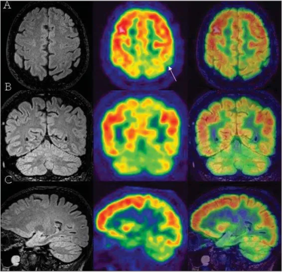
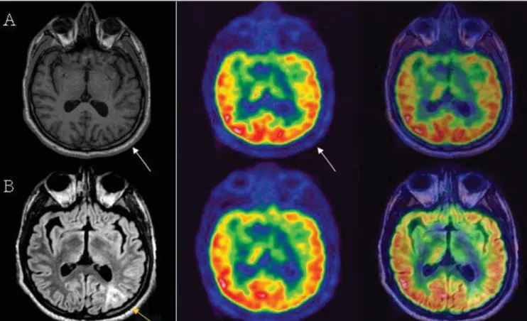
About 60–70% of patients with mild cognitive impairment and presenting mild to severe association cortex hypometabo-lism (Figure 1), even with a mini mental statement examination (MMSE) scored as
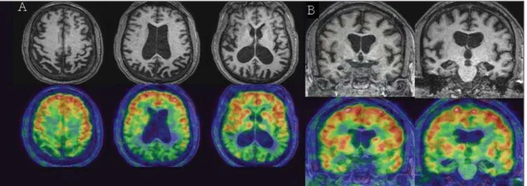
normal, may progress to dementia within two years. Early Alzheimer’s disease tends to present hypometabolism in the parietal, temporal lobes and posterior cingulate
cor-tex. This change can be easily identified at FDG-PET (Figure 2)(6).
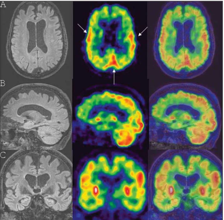
The relevance of the fusion of FDG-PET images and MRI is associated with the
identification of metabolic changes in small-sized structures that are difficult to be anatomically localized at PET, such as the hippocampus, for example (Figure 3),
Figure 3. G.M.R., a 62-year-old patient presenting progressive memory loss for one year. Mini mental statement examination score of 22 (N > 24). Lines A, B and C represent MRI and PET images and PET/MRI coregistration. Both hippocampi present a subtle metabolism decrease (arrows on A and B). Additionally, a mild hypometabolism can be observed in the frontotemporoparie-tal cortex at right (arrowheads on A and C).
that is intimately related to a future cogni-tive impairment and Alzheimer’s disease, as well as the angular gyrus and the precu-neus.
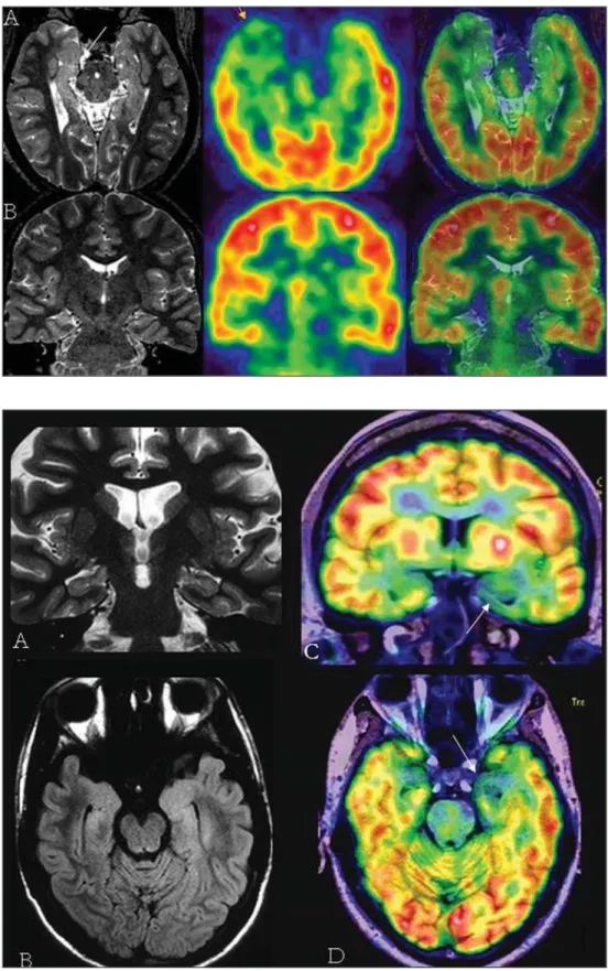
Frontotemporal dementia is not a spe-cific clinical entity, comprising a spectrum including from a classic Pick’s disease to a primary progressive aphasia. It can be identified with PET in the presence of ei-ther frontal or frontotemporal hypometabo-lism (Figure 4).
The clinical diagnosis of vascular de-mentia represents a diagnostic challenge, since a consensus is still to be reached and the convergence among specialists is poor. Additionally, about 20–40% of patients vascular dementia is associated with Alzheimer’s disease(7). Considering that MRI is highly sensitive in the evaluation of structural lesions, but poorly specific for Alzheimer’s disease, the diagnosis of vas-cular dementia is frequently tendentious.
For this reason, the fusion of metabolic PET images with structural MRI emerges as a relevant tool in the correct classifica-tion of these dementias.
PET/MRI COREGISTRATION IN EPILEPSY
The drug therapy for epilepsy is ineffec-tive in a considerable number of patients. Complete resection of the epileptogenic
focus may prevent future seizures in 90% of patients with mesial temporal lobe epi-lepsy, and in up to 70% of patients with cortical dysplasia.
MRI is the method of choice in the
in-vestigation of epileptogenic lesions. FDG-PET adds relevant prognostic information, since it is known that hypometabolism re-stricted to the temporal lobe is correlated with surgical healing in 75% of these
tients, as compared with only 45% of pa-tients with extratemporal hypometabolism (Figures 5 and 6)(8).
A relevant role is played by FDG-PET/ MRI coregistration in the identification of
cortical dysplasia (Figure 7), particularly in patients with Palmini type 1 focal cortical dysplasia, leading to an increase of 18% in the detection of this disease(9).
PET/MRI COREGISTRATION IN PARKINSONISM
Clinically differentiating among parkin-sonian syndromes may be very difficult. Morphological studies such as CT and MRI are usually used to rule out other causes that may be leading to parkinsonism. FDG-PET may be utilized in dubious cases (Fig-ure 8)(10).
Besides the already described Parkin-son’s disease, other types of parkinsonism such as progressive supranuclear palsy, multiple system atrophy and corticobasal degeneration may be investigated.
Radiotracers such as FDOPA-18F and 11C-Raclopride are more specific since they
can indicate the function and integrity of pre- and postsynaptic dopaminergic
termi-nals. However, these radiopharmaceuticals are not available yet to the Brazilian market.
CONCLUSION
Considering that the fusion of brain images with software is highly accurate, the most sensitive and specific modality for detection of metabolic changes (PET) can be associated with other modality with high spatial resolution, besides evaluating struc-tures with soft tissues signal intensity with excellent contrast resolution (MRI). There-fore, brain PET/MRI coregistration be-comes an extremely attractive noninvasive method for evaluating neurological dis-eases.
REFERENCES
1. Ishikita T, Oriuchi N, Higuchi T, et al. Additional value of integrated PET/CT over PET alone in the initial staging and follow up of head and neck ma-lignancy. Ann Nucl Med. 2010 Jan 8. [Epub ahead of print].
2. Yamamoto S, Imaizumi M, Kanai Y, et al. Design and performance from an integrated PET/MRI
system for small animals. Ann Nucl Med. 2010 Jan 8. [Epub ahead of print].
3. von Schulthess GK, Schlemmer HP. A look ahead: PET/MR versus PET/CT. Eur J Nucl Med Mol Imaging. 2009;36 Suppl 1:S3–9.
4. West J, Fitzpatrick JM, Wang MY, et al. Compari-son and evaluation of retrospective intermodality brain image registration techniques. J Comput Assist Tomogr. 1997;21:554–66.
5. Petersen RC, Doody R, Kurz A, et al. Current con-cepts in mild cognitive impairment. Arch Neurol. 2001;58:1985–92.
6. Silverman DH, Small GW, Chang CY, et al. Positron emission tomography in evaluation of dementia: regional brain metabolism and long term outcome. JAMA. 2001;286:2120–7.
7. Jellinger KA. The pathology of ischemic-vascu-lar dementia: an update. J Neurol Sci. 2002;203-204:153–7.
8. Goffin K, Dedeurwaerdere S, Van Laere K, et al. Neuronuclear assessment of patients with epi-lepsy. Semin Nucl Med. 2008;(38):227–39.
9. Salamon N, Kung J, Shaw SJ, et al. FDG-PET/ MRI coregistration improves detection of corti-cal dysplasia in patients with epilepsy. Neurology. 2008;71:1594–601.
10. Eidelberg D, Moeller JR, Ishikawa T, et al. Early differential diagnosis of Parkinson’s disease with
18F-fluorodeoxyglucose and positron emission