ABSTRACT
compared to two other all-ceramic systems
12 3
1- DDS, HDD, MDSc, PhD Candidate, Department of Conservative Dentistry, Faculty of Dentistry, University of Malaya, Kuala Lumpur, Malaysia. 2- DDS, MSc, PhD, Professor, Head, Biomaterials and Dental Technology Unit, Faculty of Dentistry, University of Malaya, Kuala Lumpur, Malaysia. 3- DDS, MDSc, PhD, Professor, Dean, Faculty of Dentistry, Universiti Teknologi MARA, Shah Alam, Malaysia.
Corresponding address: Bandar M.A. Al-Makramani, DDS, HDD, MDSc - Department of Conservative Dentistry - Faculty of Dentistry - University of Malaya - 50603 - Kuala Lumpur - Malaysia - Phone: 0060193024135 - Fax: 0060379674533 - e-mail: [email protected]
!"#$##% &' !()'*$##%+'!()'""$##%
A
dvances in all-ceramic systems have established predictable means of providing metal-free aesthetic and biocompatible materials. These materials must have !" # $ % % # of 10 disc-shaped specimens (16 mm diameter x 1.2 mm thickness - in accordance with ISO-6872, 1995) were made from the following ceramic materials: Turkom-Cera Fused Alumina [(Turkom-Ceramic (M) Sdn Bhd, Puchong, Selangor, Malaysia)], In-Ceram (Vita Zahnfabrik, Bad Säckingen, Baden-Württemberg, Germany) and Vitadur-N (Vita Zahnfabrik, Bad Säckingen, Baden-Württemberg, Germany), which were sintered according to the & " strength test in an universal testing machine at a crosshead speed of 0.5 mm/min. The was calculated from an equation in accordance with ISO-6872. Results: The mean biaxial # #'*+343;*<%=>? #@D;D4E33@ MPa and Vitadur-N: 128.7±12.72 MPa. The results were analyzed by the Levene’s test and Dunnett's T3 post-hoc test (SPSS software V11.5.0 for Windows, SPSS, Chicago, IL, USA ) 'Q X Y=\***<^ $ Y=\**'^ > $? _` # $ ? and Vitadur-N ceramic core materials.Key words: Ceramics. Strength. Dental materials.
INTRODUCTION
In the last few years, numerous new dental restorative materials have been introduced in response to an increasing demand for esthetic and biocompatible materials1.
The high-strength all-ceramic materials that are currently used in dentistry consist of alumina, zirconia, pressed, castable or machinable glass ceramics. Several developments have taken place in these areas resulting in the production of ceramic materials for clinical use. These include the aluminous porcelain crown (Vitadur, Vita Zahnfabrik, Bad Säckingen, Baden-Württemberg, Germany),
the non shrink ceramic crown (Cerestore, Johnson and Johnson, East Windsor, NJ, USA), the castable mica glass-ceramic crown (Dicor, Caulk/Dentsply, Milford, DE, USA) and the leucite-reinforced glass ceramics (IPS Empress, Ivoclar Vivadent, Schaan, Liechtenstein)8,20,22,26,32. All these all-ceramic $ Y<**<'* MPa), which make them at risk of failure when used for the construction of either posterior crowns or
12,21,23.
for the fabrication of such crowns17. A veneer porcelain placed on a core containing approximately 50% fused alumina crystals, compared to the conventional feldspathic porcelain level of about <~Q> strength from 100 to 130 MPa14.
The popularity of high-strength ceramic systems is increasing, and the range of their clinical indications is expanding constantly. Lithium disilicate ceramics (IPS Empress 2, Ivoclar Vivadent, Schaan, ^> Y? Ceram Alumina, Vita Zahnfabrik, Bad Säckingen, Baden-Württemberg, Germany; Turkom-Cera Fused Alumina, Turkom-Ceramic (M) Sdn. Bhd., Puchong, Selangor, Malaysia), densely sintered aluminum oxide ceramic (Procera), and zirconium oxide ceramic (Procera AllZirkon, Nobel Biocare, Göteborg, Sweden; Lava, 3M ESPE, St. Paul, MN, USA) are popular high-strength ceramic materials that offer favorable esthetic characteristics, mechanical properties and biocompatibility3,9.
In-Ceram Alumina has a high strength ceramic core fabricated through the slip-casting technique15,24. A slurry of densely packed Al
2O3 (80-82 wt%) is applied and sintered to a refractory die at 1120°C for 10 h. This produces a porous skeleton $ <<**D h. This system is conventionally used as a core material in conjunction with a more translucent ceramic to enhance esthetic properties5,8,15.
Advances in dental ceramics include the introduction of a high-strength all-ceramic core material (Turkom-Cera) containing primarily aluminum oxide (99.98%). A stone die is covered by a 0.1-mm-thick red plastic foil and dipped in the Turkom-Cera Alumina Gel (99.98%) following the manufacturer’s instructions. After drying of the alumina gel, the coping with the red plastic foil is removed from the stone die and sintered for 5 min at 1150°C. The sintered coping is crystal hardened in a second firing process using Turkom-Cera crystal powder for 30 min at 1150°C. Like all other > " of thermal expansion (6.5-7.2).
Many ceramics are currently available and marketed for use as dental crown and bridge materials. It has not been ascertain whether the properties of the newer dental materials enable their clinical use to be extended to crowns and bridges in the posterior region. The maximum biting forces that may occur in the posterior area vary between 300 and 880 N2,4,10,13,19. Therefore, it is important for the posterior restorations to be able to withstand the maximum biting forces created in this region.
Although long-term clinical studies constitute
the ultimate basis on which to reliably predict the long-term performance of such restorations, several physical and mechanical properties are essential to support the correct indication of these materials29. Because of different compositions and manufacturing techniques, dental ceramics vary in their physical and mechanical properties6. One important property is the strength of the materials, $ > brittle nature of ceramics18. Therefore, the aim of $ Turkom-Cera compared to two other all-ceramic materials.
MATERIAL AND METHODS
Materials
Three different types of ceramic materials, Turkom-Cera [(Turkom-Ceramic (M) Sdn. Bhd., Puchong, Selangor, Malaysia)], In-Ceram (Vita Zahnfabrik, Bad Säckingen, Baden-Württemberg, Germany) and Vitadur-N (Vita Zahnfabrik) were used in this study.
Preparation of disc-shaped specimens = 16 mm diameter and 2 mm thickness was used for the preparation of the Turkom-Cera disc specimens. The Turkom-Cera Alumina Gel was mixed to an optimum consistency and placed into the disc-shaped perspex mold. The Turkom-Cera Alumina Gel was left in the mold for 24 h. After drying of the alumina gel, the discs were taken from the Y ^ Y? Vivadent, Programat p300, Ivoclar Vivadent AG, Schaan, Liechtenstein) for 5 min at 1150°C. The Turkom-Cera Crystal Powder was mixed with water and the sintered discs were crystal-hardened in a @* <<'* > $ removed with a diamond bur. A total of 10 Turkom-Cera discs (16 mm diameter and 2 mm thickness) were fabricated.
the same furnace for 30 min at 200°C and 4 h at 1100°C. Excess glass was removed with a diamond bur. A total of 10 In-Ceram discs (16 mm diameter and 2 mm thickness) were fabricated.
According to the results of a preliminary study, Vitadur-N porcelain discs of initial diameter 18 mm <''<+ > openings of 18 mm diameter was used for the preparation of the Vitadur-N disc specimens. Vitadur-N aluminous core porcelain powder was mixed with Vita modeling liquid P to an optimum slurry consistency. The slurry was placed into the disc-shaped brass mold and vibrated to reduce air bubbles. A brass compactor was also machined and used to condense the slurry into the mold in order $ in the mold for 30 min and excess liquid was blotted away with absorbent tissue. A layer of Vita Modisol (Vita Zahnfabrik) separating medium was applied to the mold before the porcelain mixture was poured to facilitate removal of the set porcelain without $ according to the manufacturer’s recommendation in a Multimat-Touch vacuum furnace (Dentsply, Dreieich, Hessen, Germany). The furnace was programmed to give a temperature of 1120°C for 60 s under vacuum followed by a further 60 s at atmospheric pressure. A total of 10 Vitadur-N aluminous core porcelain discs with 15.5-16 mm diameter were fabricated.
Grinding of specimens
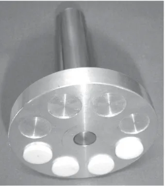
In order to meet the exact requirements of the biaxial testing protocol recommended by ISO16 (1995), all specimens were subsequently grinded to a parallel shape using the grinder/polisher machine (Metaserv 2000, Buehler, Coventry, West Midlands, UK). A custom made specimen holder made from aluminum was designed and used for the grinding purpose. Eight specimens were fixed into the specimen holder using modeling wax (Figure 1). The initial grinding was performed under running water using a diamond grinding disc with a grain size of ;*> $ @* > with a 15 μm diamond polishing paste on a polishing cloth for two min. They were then rinsed thoroughly with running water for 20 s and dried in air.
In compliance with ISO16 (1995), the specimens were trimmed to 1.2±0.2 mm in thickness with parallelism of ±0.05 mm measured using the digital caliper (Mitutoyo Corp, Tokyo, Japan).
%LD[LDOÀH[XUDOVWUHQJWKWHVWLQJ
Piston-on-three-ball test was used for the testing. In order to carry out the test, a loading
pin and mounting jig were designed and used with the Instron Testing Machine (Instron 4302, Instron Corporation, England). The loading pin was cylindrical in shape with a diameter of 1.6 mm. The mounting jig had a circular opening of 16 mm in diameter with three depressions positioned at equal
Figure 1- Specimens mounted in the specimen holder
distances from each other (120° apart) and 5 mm from the center forming a tripod. These depressions were the sites for the 3.2 mm stainless steel ball bearing supports.
The specimens were placed in the mounting jig which ensured the same relation between the supports and the applied load for all specimens. The 1.6 mm diameter loading pin was mounted to the crosshead of the Instron Testing Machine and applied the load at the center of each specimen (Figure 2). The test was carried out at a crosshead *' was recorded for each specimen and the biaxial
equation16# *E@3
7P(X-Y)/d2;Y<^Y 2/r3)
2Y<^EY 2/r3)
2; Y<^<Y1/r3^EY<^Y1/r3)2, where P is the Y`^>= (0.25), r1 is the radius of the support circle (5.0 mm), r2 is the radius of the loaded area (0.8 mm), r3 is the radius of the specimen (8 mm), d is the specimen thickness at the origin of fracture (mm).
Statistical analysis
The results of the study were statistically analyzed with the SPSS software (v. 11.5.0 for Windows, SPSS, Chicago, IL, USA) using Levene’s test and Dunnett’s T3 post-hoc test at a preset 'Q differences between tests groups were related to the ceramic material used for each group.
RESULTS
The objective was to test if the mean biaxial
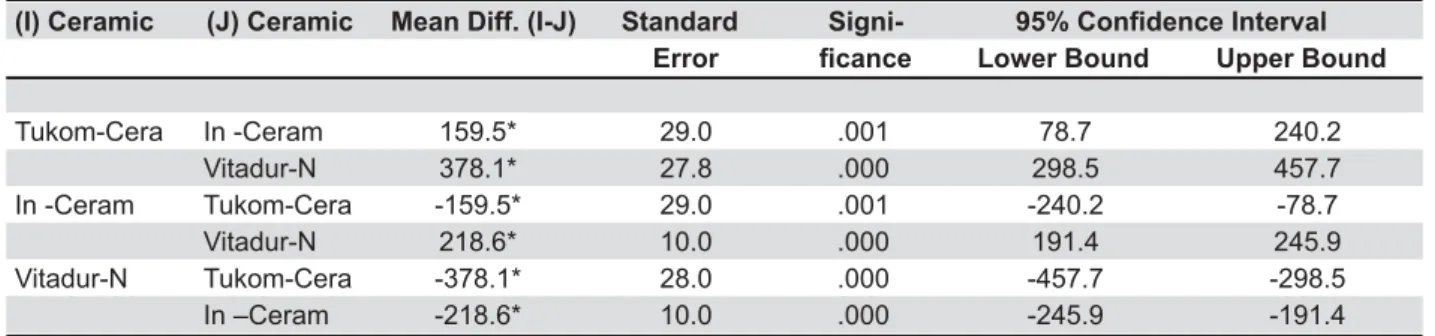
>? Vitadur-N differ from each other. The mean biaxial specimens were calculated for each of the three groups tested (Table 1). Because of violation of the assumption of homogeneous variances (Levene’s <@E<E> =\**'^> were performed with Dunnett’s T3 post-hoc test at 'QY E^
The mean biaxial flexural strength values for Turkom-Cera (506.8±87.01 MPa), In-Ceram (347.4±28.83 MPa) and Vitadur-N (128.7±12.72 %=^ $ Y 2) (P<0.05).
DISCUSSION
Strength is an important mechanical property that can assist in predicting the performance of
brittle materials33
tests, including three-point, and four-point bending tests, and biaxial bending tests are the most commonly applied methods for evaluating the strength of dental restorations27,32,33,34. For uniaxial > lower surfaces of the specimens is tensile, and it is usually responsible for crack initiation in brittle materials. However, undesirable edge fracture, which can increase the variance of the failure stress value, might occur.
The method adapted in this study was the one recommended by ISO16 (1995) since the test standardizes specimen thickness, diameter, shape and roughness. In addition, the measurement of the strength of brittle materials under biaxial ?E%GH? &'<
Lower Bound Upper Bound
Turkom-Cera 10 506.8 87.01 444.59 569.08
In-Ceram 10 347.4 28.83 326.73 367.98
Vitadur N 10 128.7 12.72 119.64 137.84
Table 1-
KM?KNM?E<<PKNM'(%GH? &' Error & Lower Bound Upper Bound
Tukom-Cera In -Ceram 159.5* 29.0 .001 78.7 240.2
Vitadur-N 378.1* 27.8 .000 298.5 457.7
In -Ceram Tukom-Cera -159.5* 29.0 .001 -240.2 -78.7
Vitadur-N 218.6* 10.0 .000 191.4 245.9
Vitadur-N Tukom-Cera -378.1* 28.0 .000 -457.7 -298.5
In –Ceram -218.6* 10.0 .000 -245.9 -191.4
Table 2- Multiple comparisons between the three all-ceramic systems tested
! "#"$
because the maximum tensile stresses occur within the central loading area and edge failures are > less variation in the strength data28,31.
According to ISO16 Y<~~'^> strength is determined by support of a disc specimen on three metal spheres positioned at equal distances from each other and from the center of the disc. The load is applied to the center of the $ can be easily made under typical restorative > specimen can be easily controlled by conventional metallographic polishing methods and typical dental X
Different researchers have studied the biaxial ? $ strength of In-Ceram has been found to be 337.5
MPa on the average20,28,32
strength value for In-Ceram (347.4 MPa) obtained in the present study is in agreement with this result.
_` material has been investigated using the same methods as the current study and found to vary from 141.2 to 155 MPa11,12,23. The mean biaxial _`Y<E3;%=^ achieved in the current study is in agreement with these results.
The higher flexural strength obtained with Turkom-Cera and In-Ceram may be attributed to the followings7,14,25,28,30: 1. Decrease of the total $ $ Y ^ Cera alumina gel and In-Ceram alumina slip; 2. The alumina particles increase the strength of the material and limit potential sites for crack propagation; 3. Prevention of the growth of cracks by crack bridging, as the crystals and glass powders in combination with alumina may bridge the opening created by a crack after the crack front passes; 4. Compressive stresses, which further improve the strength, are also introduced due to the differences in the coefficient of thermal expansion of the alumina and crystals/glass.
Despite the high strength reported with high alumina-based ceramics, they are susceptible to fatigue failure that can considerably reduce their ? $> of fatigue in the oral cavity was not considered. Therefore, further studies are highly recommended to evaluate the fracture analysis and fatigue behavior of new dental ceramics.
CONCLUSION
? $>
three all-ceramic core materials was tested in
vitro. The new high-strength all-ceramic core
material containing primarily aluminum oxide had $ Y'*+343;*< MPa) than the other ceramic core materials (In-Ceram: 347.4±28.83 MPa and Vitadur N: 128.7±12.72 MPa).
ACKNOWLEDGEMENTS
T h i s s t u d y w a s s u p p o r t e d b y a g ra n t (P019/2006C); University of Malaya, Kuala Lumpur, Malaysia. The authors thank Turkom-Ceramic (M) Sdn Bhd, Puchong, Selangor, Malaysia for supplying the materials used for the preparation of Turkom-Cera specimens.
REFERENCES
1- Andreatta OD Filho, Bottino MA, Nishioka RS, Valandro LF, Leite FPP. Effect of thermocycling on the bond strength of a ! Sci. 2003;11(1):61-7.
2- Bakke M, Holm B, Jensen BL, Michler L, Möller E. Unilateral, isometric bite force in 8-68-year-old women and men related to occlusal factors. Scand J Dent Res. 1990;98(2):149-58. 3- Blatz MB, Richter C, Sadan A, Chiche GJ. Critical appraisal. Resin bond to dental ceramics, Part II: high-strength ceramics. J Esthet Restor Dent. 2004;16(5):324-8.
4- Braun S, Bantleon HP, Hnat WP, Freudenthaler JW, Marcotte MR, Johnson BE. A study of bite force, part 1: relationship to various physical characteristics. Angle Orthod. 1995;65(5):367-72. 5- Cehreli MC, Kökat AM, Akça K. CAD/CAM Zirconia vs. slip-cast # E$ results of a randomized controlled clinical trial. J Appl Oral Sci. 2009;17(1):49-55.
6- Charlton DG, Roberts HW, Tiba A. Measurement of select physical and mechanical properties of 3 machinable ceramic materials. Quintessence Int. 2008;39(7):573-9.
7- Clarke D. Interpenetrating phase composites. J Am Ceram Soc. 1992;75(4):739-59.
8- Conrad HJ, Seong WJ, Pesun IJ. Current ceramic materials and systems with clinical recommendations: a systematic review. J Prosthet Dent. 2007;98(5):389-404.
9- Della Bona A, Borba M, Benetti P, Cecchetti D. Effect of surface treatments on the bond strength of a zirconia-reinforced ceramic to composite resin. Braz Oral Res. 2007;21(1):10-5.
10- Ferrario VF, Sforza C, Zanotti G, Tartaglia GM. Maximal bite forces in healthy young adults as predicted by surface electromyography. J Dent. 2004;32(6):451-7.
11- Fleming GJ, Narayan O. The effect of cement type and mixing on the bi-axial fracture strength of cemented aluminous core porcelain discs. Dent Mater. 2003;19(1):69-76.
<E > %>%X=% $ induced variability on the bi-axial fracture strength of aluminous core porcelain discs. J Dent. 1999;27(8):587-94.
13- Gibbs CH, Mahan PE, Mauderli A, Lundeen HC, Walsh EK. Limits of human bite strength. J Prosthet Dent. 1986:56(2):226-9. 14- Giordano RA 2nd, Pelletier L, Campell S, Pober R. Flexural
strength of an infused ceramic, glass ceramic, and feldspathic porcelain. J Prosthet Dent. 1995;73(5):411-8.
15- Haselton DR, Diaz-Arnold AM, Hillis SL. Clinical assessment of high-strength all-ceramic crowns. J Prosthet Dent. 2000;83(4):396-401.
17- McLean JW, Hughes TH. The reinforcement of dental porcelain with ceramic oxides. Br Dent J. 1965;119(6):251-67.
18- Oilo G. Flexural strength and internal defects of some dental porcelains. Acta Odontol Scand. 1988;46(5):313-22.
19- Okiyama S, Ikebe K, Nokubi T. Association between masticatory performance and maximal occlusal force in young men. J Oral Rehabil. 2003;30(3):278-82.
20- Qualtrough AJ, Piddock V. Dental ceramics: what’s new? Dent Update. 2002;29(1):25-33.
21- Rizkalla AS, Jones DW. Mechanical properties of commercial high strength ceramic core materials. Dent Mater. 2004;20(2):207-12.
22- Santos MJMC, Francischone CE, Santos GC Jr, Bresciani E, Romanini JC, Saqueto R, et al. Clinical evaluation of two types of ceramic inlays and onlays after 6 months. J Appl Oral Sci. 2004;12(3):213-8.
E@ > > dental restorative ceramics. Dent Mater. 1990;6(3):181-4. 24- Sundh A, Sjögren G. A comparison of fracture strength of yttrium-oxide-partially-stabilized zirconia ceramic crowns with varying core thickness, shapes and veneer ceramics. J Oral Rehabil. 2004;31(7):682-8.
25- Taya M, Hayashi S, Kobayashi A, Yoon HS. Toughening of a particulate reinforced ceramic matrix composite by thermal residual stress. J Am Ceram Soc. 1990;73(5):1382-91.
26- Thompson VP, Rekow DE. Dental ceramics and the molar crown testing ground. J Appl Oral Sci. 2004;12:26-36.
27- Tinschert J, Zwez D, Marx R, Anusavice KJ. Structural reliability of alumina-, feldspar-, leucite-, mica- and zirconia-based ceramics. J Dent. 2000;28(7):529-35.
E3 >% fracture toughness of three new dental core ceramics. J Prosthet Dent. 1996;76(2):140-4.
29- Wang L, D'Alpino PHP, Lopes LG, Pereira JC. Mechanical properties of dental restorative materials: relative contribution of laboratory tests. J Appl Oral Sci. 2003;11(3):162-7.
30- Wei GC, Becher PF. Improvements in mechanical properties in SiC by the addition of TiC particles. J Am Ceram Soc. 1984;67(8):571-4.
31- Wen MY, Mueller HJ, Chai J, Wozniak WT. Comparative mechanical property characterization of 3 all-ceramic core materials. Int J Prosthodont. 1999;12(6):534-41.
32- Yilmaz H, Aydin C, Gul BE. Flexural strength and fracture toughness of dental core ceramics. J Prosthet Dent. 2007;98(2):120-8.
33- Zeng K, Odén A, Rowcliffe D. Evaluation of mechanical properties of dental ceramic core materials in combination with porcelains. Int J Prosthodont. 1998;11(2):183-9.
34- Zeng K, Odén A, Rowcliffe D. Flexure tests on dental ceramics. Int J Prosthodont. 1996;9(5):434-9.