181
Carneiro DS et al. Heterotaxy syndrome: a case report
Radiol Bras. 2013 Mai/Jun;46(3):181–183
Heterotaxy syndrome: a case report
*
Síndrome de heterotaxia: relato de caso
Daniel de Souza Carneiro1, Jamil Hussein de Arantes2, Gustavo Veloso de Souza2, Aline Santos Barreto2, Mychaell Luciano Cardoso2, Flávia Gontijo2
The present report describes the findings at chest computed tomography angiography of a 28-year-old female patient with heterotaxy syndrome. This syndrome consists of a variety of anomalies of position and morphology of thoracoab-dominal organs which do not follow the situs solitus or situs inversus arrangement. Imaging studies play a fundamental role in the individualization of the approach to the patient.
Keywords: Heterotaxy syndrome; Situs inversus; Spleen – abnormalities.
Neste trabalho são apresentados os achados na angiotomografia computadorizada do tórax de uma paciente de 28 anos com síndrome de heterotaxia. Esta consiste em diversas anormalidades de posicionamento e morfologia de ór-gãos toracoabdominais, que não se enquadram no situs solitus ou situs inversus. Os exames de imagem são fundamen-tais na individualização da abordagem do paciente.
Unitermos: Síndrome de heterotaxia; Situs inversus; Baço – anormalidades. Abstract
Resumo
* Study developed at Instituto das Pequenas Missionárias de Maria Imaculada – Hospital Madre Teresa, Belo Horizonte, MG, Brazil.
1. Master, MD, Resident of Radiology and Imaging Diagno-sis at Hospital Madre Tereza, Belo Horizonte, MG, Brazil.
2. MDs, Residents of Radiology and Imaging Diagnosis at Hospital Madre Tereza, Belo Horizonte, MG, Brazil.
Mailing Address: Dr. Daniel de Souza Carneiro. Centro de Radiologia e Diagnóstico por Imagem. Avenida Raja Gabaglia,
Carneiro DS, Arantes JH, Souza GV, Barreto AS, Cardoso ML, Gontijo F. Heterotaxy syndrome: a case report. Radiol Bras. 2013 Mai/ Jun;46(3):181–183.
0100-3984 © Colégio Brasileiro de Radiologia e Diagnóstico por Imagem
CASE REPORT
monary thromboembolism, ectatic ascend-ing aorta with descendascend-ing segment at the left, infrahepatic vena cava interruption, and azygos vein engorgement from the infra-diaphragmatic portion (Figures 1 and 2). Other findings included polysplenia and bi-lobed lungs, with the main bronchi cours-ing inferiorly to the pulmonary arteries, and the liver in centrally located, characterizing heterotaxy syndrome (Figures 3 and 4).
DISCUSSION
The habitual and orderly arrangement of the organs in the human body is determined early in the embryonic development and is based on genetic information(4). The loss of such orderly arrangement may characterize situs inversus or a disordered and variable
arrangement (heterotaxy syndrome). Hete-rotaxy syndrome presents an approximate incidence of 1:10,000 births and is slightly more prevalent in men, at a ratio of 2:1(3,5). Cardiac abnormalities are present in 50% to 100% of patients(3) and, generally, such abnormalities are accountable for the severity and mortality of patients with such a syndrome. Heterotaxy syndrome encom-passes a wide range of extracardiac vari-ants, including: urinary system, biliary tract and hepatic abnormalities, duodenal atre-World Health Organization functional class
II. At the age of two, the patient was sub-mitted to catheterization, with the follow-ing findfollow-ings: dextrocardia, functionally single atrium, corrected transposition of great vessels, persistence of the arterial canal and interventricular communication. In her adulthood, echocardiogram pre-sented left atrial isomerism and dextrocar-dia, interruption of the inferior vena cava, atrioventricular septal defect (partial pre-sentation), patent arterial duct with aorto-pulmonary shunt. Five years ago, significant pulmonary hypertension was diagnosed.
Four months ago the patient presented sudden worsening of dyspnea associated with chest pain, mild cyanosis, systolic mur-mur more audible in the pulmonary focus, with fixed split of S2, physiological vesicu-lar murmurs and oxygen saturation at 70%. A chest computed tomography angiog-raphy was requested, and demonstrated dextrocardia and cardiomegaly with pre-dominance of the right chambers, atrial septal defect, increased caliber of the monary trunk (35 mm) with no sign of
pul-INTRODUCTION
Heterotaxy syndrome (from the Greek heteros – different and taxis – arrangement)
consists of several position and morphol-ogy abnormalities of thoracoabdominal organs, which do not fit in the habitual positioning order of organs laterality (situs solitus) or its mirror image (situs
inver-sus)(1–3). It is a rare entity, and its severity
is mainly due to cardiac manifestations. Terms such as asplenia (right atrial isomerism) or polysplenia (left atrial isom-erism) are falling into disuse on account of the wide spectrum of anatomical findings, with no pathognomonic characteristic ex-isting which would allow the subclassifi-cation under such terms(3).
CASE REPORT
Female, 28-year-old patient with clini-cal signs of dyspnea since her childhood,
1002, Bairro Gutierrez. Belo Horizonte, MG, Brazil, 30441-070. E-mail: [email protected].
Received October 1st, 2012. Accepted after revision Decem-ber 3, 2012.
182
Carneiro DS et al. Heterotaxy syndrome: a case report
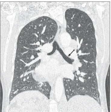
Radiol Bras. 2013 Mai/Jun;46(3):181–183 Figure 3. Chest computed tomography angiography showing bilobed lungs,
with main bronchi coursing inferiorly to the pulmonary artery (arrow).
Figure 4. Chest computed tomography angiography demonstrating polysplenia (arrow) and liver centrally located.
Figure 1. Chest computed to-mography angiography demon-strating dextrocardia, cardiome-galy and atrial septal defect (arrow).
Figure 2. Chest computed to-mography angiography showing ectatic ascending aorta, (arrow head) with descending segment at left (small arrow), engorge-ment of the azygos vein (large arrow), besides increased cali-ber of the pulmonary artery trunk (figure at right).
➤
183
Carneiro DS et al. Heterotaxy syndrome: a case report
Radiol Bras. 2013 Mai/Jun;46(3):181–183 sia, gastrointestinal malrotation and several cardiovascular alterations(6).
The presentation previously called “asplenia” generally presents duplication of structures located in the right side of the body, with trilobed lungs, left atrium with morphology corresponding to the right atrium, centrally positioned liver, left-sided aorta and inferior vena cava, besides intes-tinal malrotation.
In most such cases, death is an early event(3) as a consequence of complications from congenital heart defects, notably the presence of a single atrioventricular cham-ber. Other reported complications include immunological changes and intestinal vol-vulus(7).
In the presentation previously called “polysplenia” generally there are duplica-tion of the structures in the left side of the body, presenting bilobed lungs, right atrium anatomically identical to the left atrium, liver also centrally positioned, absence of the hepatic segment of the inferior vena
cava with continuity through the azygos or hemiazygos vein, besides intestinal malro-tation. Cardiac alterations are less fre-quently found and milder, a fact that ex-plains a higher prevalence of such findings in individuals of more advanced ages(3).
The multiplicity and diversity of find-ings in such a syndrome make the individu-alization of cases extremely valuable, as most of them do not perfectly fit in any clas-sification Thus, the radiological evaluation is indispensable for the identification and planning of the approach to patients pre-senting cardiac and immunological compli-cations or surgical conditions, allowing for the evaluation of the alterations present in each patient(8).
Acknowledgements
The authors wish to thank Dra. Cláudia Juliana Rezende and Dr. Frederico Tadeu Figueiredo Campos, as well as the patient, who promptly agreed to make the images available for the present case report.
REFERENCES
1. Kim SJ. Heterotaxy syndrome. Korean Circ J. 2011;41:227–32.
2. Kim SJ, Kim WH, Lim HG, et al. Outcome of 200 patients after an extracardiac Fontan procedure. J Thorac Cardiovasc Surg. 2008;136:108–16. 3. Applegate KE, Goske MJ, Pierce G, et al. Situs
revisited: imaging of the heterotaxy syndrome. Radiographics. 1999;19:837–52.
4. Shiraishi I, Ichikawa H. Human heterotaxy syn-drome – from molecular genetics to clinical fea-tures, management, and prognosis –. Circ J. 2012;76:2066–75.
5. Lin AE, Ticho BS, Houde K, et al. Heterotaxy: associated conditions and hospital-based preva-lence in newborns. Genet Med. 2000;2:157–72. 6. Phoon CK, Neill CA. Asplenia syndrome: insight
into embryology through an analysis of cardiac and extracardiac anomalies. Am J Cardiol. 1994;73: 581–7.
7. Mahalik SK, Khanna S, Menon P. Malrotation and volvulus associated with heterotaxy syndrome. J Indian Assoc Pediatr Surg. 2012;17:138–40. 8. Vyas HV, Greenberg SB, Krishnamurthy R. MR