Mycobacterium tuberculosis
Imparts Protection at the
Primary Site of Infection in Guinea Pigs
Priyanka Chauhan
1, P. Vineel Reddy
1, Ramandeep Singh
1¤a, Neetika Jaisinghani
2, Sheetal Gandotra
2, Anil
K. Tyagi
1∗1 Department of Biochemistry, University of Delhi South Campus, New Delhi, India, 2 CSIR-Institute of Genomics and Integrative Biology, New Delhi, India
Abstract
Background: The failure of Mycobacterium bovis Bacille Calmette-Guérin to impart satisfactory protection against adult pulmonary tuberculosis has necessitated the development of more effective TB vaccines. The assumption that the vaccine strain should be antigenically as similar as possible to the disease causing pathogen has led to the evaluation of M.tuberculosis mutants as candidate tuberculosis vaccines.
Methods/Principal Findings: In this study, we have generated a mutant of M.tuberculosis (Mtb∆mms) by disrupting 3 virulence genes encoding a mycobacterial secretory acid phosphatase (sapM) and two phosphotyrosine protein phosphatases (mptpA and mptpB) and have evaluated its protective efficacy in guinea pigs. We observed that Mtb∆mms was highly attenuated in THP-1 macrophages. Moreover, no bacilli were recovered from the lungs and spleens of guinea pigs after 10 weeks of Mtb∆mms inoculation, although, initially, the mutant exhibited some growth in the spleens. Subsequently, when Mtb∆mms was evaluated for its protective efficacy, we observed that similar to BCG vaccination, Mtb∆mms exhibited a significantly reduced CFU in the lungs of guinea pigs when compared with the unvaccinated animals at 4 weeks after challenge. In addition, our observations at 12 weeks post challenge demonstrated that Mtb∆mms exhibited a more sustainable and superior protection in lungs as compared to BCG. However, the mutant failed to control the hematogenous spread as the splenic bacillary load between Mtb∆mms vaccinated and sham immunized animals was not significantly different. The gross pathological observations and histopathological observations corroborated the bacterial findings. Inspite of disruption of phosphatase genes in MtbΔmms, the lipid profiles of M.tuberculosis and MtbΔmms were identical indicating thereby that the phenotype of the mutant was ascribed to the loss of phosphatase genes and the influence was not related to any alteration in the lipid composition.
Conclusions/Significance: This study highlights the importance of M.tuberculosis mutants in imparting protection against pulmonary TB.
Citation: Chauhan P, Reddy PV, Singh R, Jaisinghani N, Gandotra S, et al. (2013) Secretory Phosphatases Deficient Mutant of Mycobacterium tuberculosis Imparts Protection at the Primary Site of Infection in Guinea Pigs. PLoS ONE 8(10)μ e77λ30. doiμ10.1371/journal.pone.0077λ30 Editor: Jean Louis Herrmann, Hopital Raymond Poincare - Universite Versailles St. Quentin, France
Received March 30, 2013; Accepted September 6, 2013; Published October 18, 2013
Copyright: © 2013 Chauhan et al. This is an open-access article distributed under the terms of the Creative Commons Attribution License, which permits unrestricted use, distribution, and reproduction in any medium, provided the original author and source are credited.
Funding: This work was supported by a research grant from the Department of Biotechnology, Government of India, India. Council of Scientific and Industrial Research, Government of India is acknowledged for Junior/Senior research fellowships to PC. The funders had no role in study design, data collection and analysis, decision to publish, or preparation of the manuscript.
Competing interests: Co-author Prof. Anil K. Tyagi is a member of the PLOS ONE Editorial Board and this does not alter the authors' adherence to all the PLOS ONE policies on sharing data and materials.
∗ E-mailμ [email protected]
¤a Current addressμ Vaccine and Infectious Disease Research Centre, Translational Health Science and Technology Institute, Gurgaon, Haryana, India
Introduction
Tuberculosis (TB) continues to intimidate human race unabashedly and remains a major cause of morbidity and mortality throughout the world [1,2]. Every week, more than 150,000 individuals develop TB and ~30,000 human lives are lost globally due to this dreaded disease. The lethal liaison between TB and HIV infections and the emergence of various
Therefore, the need to develop a superior TB vaccine than BCG cannot be over-emphasized.
The purpose of an effective live vaccine would be best served if the vaccine strain is antigenically as similar as possible to the disease-causing pathogen in order for it to generate the host immune responses that mimic natural infection [6]. Comparative genomic studies have revealed that BCG, in comparison to M.tuberculosis, lacks 16 defined regions (RD1-16) comprising of ~150 genes, some of which are known to encode potential antigenic determinants that could increase the immunogenicity of a vaccine [7,8]. This makes the use of attenuated M.tuberculosis strains rather than BCG, for the generation of appropriate immune responses, an attractive idea [5,λ,10]. Several M.tuberculosis mutants have been evaluated in animal models and have resulted in varying degrees of success in imparting protection against TB when compared with BCG [11–15]. Immunization of mice with the ∆RD1∆panCD mutant of M.tuberculosis (an attenuated M.tuberculosis RD1 knockout and pantothenate auxotroph) resulted in 1-2 log10 CFU lower bacillary loads in the spleens, lungs and liver when compared with the BCG. However, in bull calves, no histopathological differences were observed in the lung and lymph nodes of ∆RD1∆panCD vaccinees when compared with the unvaccinated controls [14,15]. Similarly, mice vaccinated with ∆secA2 mutant (sec A2 deletion mutant of M.tuberculosis) exhibited significantly lower pulmonary and splenic CFU when compared with the BCG vaccinated group, however, the same vaccine performed as well as BCG in guinea pigs [11]. In contrast to these observations, Martin et al. demonstrated similar level of protection exhibited by SO2 strain (phoP deletion mutant of M.tuberculosis) in mice, although guinea pigs vaccinated with SO2 exhibited significantly increased survival time when compared with BCG [12]. The variable results shown by the candidate vaccines and the fact that none of the current candidates has successfully made through the clinical trials reinforce the importance of keeping the pipeline full with the new candidates [16].
Among the secretory proteins of M.tuberculosis, three phosphatases, namely, mycobacterial secretory acid phosphatase (SapM) and two phosphotyrosine protein phosphatases (MptpA and MptpB) have been shown to contribute to its pathogenicity [17–20]. SapM dephosphorylates phosphatidylinositol 3-phosphate (PI3P), a membrane trafficking regulatory lipid, resulting in the arrest of phagosome maturation [1λ]. In addition, in a study by Festjens et al, disruption of sapM locus in BCG improved its protective efficacy as a vaccine against TB [21]. The increased efficacy of the vaccine was accredited to the efficient activation and recruitment of dendritic cells to the draining lymph nodes in the absence of SapM, thus allowing successful antigen presentation and activation of the adaptive immunity by dendritic cells [21]. A recent study showed that the fbpA/sapM double mutant of M.tuberculosis was attenuated for growth and more immunogenic in macrophages as compared to M.tuberculosis [22].
MptpA has been demonstrated to block phagosome-lysosome fusion by inhibiting V-ATPase trafficking to the mycobacterial phagosome [23–25]. It has been reported that
mptpA mutant of M.tuberculosis was impaired for survival/ growth in THP-1 macrophages and phagosomes harboring the mutant strain exhibited increased phagosome-lysosome fusion [23]. It has been previously reported that M.tuberculosis devoid of MptpB activity was impaired for survival in IFN-γ activated macrophages and in guinea pigs [26]. In another study, it was shown that MptpB inhibits ERK ½, p38 signaling pathways and caspase 3 activity, thus subverting the host immune response to infection [27]. The importance of MptpB in the intracellular survival of M.tuberculosis was also demonstrated in a study in which specific inhibitors against MptpB were shown to inhibit mycobacterial survival within murine macrophages [17,27].
In this study, by deleting the function of three virulence genes, namely, mptpA (Rv2234), mptpB (Rv0153c) and sapM (Rv3310), we have developed the mutant Mtb∆mms and evaluated its protective efficacy in guinea pig model of experimental tuberculosis.
Materials and Methods
Bacterial strains and growth conditions
The bacterial strains and plasmids used in this study are listed in Table 1. M. bovis BCG (Danish strain) was obtained from BCG laboratories, Chennai, India. M.tuberculosis H37Rv (ATCC No. 25618) used for challenge was procured from Dr. J. S. Tyagi, AIIMS, New Delhi, India. Mycobacterial strains were grown to mid-log phase in MB7Hλ medium supplemented with 1X albumin-dextrose-catalase (ADC), 0.5% glycerol and 0.05% Tween 80. PBS stocks were prepared and stored at -80°C till further use. The CFU of stocks was enumerated by plating appropriate dilutions in duplicates on MB7H11 agar supplemented with 1X oleic acid-albumin-dextrose-catalase (OADC) and 0.5% glycerol. E.coli strains, XL-1 Blue (Stratagene) and HB101 (Life Technologies) were used for cloning purposes. Kanamycin and Chloramphenicol were used at 25 g/ml and 30 g/ml, respectively. Hygromycin was used at 50 g/ml for M.tuberculosis and at 150 g/ml for E.coli.
For the generation of Mtb∆mms mutant of M.tuberculosis, sapM was disrupted in Mtb∆mptpB∆mptpA. For this, we employed a modified pYUB∆sapM vector generated in our laboratory (Table 1) by replacing hygromycin resistance cassette in pYUB∆sapM with the Chloramphenicol resistance gene expressed under the mycobacterial trrn promoter, CATtrrn. Briefly, primers were designed to amplify (i) ~700 bp Table 1. Bacterial strains, plasmids, cell line and primers used in this study.
Strains/Plasmids/Cell
line/Primers Description Reference
Strains
E.coli XL-1 Blue
endA1 gyrAλ6 (nalR) thi-1 recA1 relA1 lac glnV44 F' [μμTn10 proAB + lacIq Δ (lacZ) M15] hsdR17 (rK- mK+)
Stratagene, Heidelberg, Germany
E.coli HB101
F-(gpt-proA) 62 leuB6 glnV44 ara-14 galK2 lacY1 (mcrC-mrr) rpsL20 (Strr) xyl-5 mtl-1 recA13
Life Technologies, CA, USA Mtb∆mptpB M.tuberculosis Erdman mptpB mutant [26]
Mtb∆mptpB∆mptpA M.tuberculosis mptpB and mptpA
mutant This study
MtbΔmms M.tuberculosis mptpA, mptpB and
sapM mutant This Study
M.tuberculosis H37Rv
(ATCC No. 25618) Virulent strain of M.tuberculosis
Dr. J. S. Tyagi, AIIMS, New Delhi, India
M.bovis BCG Danish Vaccine strain against tuberculosis BCG laboratories, Chennai, India Plasmids
pLIT∆A
pLitmus vector carrying the amplicons of mptpA with a deletion of 16λ bp from the central region of ORF
This study
pLIT∆AK
pLitmus vector with kanamycin resistance gene cassette flanked with mptpA amplicons
This study
pYUB∆SapM
Cloning vector with Hygromycin resistance gene cassette flanked with sapM amplicons
unpublished data
pYUB.CATtrrn∆sapM
Cloning vector with Chloramphenicol resistance gene under mycobacterial trrn promoter flanked with sapM amplicons
This study
pVR1
A derivative of pSD5 containing Chloramphenicol resistance gene under mycobacterial trrn promoter
[51]
Cell line
THP-1 The Human acute monocytic leukemia cell line
NCCS, Pune, India Primers
F-mptpA 5′-gtgtctgatccgctgcacgtcacattc-3′ This study R-mptpA 5′-tcaactcggtccgttccgcgcgagac-3′ This study F-sapM 5′-atgctccgcggaatccaggctc-3′ This study R-sapM 5′-ctagtcgccccaaatatcgg-3′ This study doiμ 10.1371/journal.pone.0077λ30.t001
amplicon comprising of ~200 bp of 5′ proximal end of sapM and ~500 bp of immediate upstream region of sapM (amplicon I) and (ii) ~700 bp amplicon comprising of ~200 bp of 3′ distal end of sapM and ~500 bp of immediate downstream region of sapM (amplicon II). Both amplicons were cloned into pYUB854 flanking the hygromycin resistance cassette to generate pYUB∆sapM. The hygromycin resistance cassette in pYUB∆sapM was replaced with CATtrrn (The CATtrrn was PCR amplified from pVR1, Table 1) resulting in pYUB.CATtrrn∆sapM. A linear Allelic Exchange Substrate (AES), ΔsapMμμCATtrrn was then excised out as KpnI/SpeI fragement and electroporated into MtbΔmptpBΔmptpA to generate the mutant which was designated as MtbΔmms (mptpA, mptpB and sapM).
Confirmation of deletion of mptpA and sapM by Southern hybridization
To confirm the deletion of mptpA in MtbΔmptpBΔmptpA, the genomic DNA was isolated from the parental strain (MtbΔmptpB) and the mutant strain (MtbΔmptpBΔmptpA) followed by the digestion of 2 µg of DNA with PvuII. The deletion of sapM in MtbΔmms was confirmed by isolating the genomic DNA from MtbΔmptpBΔmptpA and MtbΔmms, followed by digestion of 2 g of DNA with PstI. DNA was electrophoresed through 1% agarose gel followed by depurination, denaturation and neutralization of DNA within the agarose gel. DNA was then transferred onto positively charged nylon membrane by capillary transfer overnight and immobilized by UV radiation. 200 bp region at 5′ termini of mptpA and sapM were amplified for the generation of probe. The probe labeling, subsequent pre-hybridization, hybridization and detection were performed as described in the DIG High Prime DNA Labeling and Detection Starter Kit II (Roche Applied Science, IN, USA).
Lipid profile analysis
M.tuberculosis as well as MtbΔmms, suspended in diethyl ether were spotted on a thin layer chromatography (TLC) plate (Merck, TLC Aluminium sheets silica gel 60), chromatographed in hexaneμ ethylacetate (λ5μ 5, v/v) seven times and visualized by staining with 20% sulphuric acid in ethanol followed by charring.
Extraction of polar and apolar lipids. Mycobacterial lipids were extracted as described previously [30]. Briefly, 50 ml of mycobacterial cultures, grown in MB7Hλ supplemented with 1X ADC, were harvested at an A600nm of 1.0 and heat killed (λ5°C for 1 hr). Apolar lipids were extracted by adding 2 ml of methanolic solution of 0.3% sodium chloride and 1 ml of petroleum ether (60-80°C) to the cell pellet. The cell suspension was mixed end-over-end for 30 min followed by centrifugation at 2500 rpm for 10 min. The upper layer consisting of apolar lipids was collected in a separate vial and 1 ml of petroleum ether was added to the lower layer, vortexed and mixed end-over-end for 15 min. The cell suspension was again centrifuged to recollect the upper layer. The upper layers comprising of apolar lipids were pooled and dried at 60°C.
Further, the polar lipids were extracted by adding 2.3 ml of chloroformμ methanolμ 0.3% sodium chloride (λ0μ 100μ 30, v/v/v) to the bottom layer. The cell suspension was mixed end- over-end for 60 min followed by centrifugation at 2500 rpm for 10 min. Polar lipids, present in the supernatant fraction, were collected and the pellet was further treated twice with 750 l of chloroformμ methanolμ 0.3% sodium chloride (50μ 100μ 40, v/v/v), to obtain all polar lipids. The supernatants from these three extractions were pooled and further extracted with 1.3 ml of chloroform and 1.3 ml of 0.3% sodium chloride. The lower layer comprising of polar lipids was collected into a fresh glass tube and dried at 60°C. Equivalent amounts of polar and aploar lipids suspended in chloroformμ methanol (2μ 1, v/v) from both M.tuberculosis and MtbΔmms strains were then spotted on TLC plates and analysed for different lipid fractions by using different solvent system as described in Table S1. TLC plates were developed by dipping in 10% phosphomolybdate or spraying with 2% orcinol in 10% sulphuric acid (for solvent C) followed by charring.
For the detection of trehalose monomycolate (TMM), trehalose dimycolate (TDM) and sulfolipids (SL), 5 Ci of 14C-acetate was added to 10 ml of log phase culture of both M.tuberculosis and MtbΔmms strains, separately. Cultures were then harvested after 18 hrs of radioactive pulse and apolar lipids were extracted from a methanolic solution of 0.3% sodium chloride and petroleum ether as described above. The organic phase was suspended in chloroformμ methanol (2μ 1, v/v). Approximately, 25,000 counts from the samples belonging to each strain were spotted on the TLC plate followed by chromatography in the appropriate solvents (Table S1). The lipids were visualized with a Typhoon FLA 700 Phosphorimager.
Comparison of the growth of MtbΔmms and the parental strain in human macrophages
Human monocytic THP-1 cells were cultured in complete RPMI-GlutaMAXTM medium [containing 10% heat inactivated FBS and 1% antibiotic-antimycotic mix] (GIBCO Grand Island,
NY, USA) and were differentiated to macrophages by the addition of 30 nM Phorbol 12-myristate 13-acetate (PMA, Sigma) for 16 hrs at 37°C, 5% CO2. Cells were washed with complete RPMI medium and rested for 2 hrs in fresh medium without antibiotic-antimycotic mix before infection. For infection, 5 x 10⁵ macrophages were infected with 5 x 10⁵ mycobacteria to achieve an MOI of 1μ1 in 24 well plates for 4 hrs in triplicates [31]. Following infection, the extracellular bacteria were removed by overlaying the cells with RPMI medium containing 200 g/ml amikacin for 2 hrs. At designated time points, day 0 (4 hrs), 2, 4 and 6, macrophages were lysed by the addition of 0.025% SDS and intracellular bacteria were enumerated by plating appropriate dilutions on MB7H11 agar. Colonies were counted after 4 weeks of incubation at 37°C and the data was expressed as CFU/ml.
Experimental animals
Pathogen-free outbred female guinea pigs (200-300 g) of the Duncan-Hartley strain were procured from Disease Free Small Animal House Facility, Lala Lajpat Rai University, Hissar, India. The animals were housed in individually ventilated cages and were provided with food and water ad libitum in a BSLIII facility at University of Delhi South Campus (UDSC), New Delhi, India.
Ethics statement
Guinea pig experiments included in this manuscript were reviewed and approved by the Institutional Animal Ethics Committee of University of Delhi South Campus, New Delhi, India (Ref. No. IAEC/AKT/Biochem/UDSC/24.08.2010). All animals were routinely cared for, according to the guidelines of CPCSEA (Committee for the Purpose of Control and Supervision of Experiments on Animals), India. Guinea pigs were vaccinated intradermally with mycobacterial strains by injecting not more than 100 l and were euthanized, whenever required, by CO2 asphyxiation and all efforts were made to ameliorate animal suffering.
Influence of deletion of phosphatase genes on the pathogenicity of M.tuberculosis
To evaluate whether the Mtb∆mms mutant was sufficiently attenuated for its use as a vaccine, animals (n=6) were inoculated intradermally (i.d.) with 5 x 10⁵ bacilli of either
load. The specific portions of lungs and spleen were weighed and homogenized separately in 5 ml saline by using a polytron homogenizer. Appropriate dilutions of the homogenates were plated on to MB7H11 agar plates in duplicates and incubated at 37°C for 3-4 weeks. The number of colonies was counted and expressed as mean log10 CFU/organ.
Evaluation of protective efficacy of MtbΔmms against M.tuberculosis infection
Guinea pigs were divided into 3 groups (n=8) and the animals were immunized intradermally with 5 x 10⁵ CFU of either (i) BCG or (ii) Mtb∆mms in 100 l of saline. In the control group, guinea pigs were injected with 100 l of saline. Twelve weeks post immunization, guinea pigs were infected with a low dose of virulent M.tuberculosis via the respiratory route in an aerosol chamber (Inhalation Exposure System, Glascol Inc.), pre calibrated to deliver 10-30 bacilli in lungs per animal. Guinea pigs were euthanized at 4 weeks and 12 weeks after challenge and evaluated for bacterial load, gross pathological and histopathological changes in various organs as described in the previous section. A significant reduction in these parameters in vaccinated animals was considered as a protective effect of the vaccine.
Statistical analyses
For comparison between the groups, Non-parametric Kruskal–Wallis test followed by Mann-Whitney U-test, One-way analysis of variables (ANOVA) with Tukey post-test, Two-way ANOVA with Bonferroni multiple comparison test and student’s t-test were employed, wherever appropriate. Differences were considered significant when p<0.05. For statistical analyses and generation of graphs, Prism 5 software (Version 5.01; GraphPad Software Inc., CA, USA) was used.
Results
Functional disruption of mptpA and sapM in Mtb∆mptpB and characterization of the multigene mutant
To generate triple gene mutant of M.tuberculosis, we first disrupted mptpA in Mtb∆mptpB (published from our laboratory previously, [26]) to generate Mtb∆mptpB∆mptpA (Figure 1A). Deletion of mptpA was confirmed by three approaches (1). PCR by using mptpA gene specific primers (Table 1, Figure 1B). In the case of Mtb∆mptpB, a 0.5 kb amplicon representing the complete mptpA gene was amplified as expected, while in the case of Mtb∆mptpB∆mptpA, an amplicon of 2.0 kb was observed indicating the disruption of mptpA by Kanamycin resistance cassette (2). Southern hybridization. In the case of MtbΔmptpB strain, the probe hybridized to a 0.5 kb (lane 1) PvuII fragment whereas disruption of mptpA gene by Kanamycin resistance cassette resulted in a signal at 2.4 kb (lane 2) in the MtbΔmptpBΔmptpA strain (Figure 1C) (3). Nucleotide sequencing. Both 0.5 kb and 2.0 kb amplification products were DNA sequenced that further confirmed the disruption of mptpA in MtbΔmptpBΔmptpA. Further, the sapM gene was deleted in MtbΔmptpBΔmptpA by employing linear
AES to generate MtbΔmms (Figure 1D). The triple gene mutant was confirmed by PCR by employing sapM gene specific primers (Table 1, Figure 1E). The primers yielded an amplicon of 0.λ kb in Mtb∆mptpB∆mptpA, however, the deletion of sapM gene resulted in a PCR amplicon of 1.5 kb in Mtb∆mms (Figure 1E) (2). Southern hybridization. The probe in the MtbΔmptpBΔmptpA hybridized to a 3.0 kb (lane 1) PvuII fragment whereas disruption of sapM gene by Chloramphenicol resistance cassette resulted in a signal at 1.5 kb (lane 2) in the MtbΔmms strain (Figure 1F) (3). Nucleotide sequencing. Both 0.λ kb and 1.5 kb amplification products were DNA sequenced that further confirmed the disruption of sapM in MtbΔmms. Deletion of mptpA and sapM was further confirmed by immunoblot analysis by using polyclonal antibodies raised against MptpA and SapM. As shown in Figure 1G and Figure 1H, we did not observe any expression of MptpA and SapM in MtbΔmms.
Disruption of Phosphatases Does Not Alter the Lipid Profile of MtbΔmms
To ascertain whether the disruption of phosphatase genes had any influence on the lipid composition of the mutant, we performed a total lipid analysis of the parental as well as the mutant strain by TLC. M.tuberculosis and MtbΔmms were analysed for the well known characterstic lipids of the tubercle bacillus. The apolar and polar lipid fractions were extracted and assayed for phthiocerol dimycocerosate (PDIM), triacylglycerol (TAG), mycolic acids, free fatty acids, diacylglycerol (DAG), diacyltrehalose (DAT), trehalose monomycolate (TMM), trehalose dimycolate (TDM), glucose monomycolate (GMM), sulfolipids (SL), phosphatidylinositol (PI) and phosphatidylinositol mannoside (PIMs) and phospholipids (P) by TLC. Equivalent amounts of apolar as well as polar lipids from both M.tuberculosis and MtbΔmms were spotted on TLC plates and analysed for different lipid fractions (Table S1).
TLC analysis of the lipids of M.tuberculosis and MtbΔmms exhibited a similar and usual lipid profile with respect to the mycobacterial lipid components. M.tuberculosis produces three classes of mycolic acidsμ alpha-, keto- and methoxy- mycolic acids [33]. Analysis of total mycolic acids extracted from both M.tuberculosis and MtbΔmms by single dimension TLC exhibited that there was no significant difference in total or alternate types of mycolic acids (Figure 2A). In addition, we observed similar accumulation of structural variants of DIM and TAGs as described by Giovannini et al [34] (Figure 2B). Two dimensional TLC indicated the equivalent presence of both apolar (DAG, TMM, TDM, SL, GMM, DAT) and polar lipids (PIMs, PI and P) in both M.tuberculosis and MtbΔmms as described previously by Bhatt et al [35] (Figure 2C, Figure 2D and Figure 2E). Hence, our observation demonstrated that the lipid profile of M.tuberculosis and MtbΔmms was similar with no notable differences.
Mtb∆mms exhibits a severe growth defect in human THP-1 macrophages
Figure 1. Construction and characterization of Mtb∆mms. (A) Disruption of mptpA in MtbΔmptpB by homologous recombination. The figure depicts the deletion of mptpA by mptpAμμkan AES to generate MtbΔmptpBΔmptpA. Solid bar (a) depicts the region of probe binding for Southern hybridization. kan- Kanamycin resistance cassette. (B) Confirmation of disruption of mptpA in Mtb∆mptpBmptpA was carried out by PCR by employing mptpA gene specific primers. A 0.5 kb amplification product was obtained with Mtb∆mptpB DNA as template (lane 2) and 2.0 kb amplification product was obtained with MtbΔmptpBΔmptpA DNA as template (lane 3). 100 bp ladder (lane 1) and HindIII ladder (lane 4). (C) Confirmation of mptpA deletion in MtbΔmptpB∆mptpA by Southern hybridization. 200 bp at 5′ termini of mptpA was used as probe (a). Genomic DNA of MtbΔmptpB as well as MtbΔmptpBΔmptpA was digested with PvuII, separated on a 1.0% agarose gel, transferred to nylon membrane and probed with DIG-labeled DNA. The MtbΔmptpB strain (lane 1) showed a hybridization signal at 0.5 kb as expected. The presence of 2.4 kb band in the case of MtbΔmptpBΔmptpA (lane 2) confirmed that allelic exchange had occurred at the mptpA locus. (D) Disruption of sapM in Mtb∆mptpB∆mptpA by homologous recombination. The figure depicts the deletion of sapM by sapMμμCATtrrn AES to generate MtbΔmms. Solid bar (b) depicts the region of probe binding for Southern hybridization. CATtrrn-Choloramphenicol resistance cassette. (E) Confirmation of disruption of sapM in Mtb∆mms was carried out by PCR by employing sapM gene specific primers. A 0.λ kb amplification product was obtained with Mtb∆mptpB∆mptpA DNA as template (lane 2) and 1.5 kb amplification product was obtained with MtbΔmms mutant DNA as template (lane 3). 100 bp ladder (lane 1) and HindIII ladder (lane 4). (F) Confirmation of sapM deletion in MtbΔmms by Southern hybridization. 200 bp at 5′ termini of sapM was used as probe (b). Genomic DNA of MtbΔmptpBΔmptpA as well as MtbΔmms strain was digested with PstI, separated on a 1.0% agarose gel, transferred to nylon membrane and probed with DIG-labeled DNA. The MtbΔmptpBΔmptpA strain (lane 1) showed a hybridization signal at 3.0 kb as expected. The presence of 1.5 kb band in the case of MtbΔmms strain (lane 2) confirmed that allelic exchange had occurred at the sapM locus. (G) Confirmation of mptpA deletion in Mtb∆mms by immunoblot analysis. 50 g of culture filtrate proteins of Mtb∆mptpB (lane 1) and MtbΔmms (lane 2) were loaded onto a 12.5% polyacrylamide gel and subjected to electrophoresis. MptpA was detected by immunoblot analysis by using anti-MptpA polyclonal antisera. MptpA protein migrated as an 18 kDa protein band (lane 1). Disruption of mptpA in MtbΔmms was confirmed by the absence of this 18 kDa band (lane 2). (H) Confirmation of sapM deletion in Mtb∆mms by immunoblot analysis. 50 g of culture filtrate proteins of Mtb∆mptpB (lane 1) and MtbΔmms (lane 2) were loaded onto a 12.5% polyacrylamide gel and subjected to electrophoresis. SapM was detected by immunoblot analysis by using anti-SapM polyclonal antisera. SapM protein migrated as a 28 kDa protein band (lane 1). Disruption of sapM in MtbΔmms was confirmed by the absence of this 28 kDa band (lane 2).
the growth characteristics of MtbΔmms and the parental strain in MB7Hλ, however, a significant difference was observed in the growth kinetics between these two strains in THP-1 macrophages. We observed that Mtb∆mms displayed a significantly reduced ability (~2.8λ fold difference) to infect macrophages in comparison to the parental strain. Moreover, while M.tuberculosis continued to grow normally for 6 days, Mtb∆mms exhibited no sign of growth during this time period demonstrating that the deletion of 3 phosphatases rendered the mutant completely incapable of growing in the macrophages (∗∗∗p<0.001) (Figure 3B). These results demonstrate the importance of mptpA, mptpB and sapM in the growth and survival of M.tuberculosis in the human macrophages.
Deletion of phosphatase genes leads to the attenuation of M.tuberculosis
To evaluate whether the deletion of three phosphatases had rendered the Mtb∆mms mutant sufficiently attenuated for its use as a vaccine, animals were inoculated with either M.tuberculosis or BCG or Mtb∆mms strain (Figure 4A). At 4 weeks post inoculation, we observed the maximum bacillary load of 3.λ6 log10 CFU in the lungs of M.tuberculosis infected animals, as compared to negligible bacillary load of 0.28 log10 CFU in the lungs of BCG treated animals. No bacilli were detectable in the lungs of Mtb∆mms inoculated animals (Figure 4B). However, when the splenic bacillary counts were analyzed, we observed 5.68 log10 CFU, 0.36 log10 CFU and 3.λλ log10 CFU in the animals inoculated with M.tuberculosis, BCG and Mtb∆mms, respectively (Figure 4C). Hence, during this initial phase, Mtb∆mms showed some growth in the spleens of animals, although it was ~70 fold less as compared to the parental strain. At 10 weeks post inoculation, a bacillary
Figure 2. Lipid analysis of M.tuberculosis and MtbΔmms. (A) Total mycolic acids extracts from M.tuberculosis (Mtb) and MtbΔmms separated by TLC in hexaneμ ethyacetate (λ5μ 5 v/v) seven times and detected with 20% sulfuric acid in ethanol followed by charring at 100°C. Single dimension TLC depicting alpha-, methoxy- and keto- types of mycolic acids. (B) Apolar lipid extracts from Mtb and MtbΔmms separated by TLC in petroleum etherμ diethyl ether (λ0μ 10 v/v) and detected with 10% phosphomolybdic acid in ethanol followed by charring at 100°C showing PDIMs and TAG. (C) Lipid extracts from Mtb and MtbΔmms separated by two dimensional TLC in chloroformμ methanolμ water (60μ 30μ 6 v/v/v) in 1st direction and chloroformμ acetic acidμ methanolμ water (40μ 25μ 3μ 6 v/v/v/v) in 2nd direction. PIMs, PI and P were detected with 2% orcinol in 10% sulphuric acid followed by charring. (D) Lipid extracts from Mtb and MtbΔmms separated by two dimensional TLC in chloroformμ methanol (λ6μ 4 v/v) in 1st direction and tolueneμ acetone (80μ 20 v/v) in 2nd direction. DAG, free fatty acids and free mycolic acids were detected with 10% phosphomolybdic acid in ethanol followed by charring. (E) TLC analysis of lipid extracts from [14C] acetate-labeled bacterial cell of Mtb and MtbΔmms. Lipid extracts were dissolved in solvent and run in chloroformμ methanolμ water (100μ 14μ 0.8 v/v/v) in 1st direction and chloroformμ acetoneμ methanolμ water (50μ 60μ 2.3μ 3 v/v/v/v) in 2nd direction. GMM, SLIII, DAT, TMM and TDM were visualized with a Typhoon FLA 700 Phosphorimager. PDIM- phthiocerol dimycocerosate, TAG- triacylglycerol, DAG- diacylglycerol, GMM- glucose monomycolate, SL- sulfolipids, DAT- diacyltrehalose, TMM- trehalose monomycolate, TDM- trehalose dimycolate, PIM-phosphatidylinositol mannoside, PI- PIM-phosphatidylinositol, P- phospholipids. TLC analysis of the lipids of M.tuberculosis and MtbΔmms exhibited a similar and usual lipid profile with respect to the mycobacterial lipid components.
load of 4.6λ log10 CFU was observed in the lungs of
M.tuberculosis infected animals. However, at this time point, no bacilli were recovered from the lungs of the animals inoculated with either Mtb∆mms or BCG (Figure 4D). In spleens, we observed a bacillary count of 5.45 log10 CFU in M.tuberculosis infected animals. However, no bacilli were recovered from the spleens of animals inoculated with either Mtb∆mms or BCG (Figure 4E).
Although, Mtb∆mms exhibited some growth in the spleens of the inoculated animals, the bacilli were recovered only during the initial phase (4 weeks post inoculation) and the bacillary load was only 1.4% of that observed in the case of M.tuberculosis infected animals (70 fold fewer bacilli in Mtb∆mms inoculated animals). Further, on extending the time post inoculation, no Mtb∆mms bacilli were recovered in spleens as well as in lungs. Thus, based on these observations, it appeared that as a result of deletion of the phosphatase genes, Mtb∆mms was sufficiently attenuated for growth in the host tissues and could be safely used as a vaccine candidate.
Deletion of phosphatase genes renders M.tuberculosis incapable of causing pathology in guinea pigs at 10 weeks post inoculation
The gross pathological changes observed in the organs of the animals at 4 weeks and 10 weeks post inoculation with Mtb∆mms were commensurate with the bacillary load observed. At 4 weeks post inoculation, the extent of damage observed in the case of M.tuberculosis inoculated animals was found to be maximum amongst all the groups with numerous small sized tubercles along with scattered areas of necrosis in
all the organs (scoreμ 2 in lungs and liver and 3 in spleen), indicating progressive pulmonary and extra-pulmonary disease (Figure 5A). However, in the case of BCG inoculation, no pathology was observed in the lungs, liver or spleen as expected (scoreμ 1 in all the organs). In the case of inoculation with Mtb∆mms, most of the animals displayed negligible lung and hepatic pathology (scoreμ 1) with predominantly scanty and extremely small necrotic lesions. However, in the case of spleen, Mtb∆mms inoculated animals were allotted intermediate score (scoreμ 2) in comparison to other two groups. This indicated that Mtb∆mms inoculation resulted in some pathological damage to spleens, although, the damage was considerably less in comparison to the M.tuberculosis infected animals (Figure 5A). When the animals were evaluated at 10 weeks post inoculation, in the case of M.tuberculosis infected animals, as expected, the extent of damage was more than that observed at 4 weeks (scoreμ 3 in lungs, 4 in liver and 3-4 in spleen) with extensive involvement and numerous large sized tubercles effacing the entire organs (Figure 6A). However, the animals inoculated with either BCG or Mtb∆mms displayed normal lungs, liver and spleen phenotype (scoreμ 1) with no pathological damage (Figure 6A).
To evaluate the histopathological changes in the lungs and liver of guinea pigs inoculated with M.tuberculosis, BCG or Mtb∆mms, the tissue sections were stained with haematoxylin and eosin. At 4 weeks post inoculation, the lungs of M.tuberculosis infected animals exhibited granulomatous infiltration with caseating necrotic granulomas effacing the pulmonary parenchyma (Figure 5B). Inoculation with either BCG or Mtb∆mms resulted in a negligible granulomatous
Figure 3. Influence of deletion of phosphatase genes on the growth and survival of M.tuberculosisinvitro. (A) Growth of M.tuberculosis and MtbΔmms in broth culture. M.tuberculosis and MtbΔmms strains were cultured separately in MB7Hλ medium at 37°C/200 rpm for 13 days. M.tuberculosis and MtbΔmms exhibited similar growth pattern in MB7Hλ broth culture. (B) Growth of M.tuberculosis and MtbΔmms in human THP-1 macrophages. THP-1 cells were infected with M.tuberculosis or Mtb∆mms mutant separately at an MOI of 1μ1. The number of intracellular viable bacteria was determined for 6 days. The figure represents the CFU data of M.tuberculosis and MtbΔmms obtained on MB7H11 plates. Mtb∆mms mutant exhibited a significant attenuation in its growth when compared with M.tuberculosis, till 6 days post infection. The experiment was repeated thrice with three independent samples each time. ∗∗∗p<0.001 (Two-way ANOVA). The values are represented as the means (±SE).
infiltration in lungs when compared with M.tuberculosis infected animals. Liver of M.tuberculosis infected animals displayed moderate involvement of tissues with few granulomatous lesions. However, BCG or Mtb∆mms inoculated animals exhibited a normal hepatic tissue organization with clearly visible portal triad (Figure 5B). At 10 weeks post inoculation, the lungs of M.tuberculosis infected animals exhibited advanced stage granuloma with extensive coalescence of multiple granulomas. The lung granulomas were characterized by extensive necrosis resulting in the loss of lung micro-architecture (Figure 6B). However, in the case of inoculation with either BCG or Mtb∆mms, the alveolar and bronchiolar structures of the lungs were well preserved with no sign of active disease. Liver of M.tuberculosis infected animals exhibited extensive pathological damage with multiple coalescing granulomas effacing the entire hepatic tissues, where as BCG or Mtb∆mms inoculated animals exhibited
Figure 4. Bacillary load in the organs of guinea pigs post inoculation with various strains. (A) Experimental protocol for evaluating the attenuation of MtbΔmms. (B, C) Bacillary load in the lungs and spleens of guinea pigs inoculated with M.tuberculosis (Mtb), BCG or Mtb∆mms at 4 weeks post inoculation. (D, E) Bacillary load in the lungs and spleens of guinea pigs inoculated with Mtb, BCG or Mtb∆mms at 10 weeks post inoculation. At 10 weeks post inoculation, no bacterial colony was obtained from the lung and spleen homogenates of BCG or Mtb∆mms inoculated animals on MB7H11 agar. Each data point represents the Log10 CFU value for an individual animal (n=6) and the bar depicts mean (±SE) for each group. ∗∗∗p<0.001 (One way ANOVA).
doiμ 10.1371/journal.pone.0077λ30.g004
normal hepatic tissue organization with clearly visible hepatic lobules (Figure 6B).
Figure 5. Gross pathology and histopathology of guinea pig organs inoculated with various strains at 4 weeks. (A) Representative photographs of lungs, liver and spleen of individual animal (n=6) euthanized at 4 weeks post inoculation with either M.tuberculosis (Mtb), BCG or MtbΔmms. The graphical representation of gross scores of various organs is also shown alongside. The bar depicts median for each group. ∗p<0.05; ∗∗p<0.01 and ∗∗∗p<0.001 (Kruskal Wallis test). (B) The representative photomicrographs of the lung and liver tissues stained with Haematoxylin-Eosin staining (H&E). The figure depicts 20x magnification of representative photomicrographs of the organs of animals inoculated with either Mtb, BCG or MtbΔmms euthanized at 4 weeks post inoculation. The scale bars depict 500 m for the lung as well as liver sections. MtbΔmms inoculated animals displayed negligible lung and hepatic pathology. However, in the case of spleens, Mtb∆mms inoculation resulted in some pathological damage, although, the damage was considerably less in comparison to the M.tuberculosis infected animals.
Mtb∆mms vaccination limits M.tuberculosis multiplication in the lungs of guinea pigs
As the Mtb∆mms mutant appeared to be safe for its use as a vaccine candidate, we next evaluated its protective efficacy against M.tuberculosis challenge. For this, guinea pigs were vaccinated with either BCG or Mtb∆mms and were infected with 10-30 M.tuberculosis bacilli by aerosol route at 12 weeks post vaccination. As a control, one group of guinea pigs was sham immunized. Following 4 and 12 weeks after challenge,
Figure 6. Gross pathology and histopathology of guinea pig organs inoculated with various strains at 10 weeks. (A) Representative photographs of lungs, liver and spleen of individual animal (n=6) euthanized at 10 weeks post inoculation with either M.tuberculosis (Mtb), BCG or MtbΔmms. The graphical representation of gross scores of various organs is also shown alongside. The bar depicts median for each group. ∗∗∗p<0.001 (Kruskal Wallis test). (B) The representative photomicrographs of the lung and liver tissues stained with Haematoxylin-Eosin staining (H&E). The figure depicts 20x magnification of representative photomicrographs of the organs of animals inoculated with either Mtb, BCG or MtbΔmms euthanized at 10 weeks post inoculation. The scale bars depict 500 m for the lung as well as liver sections. MtbΔmms inoculated animals displayed complete restoration of pulmonary and hepatic parenchyma with negligible pathological damage to the tissues at 10 weeks post inoculation.
doiμ 10.1371/journal.pone.0077λ30.g006
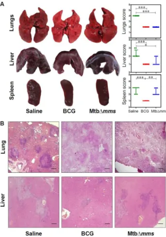
animals were euthanized and bacillary load in the lungs and spleens was determined (Figure 7A). At 4 weeks after challenge, the sham immunized animals exhibited 6.30 log10 CFU in lungs. The BCG vaccinated animals exhibited a significantly reduced CFU in lungs (4.43 log10 CFU) indicating 1.87 log10 CFU reduction (∗p<0.05) as compared to the sham-immunized animals (Figure 7B). Mtb∆mms vaccinated animals exhibited a bacillary load of only 3.60 log10 CFU in lungs indicating that the mutant also significantly reduced the pulmonary load by 2.70 log10 CFU (∗∗∗p<0.001) in comparison to the sham immunized animals (Figure 7B). Further, the sham immunized animals exhibited a splenic bacillary load of 5.37 log10 CFU (Figure 7C), while, the BCG vaccinated animals exhibited a splenic bacillary load of only 1.62 log10 CFU. This significant reduction in splenic bacillary load by 3.75 log10 CFU (∗∗∗p<0.001) demonstrated a tight control of hematogenous spread of bacilli by BCG. The splenic bacillary load in the case of Mtb∆mms vaccinated animals (4.73 log10 CFU) was 0.64 log10 CFU less when compared with the sham immunized animals but the difference was not significant (Figure 7C).
On extending the time period between challenge and euthanasia to 12 weeks, the sham immunized animals exhibited a bacillary load of 6.15 log10 CFU in lungs (Figure 7D). Immunization with BCG resulted in 4.57 log10 CFU in lungs as compared to the sham immunized animals, however, the difference in the pulmonary bacillary load between BCG and sham immunized animals was statistically not significant and with the extension of time, the ability of BCG to impede bacillary multiplication met with a considerable decline. In contrast, Mtb∆mms vaccinated animals exhibited only 3.16 log10 CFU in lungs, thus indicating a significantly reduced bacillary load by 2.λλ log10 CFU (∗p<0.05) in comparison to the sham immunized animals. These observations demonstrated that Mtb∆mms exhibited a more sustainable and superior protection as compared to BCG. The splenic bacillary load in the case of sham immunized animals was 4.λ2 log10 CFU (Figure 7E). Although, the splenic bacillary loads in the cases of BCG vaccination and Mtb∆mms vaccination were 3.30 log10 CFU and 4.21 log10 CFU, respectively, these were not significantly different in comparison to the splenic bacillary load observed in the sham immunized animals. Thus, our observations indicated that Mtb∆mms was not able to exhibit a significant control on the hematogenous spread at either 4 weeks or 12 weeks after challenge.
Mtb∆mms vaccination imparts protection from pathological damage in lungs
and spleen (scoreμ 2 in both the organs) with minimal hepatic tissue destruction (scoreμ 1) (Figure 8A). On extending the period between challenge and euthanasia to 12 weeks, we observed an overall increase in the gross pathological damage to the organs of sham immunized animals as characterized by extensive involvement of tissue with numerous large tubercles and scattered areas of necrosis in both lungs and liver (scoreμ 3-4). In addition, a marked discoloration of spleen with numerous large and small sized tubercles and occasional attrition of capsular structure was also observed in most of the sham immunized animals (scoreμ 4) (Figure λA). BCG immunized guinea pigs exhibited moderate involvement of lung and splenic tissues with small sized tubercles effacing the entire tissues (scoreμ 2-3 in lungs and 1-2 in spleen). However, liver of these animals exhibited normal architecture (scoreμ 1). In case of immunization with Mtb∆mms, the animals exhibited a lung phenotype similar to BCG immunized animals (scoreμ 2-3). However, in the case of spleen and liver, vaccination with Mtb∆mms resulted in an enhanced pathology when compared
Figure 7. Determination of bacillary load in guinea pig organs after immunization with various strains. (A) Experimental protocol for evaluating the protective efficacy of MtbΔmms against M.tuberculosis infection. (B, C) The bacillary load in the lungs and spleens of guinea pigs (n=8) vaccinated with BCG, Mtb∆mms or saline at 4 weeks after M.tuberculosis challenge. (D, E) The bacillary load in the lungs and spleens of guinea pigs (n=8) vaccinated with BCG, Mtb∆mms or saline at 12 weeks after challenge. Mtb∆mms imparted a significant protection as compared to the sham immunized animals in the lungs of vaccinated animals. Each data point represents the Log10 CFU value for an individual animal and the bar depicts mean (±SE) for each group. ∗p<0.05 and ∗∗∗p<0.001 (One-way ANOVA).
doiμ 10.1371/journal.pone.0077λ30.g007
with the BCG vaccination (scoreμ 2-3 in spleen and 3-4 in liver) (Figure λA).
Histopathological analyses of lung and liver sections further substantiated the gross pathological observations. At 4 weeks after challenge, sham immunized animals exhibited several discrete necrotic tubercles occupying 30-40% of the lung sections (Figure 8B). Vaccination with BCG or Mtb∆mms prevented pulmonary damage as was evident from the presence of only a moderate granulomatous infiltration and well preserved alveolar spaces, when compared with the sham immunized animals. Sham immunized animals displayed inflammation of hepatic tissues with scattered areas of cellular infiltration. BCG vaccinated animals exhibited minimal involvement of hepatic tissues; however, Mtb∆mms immunized guinea pigs exhibited moderate involvement with granulomatous infiltration (Figure 8B). Further, at 12 weeks after challenge, as expected, lungs of the sham immunized animals exhibited extensive granulomatous infiltration with multi-focal coalescing granulomas along with prominent central coagulative necrosis (Figure λB). BCG immunized animals exhibited the presence of scattered areas of inflammation with discrete granulomas along with necrotic centre in lungs. Mtb∆mms vaccinated animals exhibited moderate inflammation in lungs similar to BCG vaccinated animals. On comparing the pathological changes in liver (Figure λB), sham immunized animals exhibited effacement of a large proportion of hepatic parenchyma due to multiple coalescing foci of necrotic granulomas. Immunization with BCG resulted in a significant reduction in hepatic damage with only a negligible granulomatous infiltration. However, in the case of Mtb∆mms immunization, the hepatic lobules displayed an extensive granulomatous infiltration (Figure λB).
Discussion
The development and widespread administration of the BCG vaccine since the early 1λ20s was originally hailed as a major breakthrough with the promise to eradicate the scourge of TB from the world. However, the early promise was not realized and with the growing incidence of TB cases and inconsistent protective efficacy of BCG, it became evident that the BCG vaccine, in its existing form is of limited use in controlling the disease particularly in the elderly [36]. The availability of complete M.tuberculosis genome sequence and an increased understanding of the genes involved in M.tuberculosis virulence and immune responses has led to a renewed optimism that it should be possible to develop more efficient TB vaccines than the existing BCG [37,38].
Figure 8. Gross pathology and histopathology of vaccinated guinea pig organs at 4 weeks after challenge. (A) Representative photographs of lungs, liver and spleen of individual animal (n=8) vaccinated with either BCG, MtbΔmms or saline and euthanized at 4 weeks after challenge. The graphical representation of gross scores of various organs is also shown alongside. The bar depicts median for each group. ∗p<0.05; ∗∗p<0.01 and ∗∗∗p<0.001 (Kruskal Wallis test). (B) The representative photomicrographs of the lung and liver tissues stained with Haematoxylin-Eosin staining (H&E). The figure depicts 20x magnification of representative photomicrographs of organs of the animals vaccinated with either BCG, MtbΔmms or saline and euthanized at 4 weeks after challenge. The scale bars depict 500 m for lung as well as liver sections. Unvaccinated animals show severe pathology in lungs characterized by the presence of numerous large and small sized tubercles. However, hepatic and splenic tissues show moderate involvement. In the case of immunization with Mtb∆mms, the animals show moderately inflamed lungs with minimal hepatic tissue destruction similar to that observed in the case of BCG vaccination. However, spleens of Mtb∆mms vaccinated animals exhibit pathological damage similar to sham immunized animals.
doiμ 10.1371/journal.pone.0077λ30.g008
Figure 9. Gross pathology and histopathology of vaccinated guinea pig organs at 12 weeksafter challenge. (A) Representative photographs of lungs, liver and spleen of individual animal (n=8) vaccinated with either BCG, MtbΔmms or saline and euthanized at 12 weeks after challenge. The graphical representation of gross scores of various organs is also shown alongside. The bar depicts median for each group. ∗p<0.05 and ∗∗p<0.01 (Kruskal Wallis test). (B) The representative photomicrographs of the lung and liver tissues stained with Haematoxylin-Eosin staining (H&E). The figure depicts 20x magnification of representative photomicrographs of organs of the animals vaccinated with either BCG, MtbΔmms or saline and euthanized at 12 weeks after challenge. The scale bars depict 500 m for lung as well as liver sections. Unvaccinated animals show characteristic presence of multiple coalescing granulomas with necrosis in both lungs and liver. In addition, the animals show a marked discoloration of spleen with numerous large and small sized tubercles. Mtb∆mms immunized animals, on the other hand show reduced granulomatous infiltration with only a few small and discrete granulomas in lungs. However, liver and spleen of Mtb∆mms vaccinated animals exhibit pathological damage similar to sham immunized animals.
for their use for generating mutants or for challenging the animals.
We have evaluated the vaccine efficacy of the resulting mutant against M.tuberculosis challenge in guinea pigs. To ensure that the mutated genes were essential only for the growth of the pathogen in the host and not in the broth culture, we selected the genes implicated in the host-pathogen interaction. As expected, MtbΔmms grew in broth culture similar to the parental strain, however, it displayed a significantly reduced ability to infect and grow inside the human THP-1 macrophages emphasizing that the phosphatases are vital for the growth of the pathogen in macrophages.
In addition, our studies in guinea pigs also provide evidence that MtbΔmms is highly attenuated for its growth and ability to cause pathology in the host. On inoculation of guinea pigs with Mtb∆mms, no bacilli were recovered from the lungs of the animals at any time point of the study. From the spleens of these animals, bacilli were recovered during early phases after the inoculation (4 weeks) but no bacilli were recovered after this initial period. This demonstrated that MtbΔmms could survive only for a short while; therefore, at 10 weeks post inoculation, the organs of MtbΔmms as well as BCG inoculated animals appeared to be similar with no apparent damage indicating that the mutant was safe to be used as a vaccine candidate.
For the evaluation of protective efficacy, we employed guinea pig model of experimental tuberculosis. The guinea pig model of low dose aerogenic infection with virulent M.tuberculosis has been preferentially used to elucidate the events in the pathogenesis of pulmonary tuberculosis [45]. Guinea pigs are more susceptible to tuberculosis infection and have the advantage over mice in that the pathology of the disease in this model is closer to human tuberculosis. Thus, it serves as an effective model to evaluate vaccine efficacy. When guinea pigs are infected with less than 10 CFU of virulent M.tuberculosis, it has been observed that the pathogen disseminates from lungs to the pulmonary lymph nodes via hematogenous spread and then appears in spleens within ~3 weeks post infection [46,47]. Bacilli reseed the lung by ~4 weeks to form secondary granulomas. The protective efficacy of Mtb∆mms was evaluated based on its ability to reduce the bacillary load in lungs and spleens of guinea pigs post M.tuberculosis infection as well as to control the pathological damage.
Our study shows that Mtb∆mms vaccination was able to restrict bacterial multiplication at the primary site of infection leading to reduction in the pulmonary bacillary load and this bacillary load reduction by Mtb∆mms as well as by BCG was comparable during early phase (4 weeks) after infection. Unlike in the case of BCG vaccination, however, Mtb∆mms was not able to control hematogenous spread. A number of studies have reported the examples of vaccines which fail to provide consistent protection in all the organs uniformly [12,48,4λ]. For example, it has been reported that a recombinant BCG expressing ESAT-6 provided significant protection in both mice and guinea pigs against dissemination at extra-pulmonary site but failed to protect against pulmonary form of the disease [4λ]. On the other hand, vaccination of guinea pigs with DNA
encoding the mycobacterial antigen MPB83 influenced the pulmonary pathology but not the hematogenous spread following the aerogenic infection with Mycobacterium bovis [48]. Also, in the case of vaccination of guinea pigs with the ∆phoP mutant, a significant reduction in the bacillary load in lungs but not in the spleens was observed as compared to the unvaccinated animals [12].
On extending the period between the M.tuberculosis challenge and euthanasia to 12 weeks, although BCG appeared to lose the control of bacillary multiplication in the pulmonary tissue, Mtb∆mms was still very effective in controlling the lung infection. At this time point, however, neither BCG nor Mtb∆mms exhibited any significant control over the hematogenous spread. The pathological damage in the animals from various groups corroborated the CFU data. From this, we could infer that Mtb∆mms imparted as much or better control of the disease than BCG at the pulmonary site. However, immunization with the phosphatase mutant did not show any superior control over the bacillary multiplication in spleens, when compared with the sham immunized animals.
As phosphatases play an important role in the lipid metabolism, we evaluated the lipid profiles of M.tuberculosis and MtbΔmms to ascertain whether disruption of phosphatase genes might result in the altered lipid profile in MtbΔmms. However, our observations demonstrated that the lipid profiles of both the strains were identical inspite of disruption of phosphatase genes in MtbΔmms indicating thereby that the phenotype of the mutant was ascribed to the loss of phosphatase genes and the influence was not related to any alteration in the lipid composition.
To summarize, we demonstrate that mutation of genes encoding the signal transduction associated phosphatases of M.tuberculosis provides optimism for the generation of novel potential vaccine candidates against tuberculosis. The Mtb∆mms was not only significantly attenuated for growth in macrophages and guinea pigs, it also imparted an enhanced protection against pulmonary TB. However, further modifications would be required in order for Mtb∆mms to elicit more appropriate immune responses for imparting superior protection including the control of hematogenous spread. Moreover, due to increasing concern about the emergence of antibiotic resistance in human pathogens, use of antibiotic-resistant genes in recombinant vaccines meant for use in humans is not permissible [50]. Hence, the antibiotic resistance genes from MtbΔmms would have to be removed before any possibility of its use in human clinical trials. Our future efforts would focus on addressing these issues.
Supporting Information
Table S1. Solvent systems employed for lipid analyses.
(DOC)
Acknowledgements
Dr. Bappaditya Dey for useful discussions during the designing of the animal experiments and writing of this manuscript. Stuti Sharma and Dr. Garima Khare are acknowledged for purifying SapM and raising antibodies. Anti-mptpA antibody was provided by Dr. Yogendra Singh, IGIB, New Delhi. Sandeep Kumar is acknowledged for his help with the animal experiments. Priti Singh and Tannupriya Gosain are acknowledged for technical assistance. Bahadur Singh, Devender Singh and Jeevan Rawat are acknowledged for looking after the animals and cleaning of the glasswares etc.
Author Contributions
Conceived and designed the experimentsμ PC AKT. Analyzed the dataμ PC PVR. Wrote the manuscriptμ PC AKT PVR RS. Provided overall supervision throughout the studyμ AKT. Generated Mtb∆mptpB∆mptpA strainμ RS. Generated Mtb∆mms strainμ PVR. Performed all the experiments related to mutantsμ PC. Lipid analysisμ NJ PC SG.
References
1. Walker KB, Brennan MJ, Ho MM, Eskola J, Thiry G et al. (2010) The second Geneva Consensusμ Recommendations for novel live TB vaccines. Vaccine 28μ 225λ-2270. doiμ10.1016/j.vaccine.200λ.12.083. PubMedμ 20074686.
2. WHO (2012) Global tuberculosis report. Retrieved onpublished at whilst December year 1111 from httpμ//wwwwhoint/tb/publications/ global_report/gtbr12_mainpdf. Accessed 31st August 2013.
3. Gandhi NR, Nunn P, Dheda K, Schaaf HS, Zignol M et al. (2010) Multidrug-resistant and extensively drug-resistant tuberculosisμ a threat to global control of tuberculosis. Lancet 375μ 1830-1843. doiμ10.1016/ S0140-6736(10)60410-2. PubMedμ 20488523.
4. Velayati AA, Masjedi MR, Farnia P, Tabarsi P, Ghanavi J et al. (200λ) Emergence of new forms of totally drug-resistant tuberculosis bacilliμ super extensively drug-resistant tuberculosis or totally drug-resistant strains in Iran. Chest 136μ 420-425. doiμ10.1378/chest.08-2427. PubMedμ 1λ34λ380.
5. McShane H (2011) Tuberculosis vaccinesμ beyond bacille Calmette-Guerin. Philos Trans R Soc Lond B Biol Sci 366μ 2782-278λ. doiμ 10.10λ8/rstb.2011.00λ7. PubMedμ 218λ3541.
6. Brodin P, Majlessi L, Brosch R, Smith D, Bancroft G et al. (2004) Enhanced protection against tuberculosis by vaccination with recombinant Mycobacterium microti vaccine that induces T cell immunity against region of difference 1 antigens. J Infect Dis 1λ0μ 115-122. doiμ10.1086/421468. PubMedμ 151λ5250.
7. Brosch R, Gordon SV, Garnier T, Eiglmeier K, Frigui W et al. (2007) Genome plasticity of BCG and impact on vaccine efficacy. Proc Natl Acad Sci U S A 104μ 55λ6-5601. doiμ10.1073/pnas.070086λ104. PubMedμ 173721λ4.
8. Behr MA, Wilson MA, Gill WP, Salamon H, Schoolnik GK et al. (1λλλ) Comparative genomics of BCG vaccines by whole-genome DNA microarray. Science 284μ 1520-1523. doiμ10.1126/science. 284.541λ.1520. PubMedμ 10348738.
λ. Hernàndez Pando R, Aguilar LD, Infante E, Cataldi A, Bigi F et al. (2006) The use of mutant mycobacteria as new vaccines to prevent tuberculosis. Tuberculosis 86μ 203-210. doiμ10.1016/j.tube. 2006.01.022. PubMedμ 16542875.
10. Sambandamurthy VK, Jacobs WR Jr. (2005) Live attenuated mutants of Mycobacterium tuberculosis as candidate vaccines against tuberculosis. Microbes Infect 7μ λ55-λ61. doiμ10.1016/j.micinf. 2005.04.001. PubMedμ 15λ14065.
11. Hinchey J, Lee S, Jeon BY, Basaraba RJ, Venkataswamy MM et al. (2007) Enhanced priming of adaptive immunity by a proapoptotic mutant of Mycobacterium tuberculosis. J Clin Invest 117μ 227λ-2288. doiμ10.1172/JCI31λ47. PubMedμ 17671656.
12. Martin C, Williams A, Hernandez-Pando R, Cardona PJ, Gormley E et al. (2006) The live Mycobacterium tuberculosis phoP mutant strain is more attenuated than BCG and confers protective immunity against tuberculosis in mice and guinea pigs. Vaccine 24μ 3408-341λ. doiμ 10.1016/j.vaccine.2006.03.017. PubMedμ 16564606.
13. Ottenhoff TH, Kaufmann SH (2012) Vaccines against tuberculosisμ where are we and where do we need to go? PLOS Pathog 8μ e1002607. PubMedμ 2258λ713.
14. Sambandamurthy VK, Derrick SC, Hsu T, Chen B, Larsen MH et al. (2006) Mycobacterium tuberculosis ΔRD1 ΔpanCDμ a safe and limited replicating mutant strain that protects immunocompetent and immunocompromised mice against experimental tuberculosis. Vaccine 24μ 630λ-6320. doiμ10.1016/j.vaccine.2006.05.0λ7. PubMedμ 16860λ07.
15. Waters WR, Palmer MV, Nonnecke BJ, Thacker TC, Scherer CF et al. (2007) Failure of a Mycobacterium tuberculosis ΔRD1 ΔpanCD double deletion mutant in a neonatal calf aerosol M. bovis challenge modelμ comparisons to responses elicited by M. bovis bacille Calmette Guerin.
Vaccine 25μ 7832-7840. doiμ10.1016/j.vaccine.2007.08.02λ. PubMedμ 17λ31755.
16. Tuberculosis Vaccine candidates (2011). Retrieved onpublished at whilst December year 1111 from httpμ//www.stoptb.org/wg/ new_vaccines/assets/documents/TB%20Vaccine%20Pipeline_rAug %202012.pdf. Accessed 31st August 2013.
17. Beresford NJ, Mulhearn D, Szczepankiewicz B, Liu G, Johnson ME et al. (200λ) Inhibition of MptpB phosphatase from Mycobacterium tuberculosis impairs mycobacterial survival in macrophages. J Antimicrob Chemother 63μ λ28-λ36. doiμ10.10λ3/jac/dkp031. PubMedμ 1λ24007λ.
18. Saleh MT, Belisle JT (2000) Secretion of an acid phosphatase (SapM) by Mycobacterium tuberculosis that is similar to eukaryotic acid phosphatases. J Bacteriol 182μ 6850-6853. doiμ10.1128/JB. 182.23.6850-6853.2000. PubMedμ 11073λ36.
1λ. Vergne I, Chua J, Lee HH, Lucas M, Belisle J et al. (2005) Mechanism of phagolysosome biogenesis block by viable Mycobacterium tuberculosis. Proc Natl Acad Sci U S A 102μ 4033-4038. doiμ10.1073/ pnas.040λ716102. PubMedμ 15753315.
20. Wong D, Chao JD, Av-Gay Y (2013) Mycobacterium tuberculosis-secreted phosphatasesμ from pathogenesis to targets for TB drug development. Trends Microbiol 21μ 100-10λ. doiμ10.1016/j.tim. 2012.0λ.002. PubMedμ 23084287.
21. Festjens N, Bogaert P, Batni A, Houthuys E, Plets E et al. (2011) Disruption of the SapM locus in Mycobacterium bovis BCG improves its protective efficacy as a vaccine against M. tuberculosis. EMBO. Mol Med 3μ 222-234.
22. Saikolappan S, Estrella J, Sasindran SJ, Khan A, Armitige LY et al. (2012) The fbpA/sapM double knock out strain of Mycobacterium tuberculosis is highly attenuated and immunogenic in macrophages. PLOS ONE 7μ e361λ8. doiμ10.1371/journal.pone.00361λ8. PubMedμ 22574140.
23. Wong D, Bach H, Sun J, Hmama Z, Av-Gay Y (2011) Mycobacterium tuberculosis protein tyrosine phosphatase (PtpA) excludes host vacuolar-H+-ATPase to inhibit phagosome acidification. Proc Natl Acad
Sci U S A 108μ 1λ371-1λ376. doiμ10.1073/pnas.110λ201108. PubMedμ 22087003.
24. Bach H, Papavinasasundaram KG, Wong D, Hmama Z, Av-Gay Y (2008) Mycobacterium tuberculosis virulence is mediated by PtpA dephosphorylation of human vacuolar protein sorting 33B. Cell Host Microbe 3μ 316-322. doiμ10.1016/j.chom.2008.03.008. PubMedμ 18474358.
25. Koul A, Choidas A, Treder M, Tyagi AK, Drlica K et al. (2000) Cloning and characterization of secretory tyrosine phosphatases of Mycobacterium tuberculosis. J Bacteriol 182μ 5425-5432. doiμ 10.1128/JB.182.1λ.5425-5432.2000. PubMedμ 10λ86245.
26. Singh R, Rao V, Shakila H, Gupta R, Khera A et al. (2003) Disruption of mptpB impairs the ability of Mycobacterium tuberculosis to survive in guinea pigs. Mol Microbiol 50μ 751-762. doiμ10.1046/j. 1365-2λ58.2003.03712.x. PubMedμ 14617138.
27. Zhou B, He Y, Zhang X, Xu J, Luo Y et al. (2010) Targeting mycobacterium protein tyrosine phosphatase B for antituberculosis agents. Proc Natl Acad Sci U S A 107μ 4573-4578. PubMedμ 201677λ8. 28. Hou S, Li X, Feng XZ (200λ) Method to improve DNA condensation efficiency by alkali treatment. Nucleosides Nucleotides Nucleic Acids 28μ 725-735. doiμ10.1080/15257770λ031554λ3. PubMedμ 20183612. 2λ. Vilchèze C, Jacobs WR (2007) Isolation and analysis of Mycobacterium
tuberculosis mycolic acids. Curr Protoc Microbiol Pp. 10A.3μ Unit 10A. 3.1-10A.311. PubMedμ 18770604