Preconditioning against Acute Myocardial Infarction in
Male Mouse Heart
In Vivo
Virginija Jazbutyte1*, Jan Stumpner2, Andreas Redel3, Johan M. Lorenzen1, Norbert Roewer2, Thomas Thum1., Franz Kehl4.
1Institute of Molecular and Translational Therapeutic Strategies (IMTTS), Hannover Medical School (MHH), Hannover, Germany,2Department of Anesthesia and Critical Care, University of Wu¨rzburg, Wu¨rzburg, Germany,3Department of Anesthesia, University of Regensburg, Regensburg, Germany,4Department of Anesthesiology and Critical Care, Klinikum Karlsruhe, Karslruhe, Germany
Abstract
The volatile anesthetic desflurane (DES) effectively reduces cardiac infarct size following experimental ischemia/reperfusion injury in the mouse heart. We hypothesized that endogenous estrogens play a role as mediators of desflurane-induced preconditioning against myocardial infarction. In this study, we tested the hypothesis that desflurane effects local estrogen synthesis by modulating enzyme aromatase expression and activity in the mouse heart. Aromatase metabolizes testosterone to 17b- estradiol (E2) and thereby significantly contributes to local estrogen synthesis. We tested aromatase effects in acute myocardial infarction model in male mice. The animals were randomized and subjected to four groups which were pre-treated with the selective aromatase inhibitor anastrozole (A group) and DES alone (DES group) or in combination (A+DES group) for 15 minutes prior to surgical intervention whereas the control group received 0.9% NaCl
(CON group). All animals were subjected to 45 minutes ischemia following 180 minutes reperfusion. Anastrozole blocked DES induced preconditioning and increased infarct size compared to DES alone (37.94615.5% vs. 17.163.62%) without affecting area at risk and systemic hemodynamic parameters following ischemia/reperfusion. Protein localization studies revealed that aromatase was abundant in the murine cardiovascular system with the highest expression levels in endothelial and smooth muscle cells. Desflurane application at pharmacological concentrations efficiently upregulated aromatase expressionin vivoandin vitro.We conclude that desflurane efficiently regulates aromatase expression and activity which
might lead to increased local estrogen synthesis and thus preserve cellular integrity and reduce cardiac damage in an acute myocardial infarction model.
Citation:Jazbutyte V, Stumpner J, Redel A, Lorenzen JM, Roewer N, et al. (2012) Aromatase Inhibition Attenuates Desflurane-Induced Preconditioning against Acute Myocardial Infarction in Male Mouse HeartIn Vivo. PLoS ONE 7(8): e42032. doi:10.1371/journal.pone.0042032
Editor:Tobias Eckle, University of Colorado Denver, United States of America
ReceivedMay 21, 2012;AcceptedJune 29, 2012;PublishedAugust 2, 2012
Copyright:ß2012 Jazbutyte et al. This is an open-access article distributed under the terms of the Creative Commons Attribution License, which permits unrestricted use, distribution, and reproduction in any medium, provided the original author and source are credited.
Funding:The study was supported by the University Clinics of Wu¨rzburg, Germany, by Else-Kro¨ner-Fresenius Foundation (FK) and by the German Ministry for Education and Research (IFB-Tx, grant 01EO0802 for TT). The funders had no role in study design, data collection and analysis, decision to publish, or preparation of the manuscript.
Competing Interests:Co-author Thomas Thum is a PLoS ONE Editorial Board Member. This does not alter the authors‘ adherence to all the PLoS ONE policies on sharing data and materials.
* E-mail: vjazbutyte@hotmail.com
.These authors contributed equally to this work.
Introduction
Cardioprotective events following application of volatile anes-thetics prior to ischemia/reperfusion are called anesthetic precon-ditioning [1]. The mechanisms involved in cardioprotection by volatile anesthetics, such as sevoflurane and desflurane, include increased nitric oxide (NO) synthesis, activation of mitochondrial large- conductance calcium- activated potassium channel, beta1-adrenergic pathway and generation of reactive oxygen species (ROS) [2–5]. Anesthetic preconditioning is similar to and shares many common signal transduction pathways with ischemic preconditioning which is characterized by brief repeated periods of vascular occlusion prior to prolonged ischemia/reperfusion which protects ischemic myocardium [6]. Despite that, the phenomenon of anesthetic preconditioning is not fully understood and little is known about the mediators of anesthetic precondi-tioning. The major female sex hormone 17b- estradiol (E2) was
Additionally, aromatase disruption in mouse model (ArKO mice) resulted in increased adiposity, altered fatty acid metabolism and higher levels of testosterone, the features characteristic for metabolic syndrome [12]. Aromatase expression analysis in the cardiovascular system showed that aromatase mRNA was abundant in endothelial cells and also smooth muscle cells [13– 15]. Functional analysis of aromatase deficiency in the cardiovas-cular system of female mice showed that ArKO mice had reduced diastolic and mean blood pressure together with increased blood pressure variability [16]. Aromatase inhibition in healthy young men caused significant decrease in serum estrogen levels and reduced vasodilation without altering other parameters, such as serum testosterone, lipoprotein levels and endothelium- indepen-dent vasodilation [17].
In the current study, we determined whether the volatile anesthetic desflurane modulates aromatase expression and/or activity in the heart and thus may contribute to cardioprotection in an acute ischemia/reperfusion model.
Materials and Methods
1. Ethics Statement
Animal treatment and surgery were performed according to recommendations of the Guide for the Care and Use of Laboratory Animals published by the United States National Institute of Health. All experimental protocols were reviewed and approved by the Animal Care and Use Committee of the Government of Lower Franconia, Bavaria, Germany.
2. Animals and Surgery
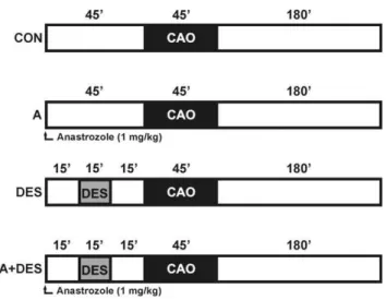
Male C57/BL6N mice (8–12 weeks) were purchased by Charles River Laboratories (Sulzfeld, Germany). Animals were housed under controlled conditions (22uC, 55%–65% humidity and artificial 12 hour light/dark regime) and had free access to standard laboratory chow and tap water. A total of forty (n = 6–10 animals/group) were randomly assigned to following experimental groups: control group (CON) which underwent ischemia/reper-fusion surgery, A group which received the aromatase inhibitor Anastrozole (ArimidexH, AstraZeneca Pharmaceuticals Group, Alderley Park, UK) prior to ischemia/reperfusion, DES group received 1 minimum alveolar concentration (MAC) of Desflurane (Baxter Deutschland GmbH) prior to ischemia/reperfusion and DES+A group obtained Anastrozole and subsequent DES treatment prior to ischemia/reperfusion. The mice were anesthe-tized with sodium pentobarbital, intubated and mechanically ventilated. Anastrozole was dissolved in 0.9% NaCl and injected intravenously at a dosage of 1 mg/kg (max. vol. 100ml). Mean arterial blood pressure and heart rate were measured via cannulation of the right carotid artery. Coronary artery occlusion (CAO) was performed by 45 minutes ligation of the left anterior descending coronary artery (LAD) using a 6-0 silk suture. Subsequently, the ligature was removed and the hearts were reperfused 180 minutes. A schematic diagram of the experimental protocol is shown in figure 1.
For aromatase gene expression regulation experiments, mice were anesthetized with sodium pentobarbital, intubated and mechanically ventilated. The mice were treated with desflurane (1 MAC) for 15 minutes, awoken from anesthesia and kept under standard conditions for 15 minutes (DES15 group) or 48 hours (DES48 group) as described at the beginning of this chapter. Afterwards, the hearts were collected, shock-frozen in liquid nitrogen and subjected to RNA isolation and western blotting assays.
3. Measurement of Myocardial Infarct Size
Area at risk (AAR) and infarct size (IS) were quantified using Evans Blue staining as described previously [18,19]. After 45 minutes of ischemia and 180 minutes reperfusion, the LAD was reoccluded and 1 ml of Evans blue (Sigma- Aldrich, St. Louis, USA) was injected into the carotid artery. The hearts were explanted, cut into 1 mm thick transversal slices, incubated with 2% triphenyltetrazolium chloride (Sigma- Aldrich, St. Louis, USA), fixed overnight in 10% formalin, weighed, and digitally photographed. The photographs were then analyzed with AdobePhotoshop CS 8.0.1 software. Infarct size, area at risk and normal zone were quantified by an investigator blinded to the treatment protocol. The resulting fractions of infarct size, area at risk and normal zone of each slice were multiplied by the weight of that slice. Animals with an AAR,15% of the left ventricle were excluded from the study. Myocardial infarct size (IS) is expressed as a percentage of the left ventricular area at risk (AAR).
4. Systemic Hemodynamics
During experiments, heart rate (HR, beats/min), mean arterial pressure (MAP, mmHg) were recorded using a saline-filled PE-10 catheter connected to a pressure transducer (Combitrans, B. Braun, Melsungen, Germany) which was inserted into the right common carotid artery. The parameters were recorded at the end of the baseline period (BL), at the end of the intervention period (desflurane or corresponding time point), at the end of memory period (MEM, memory period), at the end of coronary artery occlusion ( = CAO) and 60, 120 and 180 min after the onset of reperfusion. The rate pressure product (RPP) which is an indicator of myocardial oxygen consumption was calculated using following formula: RPP = HRxMAP/1000.
Figure 1. Schematic diagram illustrating the experimental protocol.CON = control; CAO = coronary artery occlusion; A = anastro-zole (1 mg/kg BW); DES = desflurane (1 minimum alveolar concentra-tion); A+DES = anastrozole and desflurane at concentrations indicated
5. RNA Isolation and Quantitative Real-time PCR
Total RNA was extracted from mouse heart using Trizol (Invitrogen, Carlsbad, USA) according to the manufacturer’s recommendations. The cDNA was generated using First- strand cDNA synthesis kit (Biorad, Munich, Germany). Aromatase gene Cyp19a1 and GAPDH expression analysis was performed using quantitative real- time PCR method. PCR master mix and primer mix were purchased from SABiosciences (Biomol, Hamburg, Germany), PCR reactions were carried out with a DNA Engine Opticon 2 System (MJ Research, Biozym, Oldendorf, Germany) and the data were analyzed using delta delta C(t) method (DDC(t)).
6. Cell Culture
Human umbilical cord endothelial cells (HUVEC) were extracted from umbilical cords using Trypsin-EDTA (Sigma-Aldrich, St. Louis, USA) digestion and cultivated in M199 medium (Gibson- Invitrogen, Carlsbad, USA) supplemented with 20% FCS, endothelial cell growth supplement (ECGF) (BD Biosciences, Bedford, USA), heparin (LiqueminHN, Hoffmann-La Roche AG, Grenzach-Wyhlen, Germany) and penicillin/ streptomycin (Sigma- Aldrich, St. Louis, USA). Briefly, the umbilical cords were washed several times with PBS and
endothelial cells were extracted following trypsin digestion. The cells were cultivated in Medium 199 with supplements and subjected for in vitro experiments. For in vitro experiments, HUVECs were treated with oxygen (1 liter/h, control group, CON) or oxygen: desflurane mixture containing 7.5 vol. % and comprising 1 mean alveolar concentration (MAC) of desflurane for 15 minutes in a closed chamber (DES group). Following 15 minutes treatment, cells were transferred to the incubator and further kept under standard conditions (5% CO2, 37uC). Cells were collected 15 minutes (DES 0.25), 24 hours (DES24) or 48 hours (DES48) later and subjected to further analysis. Cells from the native group were kept under standard conditions and collected together with control and desflurane-treated cells.
Primary rat aortic smooth muscle cells were cultivated in DMEM (Sigma- Aldrich, St. Louis, USA) containing 10% FCS. The cells were provided by Paula Anahi Arias- Loza from the Department of Cardiology, University Clinics Wu¨rzburg, Wu¨rz-burg, Germany.
7. Immunostaining
Mouse heart tissue and the aorta were frozen in Tissue- Tec OCT compound (Sakura, Alphen an den Rijn, The Nether-lands) and cut in 5mm thick cryosections. The sections were fixated with 4% PFA, permeabilized with 0.1% Triton X-100 and subjected to immunostaining. Aromatase was detected using rabbit- anti- aromatase antibody (Biocat, Heidelberg, Germany), smooth muscle cells were visualized by Cy3- labelled mouse anti- smooth muscle actin antibody (Sigma- Aldrich, Hamburg, Germany), endothelial cells were stained using rat anti- CD31 (PECAM1) antibody (BD Biosciences/Pharmingen, Heidelberg, Germany) and cell nuclei were visualized using goat- anti- lamin A/C antibody (N-18) (Santa Cruz biotechnology, Santa Cruz, USA). Unspecific background was blocked using donkey serum diluted in PBS (Sigma- Aldrich, Hamburg Germany). Specific signals for immunofluorescence were visualized using AlexaFluor labelled donkey rat, donkey- rabbit and donkey- anti-goat antibodies purchased from Molecular Probes (now Invitro-gen, Carlsbad, USA). In immunohistochemical staining, specific signal was detected using HRP- labelled secondary antibodies in combination with streptavidin- horse reddish peroxidase (HRP) and visualized by diaminobenzydine (DAB) system as described by manufacturers (Vector Laboratories, Burlingame, USA). Fluorescence imaging was performed using confocal laser scanning microscope MRC-1024 and the picture acquisition was performed using the Lasersharp2000 software (Biorad, Munich, Germany).
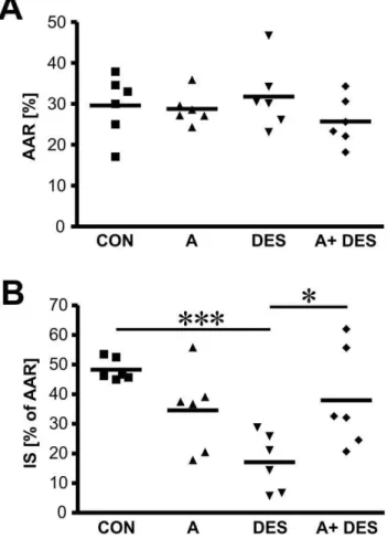
Figure 2. Summary charts of area at risk and infarct size measurements in male mice hearts.After hemodynamic measure-ments, the animals were perfused with triphenyltetrasolium (TPP) and Evans Blue to assess perfused myocardium, area at risk and infracted (necrotic) tissue. Myocardial infarct size (IS) was expressed as a percentage of area at risk (AAR). Values are mean percent6 SEM, n = 6 animals/group. A Pvalue ,0.05 was considered as statistically
significant.
doi:10.1371/journal.pone.0042032.g002
Table 1.The results of area at risk and infarct size measurements in cardiac ischemia/reperfusion model.
Group n Area at risk [%] Infarct size [% of AAR]
CON 6 29.662.8 48.361.4* A 6 28.861.5 34.665.21
DES 6 31.863.1 17.163.6*#1
A+DES 6 25.762.2 37.9615.5#
Data: Mean [%]6SEM. *,#
,1
- statistically significant differences, p,0.05.
CON = control group; A = Anastrozole group; DES = desflurane group; A+DES = anastrozole+desflurane.
Table 2.Invasive hemodynamics results.
BL 159 DES 159 MEM 159 CAO 459 Reperfusion
HR (min21) 60 min 120 min 180 min
CON 447 619 464 625 463 629 458 621 450 613 465 618 460 613
A 467 621 466 619 464 618 448 614 467 614 477 613 477 630
DES 473 617 417 611 480 619 432 623 452 613 447 66 463 615
DES+A 468 619 448 617 460 621 495 626 483 618 489 627 455 619
MAP(mmHg)
CON 71 63 71 63 66 65 60 61 67 64 63 64 66 65
A 67 66 61 65 58 64 50 65 61 67 58 67 68 66
DES 72 62 73 65 69 63 61 61 67 61 67 62 62 62
DES+A 74 63 80 62 74 67 66 67 69 68 58 67 58 68
RPP(HRxMAP/1000)
CON 32 63 33 63 30 63 27 61 30 62 30 62 31 63
A 30 63 29 63 27 63 23 63 29 64 28 63 33 64
DES 34 62 30 62 33 62 26 61 31 61 30 61 29 62
DES+A 36 62 36 62 34 64 34 64 34 64 29 64 27 64
BL 159, baseline; DES 159, desflurane; MEM, memory; CAO, coronary artery occlusion; HR, heart rate; MAP, mean arterial pressure; RPP, rate pressure product. Hemodynamic measurements were performed 15 minutes following equilibration period (baseline), at the end of desflurane application, at the end of memory phase (15 min.), at the end of 45 minutes of coronary artery occlusion and three times (at 60, 90 and 180 minutes) during reperfusion.
doi:10.1371/journal.pone.0042032.t002
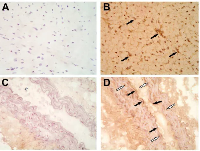
Figure 3. Aromatase immunostaining in mouse heart and aorta.Aromatase protein was abundant in mouse myocardium (B, solid arrows) and in the endothelium (solid arrows) as well as in media (white arrows) in the aorta (D). Aromatase signal was visualized by diaminobenzidine substrate (DAB) combined with hematoxylin/eosin staining. Primary antibody was omitted in negative control staining (A and C).
8. Western Blotting
Protein samples were prepared from untreated HUVEC cells (Native group), treated with oxygen: desflurane (DES group) or with pure oxygen (CON group). Briefly, cells were washed with ice- cold PBS and lysed for 15 minutes in cold RIPA buffer containing Complete protease inhibitor cocktail (Roche, Penzberg, Germany). Protein samples were electrophoretically separated and transferred overnight at 100 mA to nitrocellulose membrane. Next day, the membranes were blocked with 5% skimmed milk in PBS and aromatase protein expression was detected using rabbit- anti-aromatase antibody (Biocat, Heidelberg, Germany) at concentra-tion 1mg/ml in blocking solution. Aromatase signal was visualized using HRP - conjugated goat- anti- rabbit secondary antibody (dilution 1:10000) (Cell signaling, Beverly, MA, USA) and Amersham ECL Plus western blotting detection reagent (GE Healthcare, Pittsburgh, PA, USA). We used GAPDH protein as an internal loading control, which was detected on the same membranes using mouse anti- GAPDH antibody (Chemicon, Billerica, MA,USA) at concentration 0.5mg/ml, visualized using HRP- conjugated goat anti- mouse antibody (Cell signaling, Beverly, MA, USA) in combination with ECL Plus substrate. Aromatase and GAPDH signal was developed using X- ray film and signal intensity was quantified using Scion Image freeware (Scion corp). Signal intensity ratio between aromatase and GAPDH was statistically analyzed and the results are presented as mean values of four independent experiments.
9. Statistics
Statistical analysis of data within and among groups was performed with one-way analysis of variance (ANOVA) followed bypost hoc Student-Newman-Keuls method test using SigmaStat 2.0 software. A Pvalue less than 0.05 was considered as statistically significant.
Results
Area at Risk and Infarct Size Measurements
Area at risk and myocardial infarct size were measured to identify the levels of cardiac injury following ischemia/reperfusion. Area at risk values were comparable among all treatment groups (figure 2, panel A) whereas infarct size measurements demonstrat-ed that desflurane alone (DES group) was superior in rdemonstrat-eduction of myocardial infarction size in male mice compared to the control
group (CON) which underwent ischemia/reperfusion
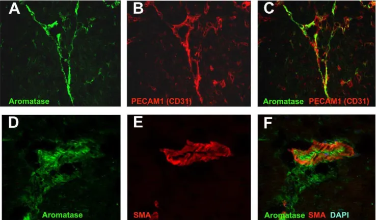
(17.163.62% vs. 48.361.4%, p,0.001) (figure 2, panel B). Interestingly, aromatase inhibition with anastrozole resulted in marginal decrease in infarct size (group A in figure 2B) but anastrozole application prior to desflurane treatment (group A+DES) significantly increased infarct size compared to DES alone (37.94615.5% vs. 17.163.62%). Therefore, we conclude that DES- induced cardioprotection was at least partially mediated by endogenous locally synthesised estrogens in male mice in our experimental model. The data of area at risk and myocardial infarction size measurements are summarized in table 1. Figure 4. Aromatase co- localization with endothelial cells and smooth muscle cells in mouse heart studies using confocal microscopy.Aromatase was detected by immunostaining using specific antibody and visualized by AlexaFluor488- coupled secondary antibody (A and D). Endothelial cell specific marker PECAM1 (CD31) (B) and smooth muscle markera-smooth muscle actin (SMA) (E) were detected using specific primary antibodies and visualized using AlexFluor594-coupled secondary antibodies (B and E, respectively). Aromatase and PECAM1 co- localization is shown in section C, whereas aromatase anda-smooth muscle actin co- expression in the heart is demonstrated in section F.
Systemic Hemodynamics
Anastrozole application did not influence cardiac function and there were no significant differences between the treatment groups. Hemodynamic data are summarized in table 2.
Aromatase Localization in Male Mouse Heart
We performed aromatase localization studies to assess aroma-tase protein expression and its localization pattern in the cardiovascular system of male mice. Immunostaining results showed that aromatase was highly expressed in the myocardium and in the aorta (Fig. 3 B and D). Aromatase protein was abundant in cardiac vasculature and, to lesser extent, in cardiomyocytes (Fig. 3B). Aromatase was also detectable in intima and media in the aorta (Fig. 3D, solid arrows and white arrows, respectively). Using confocal imaging technique, we could confirm immunohistochemistry data and show that aromatase was
expressed in the murine heart (Fig. 4 A and D) and co-localized with endothelial cells which were detected using endothelial cell marker PECAM1 (CD31) specific antibody (Fig. 4, B and C). Additionally, we observed partial aromatase co- localization with smooth muscle cells, which were visualized using smooth muscle actin- specific antibody (Fig. 4, E and F). Further cellular localization studies revealed strong aromatase expression in the cytosol of both, human endothelial (Figure 5, A and B) and aortic smooth muscle cells (Figure 5, C and D).
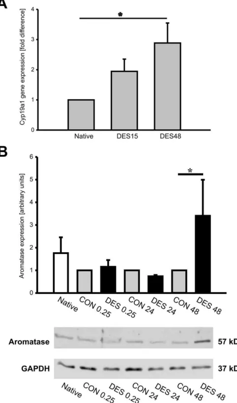
Aromatase Gene Expression is Upregulated by Desflurane
Figure 6. Aromatase upregulation by desfluranein vivoandin vitro.Male mice were treated with 1 MAC desflurane for 15 minutes, then were euthanized 15 minutes later (DES15) or 48 hours later (DES48). The hearts from untreated animals served as native control (Native). Total RNA was extracted from myocardial tissue and aromatase gene (Cyp19a1) expression was analyzed by quantitative real- time PCR (qRT-PCR), n = 5/group (A). Aromatase protein expressionin vitrowas studied by western blotting. Endothelial cells were treated with oxygen: desflurane (1 MAC) mixture for
15 minutes and collected 15 minutes later (DES0.25), 24 hours later (DES24) or 48 hours later (DES 48). Cells treated with oxygen alone for 15 minutes and collected 15 minutes later, 24 hours later and 48 hours later were assigned as controls (CON0.25, CON 24 and CON 48, respectively). The cells kept under standard conditions served as native control (B). The chart in panel B represents statistical analysis of aromatase signal quantification (n = 4). Aromatase signal in CON group was set to 1 (gray bars) and desflurane influence on aromatase protein expression was compared to CON group (solid bars). GAPDH protein served as internal loading control. Representative aromatase western blot is shown in lower part of panel B. The data are presented as mean6SEM and thePvalue,0.05 was considered as statistically significant.
regulation by desflurane was assessed by quantitative real- time PCR. The data showed that aromatase mRNA expression was upregulated by desflurane as early as 15 minutes following desflurane application and significant increase inCyp19a1 expres-sion levels was observed following 48 hours (figure 6A). Aromatase protein expression in the heart was comparable among the groups (data not shown). To confirm in vivo data, we treated human endothelial cells (HUVECs) with a mixture of desflurane and oxygen (1 MAC (7.5 vol%) and 92.5% oxygen) for 15 minutes, transferred the cells to the incubator with standard conditions (5% CO2, 37uC) and collected them 15 minutes, 24 hours or 48 hours later. For negative control, the cells were treated 15 minutes with 100% oxygen, transferred to the incubator with standard conditions and collected 15 minutes, 24 or 48 hours later. Western blotting analysis using aromatase- specific antibody demonstrated significant increase in aromatase protein expression in endothelial cells treated with desflurane and collected 48 hours later (Fig. 6 B).
Discussion
In this study we could show that 1) aromatase inhibition using the pharmacological agent anastrozole attenuated desflurane-induced cardioprotection, 2) aromatase is expressed in the cardiovascular system of male mice, particularly in the endothe-lium and smooth muscle cells and 3) desflurane itself can induce aromatase expression in endothelial cellsin-vitro.
To study the role of endogenous estrogens in anesthetic preconditioning in male mice, we performed pharmacological inhibition of aromatase using the highly selective aromatase inhibitor anastrozole prior to anesthetic preconditioning and subsequent ischemia/reperfusion experiments. We also investigat-ed whether desflurane can modulate aromatase gene expression and studied its localization profile in the murine heart.
Desflurane is a potent cardioprotective volatile anesthetic which protective action in acute myocardial infarction model was shown by us and other groups [4,20,21]. Experimental data are supported by several clinical studies which showed less cardiac damage in patients receiving the volatile anesthetic desflurane compared to control patients [22–24]. Another study compared the effects of sevoflurane versus total intravenous anesthesia, in terms of postoperative cardiac troponin I release in patients undergoing noncardiac surgery. In this study patients undergoing noncardiac surgery did not benefit from anesthesia based on halogenated anesthetics [25]. In another study a longitudinal study of 34,310 coronary artery bypass graft interventions performed in Italy estimated the risk-adjusted mortality ratio. The survey among 64 Italian centers showed that risk-adjusted mortality may be reduced by the use of volatile agents in patients undergoing coronary artery bypass graft surgery [26]. Another study compared volatile agents to total intravenous anesthesia on mortality in cardiac surgery. Indeed, volatile agents reduce myocardial infarction (NNT = 37) and mortality (NNT = 83) in cardiac surgery when compared to total intravenous anesthesia [27]. Although there currently is no definitive proof, it seems that volatile anesthetics have beneficial effects especially on mortality when compared with total intravenous anesthesia.
Our study confirmed that even short- term application of desflurane was sufficient to reduce efficiently cardiac damage in acute myocardial infarction model. Volatile anesthetic- mediated cardioprotection was efficient in male patients and in experimental animal studies but only several studies analyzed gender- specific differences in anesthetic preconditioning. Wang et al. observed female- gender specific reduction in infarct size and showed that isoflurane preconditioning did not produce additional
cardiopro-tection [9]. Similarly, Kitano et al. studied effects of isoflurane preconditioning of male and female mice which underwent reversible middle cerebral artery occlusion. Isoflurane- precondi-tioned young and middle- aged male mice were neuroprotected whereas female mice were not affected by isoflurane precondi-tioning [28]. The protective role of locally synthesised female sex hormones estrogens in men was analyzed by Lew et al. where it was demonstrated that local estrogen synthesis is required for normal endothelial function whereas inhibition of estrogen producing enzyme aromatase resulted in impairment of flow-mediated dilation in young healthy men [17]. Our observation that aromatase was among the genes which were upregulated by desflurane in the heart and it was abundantly expressed in endothelial and in smooth muscle cells in male mouse heart, let us hypothesize that aromatase might also be involved in desflurane-induced cardioprotection in cardiac ischemia/reperfusion model. For our study, we used a highly selective fourth generation aromatase inhibitor anastrozole [29]. We demonstrated that aromatase inhibition by anastrozole at therapeutic concentrations (1 mg/kg BW) efficiently reduced local estrogen synthesis and thus blunted desflurane- induced cardioprotection in male mice without influencing cardiac function.
Estrogen acts not only via its cognate receptors (genomic action), but can also directly influence nitric oxide generation and bioavailability (non- genomic action). Estrogens were shown to influence endothelial NO synthase (eNOS) activity and expression in rat aortic endothelial cells leading to increased NO generation and improvement of endothelial function in spontaneously hypertensive female rats [30]. In acute myocardial infarction in dogs, acute estrogen application resulted in reduced myocardial infarct size in both, male and female dogs whereas application of a NOS inhibitor abolished estrogen mediated cardioprotection [31]. Therefore, it is plausible to speculate that downstream effects of aromatase inhibition comprise ablation of estrogen synthesis leading to reduce NO production and/or stability and, subse-quently, impairment of desflurane mediated cardioprotection in ischemia/reperfusion model.
Taken together, our data indicate that anastrozole is a potent aromatase inhibitor underin vivoconditions and is applicable for pharmacological studies of aromatase function. Also, anastrozole effectively blocked volatile anesthetic desflurane-induced precon-ditioning against myocardial infarction. Therefore, we conclude that estrogens at least partially contribute to desflurane-mediated cardioprotection in male mice. It is tempting to speculate that estrogen- like substances, such as estrogen receptor isoform-specific ligands described previously [30,32,33], might help to reduce cardiac damage following acute myocardial infarction in men. Thus, our findings may have important clinical implications and future clinical studies should identify whether sex could be a predictor of cardiac protection with volatile agents.
Study Limitations
Due to application of Evans blue to assess myocardial infarction size, we could not measure estrogen and cyclic guanosine monophosphate levels (cGMP, a surrogate parameter for NO generation) in the plasma.
Acknowledgments
Author Contributions
Conceived and designed the experiments: VJ FK. Performed the experiments: VJ JS AR. Analyzed the data: VJ JS AR JL. Contributed reagents/materials/analysis tools: NR FK. Wrote the paper: VJ TT FK.
References
1. Stadnicka A, Marinovic J, Ljubkovic M, Bienengraeber MW, Bosnjak ZJ (2007) Volatile anesthetic-induced cardiac preconditioning. J Anesth 21(2): 212–219. 2. Lange M, Smul TM, Blomeyer CA, Redel A, Klotz KN, et al. (2006) Role of the
beta1-adrenergic pathway in anesthetic and ischemic preconditioning against myocardial infarction in the rabbit heart in vivo. Anesthesiology 105 (3): 503– 510.
3. Redel A, Lange M, Jazbutyte V, Lotz C, Smul TM, et al. (2008) Activation of mitochondrial large-conductance calcium-activated K+channels via protein kinase A mediates desflurane-induced preconditioning. Anesth Analg 106(2): 384–391, table of contents.
4. Smul TM, Lange M, Redel A, Burkhard N, Roewer N, et al. (2006) Desflurane-induced preconditioning against myocardial infarction is mediated by nitric oxide. Anesthesiology 105 (4): 719–725.
5. Tanaka K, Weihrauch D, Kehl F, Ludwig LM, LaDisa JF Jr, et al. (2002) Mechanism of preconditioning by isoflurane in rabbits: a direct role for reactive oxygen species. Anesthesiology 97(6): 1485–1490.
6. Murry CE, Jennings RB, Reimer KA (1986) Preconditioning with ischemia: a delay of lethal cell injury in ischemic myocardium. Circulation 74(5): 1124– 1136.
7. Kolodgie FD, Farb A, Litovsky SH, Narula J, Jeffers LA, et al. (1997) Myocardial protection of contractile function after global ischemia by physiologic estrogen replacement in the ovariectomized rat. J Mol Cell Cardiol
29(9): 2403–2414.
8. Sbarouni E, Iliodromitis EK, Bofilis E, Kyriakides ZS, Kremastinos DT (1998) Short-term estrogen reduces myocardial infarct size in oophorectomized female rabbits in a dose-dependent manner. Cardiovasc Drugs Ther 12(5): 457–462. 9. Wang C, Chiari PC, Weihrauch D, Krolikowski JG, Warltier DC, et al. (2006)
Gender-specificity of delayed preconditioning by isoflurane in rabbits: potential role of endothelial nitric oxide synthase. Anesth Analg 103(2): 274–280, table of contents.
10. Simpson E, Rubin G, Clyne C, Robertson K, O’Donnell L, et al. (1999) Local estrogen biosynthesis in males and females. Endocr Relat Cancer 6(2): 131–137. 11. Fisher CR, Graves KH, Parlow AF, Simpson ER (1998) Characterization of mice deficient in aromatase (ArKO) because of targeted disruption of the cyp19 gene. Proc Natl Acad Sci U S A 95(12): 6965–6970.
12. Jones ME, Boon WC, Proietto J, Simpson E (2006) Of mice and men: the evolving phenotype of aromatase deficiency. Trends Endocrinol Metab 17(2): 55–64.
13. Bayard F, Clamens S, Meggetto F, Blaes N, Delsol G, et al. (1995) Estrogen synthesis, estrogen metabolism, and functional estrogen receptors in rat arterial smooth muscle cells in culture. Endocrinology 136(4): 1523–1529.
14. Bayard F, Clamens S, Delsol G, Blaes N, Maret A, et al. (1995) Oestrogen synthesis, oestrogen metabolism and functional oestrogen receptors in bovine aortic endothelial cells. Ciba Found Symp 191: 122–132; discussion 132–138. 15. Nathan L, Shi W, Dinh H, Mukherjee TK, Wang X (2001) Testosterone inhibits
early atherogenesis by conversion to estradiol: critical role of aromatase. Proc Natl Acad Sci U S A 98(6): 3589–3593.
16. Head GA, Obeyesekere VR, Jones ME, Simpson ER, Krozowski ZS (2004) Aromatase-deficient (ArKO) mice have reduced blood pressure and baroreflex sensitivity. Endocrinology 145(9): 4286–4291.
17. Lew R, Komesaroff P, Williams M, Dawood T, Sudhir K (2003) Endogenous estrogens influence endothelial function in young men. Circ Res 93(11): 1127– 1133.
18. Redel A, Jazbutyte V, Smul TM, Lange M, Eckle T, et al. (2008) Impact of ischemia and reperfusion times on myocardial infarct size in mice in vivo. Exp Biol Med (Maywood) 233(1): 84–93.
19. Michael LH, Entman ML, Hartley CJ, Youker KA, Zhu J, et al. (1995) Myocardial ischemia and reperfusion: a murine model. Am J Physiol 269(6 Pt 2): H2147–2154.
20. Redel A, Stumpner J, Tischer-Zeitz T, Lange M, Smul TM, et al (2009) Comparison of isoflurane-, sevoflurane-, and desflurane-induced pre- and postconditioning against myocardial infarction in mice in vivo. Exp Biol Med (Maywood)234(10): 1186–1191.
21. Tsai SK, Lin SM, Huang CH, Hung WC, Chih CL, et al. (2004) Effect of desflurane-induced preconditioning following ischemia-reperfusion on nitric oxide release in rabbits. Life Sci 76(6): 651–660.
22. Tritapepe L, Landoni G, Guarracino F, Pompei F, Crivellari M (2007) Cardiac protection by volatile anaesthetics: a multicentre randomized controlled study in patients undergoing coronary artery bypass grafting with cardiopulmonary bypass. Eur J Anaesthesiol 24(4): 323–331.
23. Yu CH, Beattie WS (2006) The effects of volatile anesthetics on cardiac ischemic complications and mortality in CABG: a meta-analysis. Can J Anaesth 53(9): 906–918.
24. Meco M, Cirri S, Gallazzi C, Magnani G, Cosseta D (2007) Desflurane preconditioning in coronary artery bypass graft surgery: a double-blinded, randomised and placebo-controlled study. Eur J Cardiothorac Surg 32(2): 319– 325.
25. Zadrillo A, Testa V, Aldrovandi V, Tuoro A, Casiraghi G, et al. (2011) Volatile agents for cardiac protection in noncardiac surgery: a randomized controlled study. J Cardiothorac Vasc Anesth.: 25(6): 902–907.
26. Bignami E, Biondi-Zoccai G, Landoni G, Fochi O, Testa V, et al. (2009) Volatile anesthetics reduce mortality in cardiac surgery. J Cardiothorac Vasc Anesth.: Oct;23(5): 594–599.
27. Landoni G, Zambon M, Zangrillo A (2009) Reducing perioperative myocardial infarction with anesthetic drugs and techniques. Curr Drug Targets.: Sep;10(9): 858–862.
28. Kitano H, Young JM, Cheng J, Wang L, Hurn PD, et al. (2007) Gender-specific response to isoflurane preconditioning in focal cerebral ischemia. J Cereb Blood Flow Metab 27(7): 1377–1386.
29. Plourde PV, Dyroff M, Dukes M (1994) Arimidex: a potent and selective fourth-generation aromatase inhibitor. Breast Cancer Res Treat 30(1): 103–111. 30. Widder J, Pelzer T, von Poser-Klein C, Hu K, Jazbutyte V, et al (2003)
Improvement of endothelial dysfunction by selective estrogen receptor-alpha stimulation in ovariectomized SHR. Hypertension 42(5): 991–996.
31. Node K, Kitakaze M, Kosaka H, Minamino T, Funaya H, et al. (1997) Amelioration of ischemia- and reperfusion-induced myocardial injury by 17beta-estradiol: role of nitric oxide and calcium-activated potassium channels. Circulation 96(6): 1953–1963.
32. Jazbutyte V, Arias-Loza PA, Hu K, Widder J, Govindaraj V, et al. (2008) Ligand-dependent activation of ER{beta} lowers blood pressure and attenuates cardiac hypertrophy in ovariectomized spontaneously hypertensive rats. Cardiovasc Res 77(4): 774–781.