781 781781 781 781 Mem Inst Oswaldo Cruz, Rio de Janeiro, Vol. 99(8): 781-787, D ecem ber 2004
Update on Chagas D isease in Venezuela – A Review
Néstor Añez+, Gladys Crisante, Agustina Rojas
Departamento de Biología, Facultad de Ciencias, Universidad de Los Andes, Mérida, 5101, Venezuela
The present article reviews the status of Chagas disease in Venezuela based on the detection of Trypanosoma cruzi infections both in referred patients with clinical presumptive diagnosis (1988-2002) and in individuals sampled from rural localities representative of the different geographical regions of the country (1995-2002). In the former group from 306 individuals examined, 174 (56.8%) were seropositive to T. cruzi; 73 (42%) in the acute phase with 52 (71%) showing blood circulating parasites, and from these 38% were children under 10 years old. The other 101 (58%) showed chronic infection at different degrees of cardiac complication. In addition, serologic examination of 3835 individuals from rural areas revealed 11.7% seroprevalence. From these, 8.5% (38/448) were children aged from 0 to 10 years old. These figures suggest that Chagas disease may be re-emerging in Venezuela judging for the active transmission detected during the last decade. The success of the Venezuelan anti-chagasic campaign during the last 40 years is evaluated in the frame of the present results. The epidemiological situation is discussed and recommendation to consider Chagas disease as a national priority is given.
Key words: Chagas disease - re-emergence - update - Venezuela
Chagas disease, caused by the Kinetoplastida ha-emoflagellate protozoa Trypanosoma cruzi, is one of the most important endemic pathologies in America. It repre-sents the third largest parasitic disease below malaria and schistosomiasis (World Bank 1993). The fact that 18-20 million people are already suffering T. cruzi-infection and nearly 120 million inhabitants of Latin American countries remain at risk of infection appears to support the above statement. In order to establish a campaign against Chagas disease in South America, an intergovernmental program known as Southern Cone Initiative was launched in 1991 by the governments of Argentina, Bolivia, Brazil, Chile, Paraguay, and Uruguay. Due to the apparent success of the Southern Cone Initiative, two further regional initia-tives were later launched. One included the Central America countries (Belize, Costa Rica, El Salvador, Guatemala, Honduras, Nicaragua, and Panama) and the other the Andean Pact countries made up by Colombia, Ecuador, Peru, and Venezuela (WHO 1997, 1998). In all cases the basic control strategy involves avoidance of transfusional transmission and elimination of domestic populations of triatomine-bugs vectors of T. cruzi (Schofield 2000). De-spite the common objective shared by all these countries, differential results have been observed with the three ini-tiatives over the years. While Uruguay and Chile were declared free of Chagas disease transmission in 1997 and 1999 respectively, and parts of Brazil were considered to have interrupted transmission in 2000 (WHO 1998, 1999, 2000), the same trend in other countries will be doubtfully reached in the near future. The reasons for this
disap-Financial support: Fonacit grant G-99000036, CDCHT-ULA-C-1016-00-07-AA
+Corresponding author. Fax: +58-274-240.1285. E-mail:
Received 12 January 2004 Accepted 1 October 2004
pointing statement include the lack of knowledge of the real prevalence of T. cruzi-infection in most of these coun-tries, and/or the high prevalence or incidence rates de-tected in some, which can exceed 40 or 5% respectively (Schofield 2000). In addition, there are still countries in South America where acute cases have been recently de-tected suggesting an active transmission of T. cruzi -in-fection. This is particularly true in Venezuela where 59 acute phase cases were detected from 1988 to 1996 in a restricted area in the Western part of the country (Parada et al. 1997, Añez et al. 1999).
HISTORICAL REMARK OF CHAGAS DISEASE IN VENE-ZUELA
How-782 782782
782782 Chagas Disease in Venezuela • Néstor Añez et al.
ever, in a survey carried out during the 1960s comprising 8 states and a total of 10,300 inhabitants, Pifano (1974) reported 43.9% of seropositives, including 4% positive to xenodiagnosis. The analysis of these results also revealed that the age group of 0-10 years had 20.4% seropositives, indicating recent active transmission. As expected, the age group over the 50 years showed the highest preva-lence with figures of 63% of seropositives. The author pointed out that during the period from 1960 to 1973 nearly 5000 deaths were possibly attributable to Chagas disease. Pifano’s figures were supported by official reports that showed, from 1962 to 1971, values of seroprevalence of up to 51.3% and xenodiagnosis around 12% (Pifano 1974, MSAS 1970, 1972). During the 1980s, according to official reports, Chagas disease was significantly reduced as a consequence of the anti-chagasic program. In this con-text, Maekelt (1983) reported that seropositives in the age group of 0-10 years fall from 20.4%, as demonstrated by Pifano (1974), to 3.2%. More recently, Aché and Matos (2001) summarized the official effort to interrupt Chagas disease transmission in Venezuela. They made an epide-miological description based on cumulative data from 1958 to 1998, available at the national Chagas disease control program archives. The authors pointed out how sero-prevalence declined from 44.5 to 9.2% during the last four decades. They also attributed to the Chagas disease con-trol program the reduction of the original endemic area from 750,000 km2 to 365,000 km2, making an estimation of nearly 4 × 10⁶ people living at risk of infection. Despite the apparent control attributable to the effect of the cam-paign against Chagas disease in Venezuela, there is, at present, a re-emergence of the disease judging for the increasing number of reported acute cases during the last decade (Parada et al. 1996, 1997, Añez et al. 1999). This information appears to be relevant taking into consider-ation that in just one rural area eight acute chagasic pa-tients per year were diagnosed, with a mortality of 12.5% irrespective of the received treatment (Parada et al. 1996, Añez et al. 1999). In the present study, thus, we bring up to date the prevalence of Chagas disease in Venezuela, based on data from two different sources. One group is made up by 306 referred patient from clinical units located at different parts of the country. In this case patients were referred with a presumptive clinical diagnosis for Chagas disease to be confirmed in our research group. The other group consisted of 3835 individuals chosen from 75 lo-calities of 10 states of Venezuela and from whom the seroprevalence to T. cruzi-infection was estimated. The aim of the present review is to shed some light on the real estimation of the prevalence of Chagas disease in Ven-ezuela.
Sampling methods, diagnostic criteria, and statistical analysis
Samples obtained from 1995 to 2002 on 3835 individu-als from 75 endemic localities of 10 states were seropara-sitologically processed. This included estimation and/or detection of T. cruzi-infection for territorial units, sex and age group. The study also includes a sample of 306 indi-viduals referred during the period 1988-2002 from cardio-logic units to a unique diagnostic center located in
West-ern Venezuela. Most patients came from localities where Chagas disease is endemic. They were clinically exam-ined prior to be referred for parasitological and/or sero-logical diagnosis. Serosero-logical methods were standardized using samples from individuals at different phases of the infection. The methods were statistically evaluated to estimate their specificity and sensitivity, and were also compared to know the reliability and concordance among them (Añez et al. 1999). The evaluation of serology in samples from endemic areas was carried out in a similar manner as those referred samples evaluated at the labora-tory.
Blood samples for seroparasitologic examination were taken as previously indicated (Añez et al. 1999). Prior to sampling, a written consent from the patients to be in-cluded in the study protocol and the approval from the Biomedical Committee of the National Research Council, were obtained as previously indicated (Añez et al. 1999). In all cases parasitologic methods included a hemoculture of 0.5 ml of blood in NNN culture medium with insect saline solution as an overlay, and xenodiagnosis using 10 clean III instar nymphs of colonized R. prolixus kept at the laboratory. The choice of these two methodswas de-cided after a statistical correlation analysis revealed that hemoculture and xenodiagnosis were the most reliable and concordant methods when compared with fresh periph-eral blood samples, blood smears stained with Giemsa stain and subcutaneous inoculation of mice with blood samples. Patients with allergy problems were excluded or an artificial xenodiagnosis was performed (Añez et al. 1999). Serologic methods to detect circulating anti-T.cruzi
783 783 783 783 783 Mem Inst Oswaldo Cruz, Rio de Janeiro, Vol. 99(8), D ecem ber 2004
Cronbach Coefficient Alpha (ρ2). Concordance between methods was estimated by applying the Phi Coefficient (ϕ). Pairwise correlation between IgM, IgG and age was computed by Kendall’s rank correlation tau with S-Plus statistical software (Stuart & Ord 1991, Mathsoft 1999). In addition, the analysis of the 3835 samples from different areas was performed using correspondence analysis and proportions test. The former let us to group chagasic in-fection per geographical entities, clinical condition and age using bi-plot graphs. The proportions test allowed us to verify whether the observed groups accepted the hy-pothesis of equal proportions in relation to the phase of infection or age group.
RE-EMERGENCE OF CHAGAS DISEASE IN VENEZUELA
Detection of T. cruzi - infection in referred patients
A total of 306 individuals with a clinical presumptive diagnosis for Chagas disease were referred for confirm-ing diagnosis to the Center for Parasitological Research at the Faculty of Sciences, University of Los Andes, Merida, Venezuela, during the period 1988-2002. They came from 10 states of Venezuela, including Barinas (190), Merida (68), Trujillo (16), Portuguesa (14), Zulia (10), Tachira (4), Carabobo (1), Lara (1), Federal District (1), and Falcon (1). All of them were clinically controlled at local cardiovascular centers or cardiologic units located in dif-ferent parts of the country.
From the total 306 referred patients examined for chagasic infection, 174 (56.8%) showed to be positive by serologic methods, and 52 of them (29.8%) showed T. cruzi-blood circulating trypomastigotes when parasito-logic tests were applied. This indicates that the decision of referring patients to a diagnostic center to confirm a presumptive clinical diagnosis was successful in more than a half of the cases. The group of infected people was composed of 99 males (57%) and 75 females (43%), with a male to female sex ratio of 1.3:1, and a mean ± standard deviation age of 29 ± 17 years (range, 1-78 years).
The combination of clinical and seroparasitologic methods allowed the detection of 73 (42%) acute and 101 (58%) chronic cases. The fact that 52 (71%) of the acute-phase patients showed blood circulating parasites and high levels of anti-T. cruzi specific IgM, revealed that an active transmission had been occurring during the study period. The 174 individuals that resulted with T. cruzi in-fection came from villages located in seven of the ten mentioned states. The higher proportion of the infection was detected in people from Barinas, a state located in Western Venezuela, from which 116 chagasic cases were diagnosed, 70 suffering the acute phase and 46 in the chronic condition of Chagas disease. The analysis also revealed three more groups of infected people. One with the patients from Merida, a state located in the Andean region, with 17% of the total cases; a second group made up by individuals from Trujillo, Zulia, and Portuguesa with an average of 5% of chagasic infection, and the third group with two chronic cases, one from Tachira state, also lo-cated in the Andean region, and one from Carabobo state located at the central part of the country. Distribution according to the geographical origin of the Venezuelan
chagasic cases during the period 1988-2002, is presented in Table I; details on the amount of acute and chronic cases are also given. The fact that 38% of the referred acute chagasic cases occurred in children from 0 to 10 years old (Fig. 1) indicates an active transmission in some regions of Venezuela in recent years; Barinas state seems to be the more affected area, judging by the high number of acute cases detected in children of the first age group. This also indicates that the anti-chagasic campaign es-tablished in 1961 has not been efficient enough, at least in the Western part of the country. An aspect that appears to support this claim is the detection of T. cruzi-infection in people from 0 to 30 years old in the states of Barinas, Merida, Trujillo, Zulia, and Portuguesa, located in West-ern Venezuela. One more point to be considered in sup-port to the abovestatement is the fact that 66% of the individuals that resulted with T. cruzi-infection informed on the presence of triatomine-bugs colonizing indoors. In some cases engorged bugs were collected, and they showed T. cruzi infective forms when the digestive tract was microscopically examined. In addition, 6% of the in-fected people reported the presence of adult bugs attracted by light during nights, and 28% declared the absence of triatomine-bugs indoor. However, they admitted the pres-ence of palm trees close to their respective dwellings, from which infected bugs (R. prolixus and R. robustus) have been previously found (Añez et al. 1999). Although the amount of chagasic cases referred to and detected at
TABLE I
Distribution of referred acute and chronic chagasic cases diagnosed according to the state of origin in Venezuela
during the period 1988-2002
State of origin Acute cases Chronic cases Total cases
nr (%) nr (%) nr (%)
Barinas 70 (60) 46 ( 40) 116 ( 67 )
Merida 1 ( 3) 28 ( 97) 29 ( 17 )
Trujillo 0 ( 0) 11 (100) 11 ( 6 ) Zulia 1 (14) 6 ( 86) 7 ( 4 ) Portuguesa 1 (11) 8 ( 89) 9 ( 5 ) Tachira 0 ( 0) 1 (100) 1 ( 0.5) Carabobo 0 ( 0) 1 (100) 1 ( 0.5)
Total 73 (42) 101 ( 58) 174 (100 )
784 784784
784784 Chagas Disease in Venezuela • Néstor Añez et al.
the diagnostic center appears to be insufficient to con-clude on the efficiency of the anti-chagasic campaign in Venezuela, what must be taken into consideration is the fact that in a restricted area 174 cases were detected dur-ing the last 15 years, with an estimate of 11.6 ± 7 cases/ year, and that the 52.8% of the patients referred from seven states were under 30 years old. These figures, together with the reduction of the vector control due to the sus-pension of dwellings sprayings with insecticide during recent time, and the finding of infected bugs transmitting infection using different strategies, lead us to conclude that the re-emergence of Chagas disease in Venezuela is imminent in the near future. Hence it is not difficult to predict that Chagas disease in Venezuela at the beginning of the 21st century will not be so different of that casuis-tic lived during the early years of the past century, if correctives to avoid vectorial transmission are not quickly taken into consideration.
Estimation of seroprevalence to T. cruzi in people from rural localities
This part of the present study was carried out during the period 1995-2002 in an unbiasedcollection of samples in 3835 people from 75 rural localities (with an average populationof 400 inhabitants) of 10 states of Venezuela where Chagas disease is considered to be endemic (Fig. 2). The total samples were taken at local health centers, rural schools or sampling people at their own houses ac-cordingly. Although the a priori established sampling criterion considered the possibility of getting a quite simi-lar number of samples to be taken at the different territo-rial units, in some cases a reduced number of people were sampled. Two principal factors conspired against the pur-poses: distant localities and/or a few population found by the time of sampling. However, apart from the states of Anzoategui and Apure with 2% each of the total sampled people, the other studied areas contributed with propor-tions from 5 to 20% of the total analyzed samples. The analyzed samples per states included Anzoategui (n = 80), Apure (n = 72), Barinas (n = 390), Cojedes (n = 722), Fal-con (n = 627), Merida (n = 285), Monagas (n = 195),
Portuguesa (n = 425), Trujillo (n = 693), and Yaracuy (n = 346). The group was composed of 1570 males (40.9%) and 2265 females (59.1%) with a male to female sex ratio of 0.7:1, and a mean ± standard deviation age of 26.7 ± 18.6 years (range, 1-100 years).The total number of sampled people was divided into six age groups. These included 0 to10 years (926; 24.1%), 11-20 (822; 21.4%), 21-30 (671; 17.5%), 31-40 (565; 14.7%), 41-50 (369; 9.6%), and > 50 (482; 12.6%).
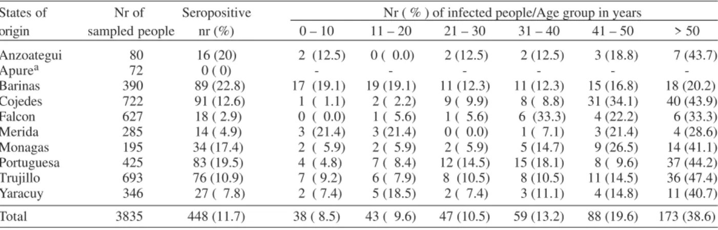
From the total people sampled, 448 (11.7%) were sero-positive for T. cruzi. Infection was detected in people with mean ± standard deviation age of 42.2 ± 21 years (range, 1-100 years). The gender composition was 195 (43.5%) males and 253 (56.5%) females, with a male:female sex ra-tio of 0.7:1. In all the states under study, except Apure, T. cruzi-infection was detected, ranging from 3.1% in Merida to 20% in Barinas and Cojedes. Once the total number of seropositive for T. cruzi was determined, the established criteria to quantify IgM and IgG levels allowed us to iden-tify 2.7% (12/448) individuals with recent or acute infec-tion, 34.4% (154/448) in chronical clinical condition and 62.9% (282) of the seropositives bearing an inapparent or occult infection. Although acute infections were only detected in three states [Barinas (7.9%), Cojedes (3.3%) and Trujillo (2.6%)], people in chronic phase and with inapparent infections were detected in localities of all the nine states where T. cruzi was circulating. Details on the proportion of infection observed at different geographi-cal entities in relation to the age group of sampled people, is presented in Table II. In addition, the proportion of people detected at different phase of chagasic infection distributed according to its respective age group is shown in Table III. The analysis of the prevalence observed in sampled people from different geographical entities in Venezuela revealed an increasing value from the young-est to the oldyoung-est age group. It is relevant to note that 8.5% of T. cruzi infection was detected in children from 0 to 10 years old. A similar figure was observed in the age groups 11-20 and 21 to 30 years. This indicates that infection has been actively occurring in Venezuela during the last three decades, affecting children in almost all the territory. This fact appears to be more relevant taking into consideration
TABLE II
Age group distribution of seroprevalence to Trypanosoma cruzi in rural populations at different states in Venezuela
States of Nr of Seropositive Nr ( % ) of infected people/Age group in years
origin sampled people nr (%) 0 – 10 11 – 20 21 – 30 31 – 40 41 – 50 > 50
Anzoategui 80 16 (20) 2 (12.5) 0 ( 0.0) 2 (12.5) 2 (12.5) 3 (18.8) 7 (43.7)
Apurea 72 0 ( 0) - - -
-Barinas 390 89 (22.8) 17 (19.1) 19 (19.1) 11 (12.3) 11 (12.3) 15 (16.8) 18 (20.2)
Cojedes 722 91 (12.6) 1 ( 1.1) 2 ( 2.2) 9 ( 9.9) 8 ( 8.8) 31 (34.1) 40 (43.9) Falcon 627 18 ( 2.9) 0 ( 0.0) 1 ( 5.6) 1 ( 5.6) 6 (33.3) 4 (22.2) 6 (33.3) Merida 285 14 ( 4.9) 3 (21.4) 3 (21.4) 0 ( 0.0) 1 ( 7.1) 3 (21.4) 4 (28.6) Monagas 195 34 (17.4) 2 ( 5.9) 2 ( 5.9) 2 ( 5.9) 5 (14.7) 9 (26.5) 14 (41.1) Portuguesa 425 83 (19.5) 4 ( 4.8) 7 ( 8.4) 12 (14.5) 15 (18.1) 8 ( 9.6) 37 (44.2) Trujillo 693 76 (10.9) 7 ( 9.2) 6 ( 7.9) 8 (10.5) 8 (10.5) 11 (14.5) 36 (47.4) Yaracuy 346 27 ( 7.8) 2 ( 7.4) 5 (18.5) 2 ( 7.4) 3 (11.1) 4 (14.8) 11 (40.7)
Total 3835 448 (11.7) 38 ( 8.5) 43 ( 9.6) 47 (10.5) 59 (13.2) 88 (19.6) 173 (38.6)
785 785 785 785 785 Mem Inst Oswaldo Cruz, Rio de Janeiro, Vol. 99(8), D ecem ber 2004
that in the states of Barinas and Merida, which are geo-graphically very different, the level of prevalence in young children was almost the same.
When the statistical correspondence analysis was applied to the nine states in which chronic infections were diagnosed, two major groups were clearly detected. One conformed by Trujillo (50%), Monagas (47.1%), Anzoategui (43.8%), and Merida (42.9%); and a second group made up by the states of Barinas (28.1%), Yaracuy (25.9%), Portuguesa (24.1%), and Cojedes (23.1%). The state of Falcon, which showed the greatest proportion of chronic infection with 77.8%, conformed an isolated point from the two groups. The use of the proportion test to corroborate whether the presence of chronic infections among states of the same group was significantly similar, revealed p values of 0.8794 and 0.9409 for both groups of states respectively. This analysis revealed that chronic T. cruzi-infection may occur in the same proportion in differ-ent geographical regions in Venezuela, which may be con-sidered a very important fact from the epidemiological point of view. This is particularly true when the states of Monagas and Anzoategui, located at low altitudes at the Eastern part of the country, were compared with Merida and Trujillo, which are located at the Andean region of Western Venezuela. In addition, the fact that Barinas, Yaracuy, Portuguesa, and Cojedes showed a similar pro-portion in the chronic infection revealed similar risk con-ditions in these bordering states located in the llanos of the West-central part of Venezuela. One additional fact that puts into evidence the general distribution of chronic
T. cruzi-infection is the finding of the major proportion of this clinical condition in Falcon, a state located in a semi-arid region in the coastal North-western part of the coun-try. These findings clearly contrast with those of Aché and Matos (2001) who stated that the chagasic endemic areas in Venezuela are confined to geographical land-scapes of piedmonts as well as patchy foci in higher moun-tain where the exclusive vector is R. prolixus.
The practice of insecticide spraying during the anti-malarial and/or antichagasic campaigns of the 1960s, to-gether with the improvements in rural housing over the last 20-30 years, have caused a significant decrease of triatomine-bugs in Venezuela. However, the information
obtained in the present work showing the presence of triatomine vectors of T. cruzi in endemic areas during the last five years, clearly contrasts with the data from the aforementioned statement. In fact, the survey carried out in 38 localities of 10 states of Venezuela, revealed that 427 out of 1388 individuals (30.7%), including children, were able to recognize triatomine-bugs, principally R. prolixus
at any developmental stage. Similarly, 419 of 1996 sampled people (20.9%) claimed to have found bugs indoor, most of them engorged with blood and near to the resting places; but what appears to be more important is that 171 of 2048 people (8.3%) declared to have been bitten by triatomine-bugs. Obviously, these figures on the presence of triatomine-bugs strongly support the argument that in rural areas of Venezuela highly risk conditions for active transmission of Chagas disease still persist.
CONCLUDING REMARKS
The analysis of the present data related to the epide-miological situation of Chagas disease in Venezuela, lead us to reach the following conclusions: there has been, during the last decade, a remarkable decrease on the ef-fect of the vector control program due to reduction or elimination of the anti-triatomine spraying activities in some geographical areas as a consequence of budget adjustment from the central or local governments. This assumption finds support in the fact that a high propor-tion of people living in areas considered endemic for Chagas disease, were able to recognize the insect vector (30%), have found bugs indoor (21%) or have been bitten by triatomine-bugs (8%); the high population of palm trees found all around the localities or houses in most of the endemic areas facilitates the presence of R. prolixus and
R. robustus species naturally inhabiting this plant, from which the local people benefit much. The fact that local people use palm trees to build up house roofs, or house accessories, may allow the establishment of bug’s do-mestic colonies favoring indoor transmission when arriv-ing infected or when encounterarriv-ing domestic and perido-mestic animals with T. cruzi infections; the regular find-ing, nearby the houses, of triatomine-bugs attracted by light during night from close palm trees enables us to interpret the dynamic transmission of chagasic infection TABLE III
Discrimination of clinical conditions per age group in asymptomatic seropositive people from endemic areas of Chagas disease in Venezuela based on anti-Trypanosoma cruzi specific immunoglobulin M and G levels
Age group T. cruzi-infected Clinical condition a
(years) people nr (%) Acute infection nr (%) Chronic infection nr (%) Inapparent infection nr (%)
0 – 10 38 (8.4) 2 (5.3) 6 (15.8) 30 (78.9)
11 – 20 43 (9.6) 1 (2.3) 8 (18.6) 34 (79.1)
21 – 30 47 (10.5) 4 (8.5) 9 (19.1) 34 (72.3)
31 – 40 59 (13.1) 0 (0.0) 19 (32.2) 40 (67.8)
41 – 50 88 (19.6) 2 (2.3) 39 (44.3) 47 (53.4)
> 50 173 (38.6) 3 (1.7) 73 (42.2) 97 (56.1)
Total 448 12 (2.7) 154 (34.4) 282 (62.9)
786 786786
786786 Chagas Disease in Venezuela • Néstor Añez et al.
in triatomine-free houses. In fact, this aspect call the at-tention for a new epidemiological situation which appears to be different from the well known traditional one with triatomine domiciliation. In this case T. cruzi-infection may be produced as a consequence of the bite and posterior defecation of visiting infected bugs, a possibility recog-nized by nearly 30% of people living under risk condi-tions.
Considering the argument given above, it is not diffi-cult to conclude that, at present, Venezuela is far from getting eradication of domestic populations of triatomine-bugs vectors of T. cruzi as expected in the Andean Pact Countries Initiative and as it was successfully obtained in the South Cone Initiative. Ecological conditions, social behavior and lack of constancy in the vector control pro-gram, conspire all against the purposes of the plan estab-lished for Venezuela to eliminate Chagas disease trans-mission.
One more aspect that strongly support the idea of a re-emergence of Chagas disease in Venezuela is the de-tection of T. cruzi-infection in 56% (174/304) of the pa-tients referred from different clinical units, located in 10 states, to our diagnostic center during the last 14 years, with 30% (52/174) of them bearing blood circulating para-sites. If this figure is impressive itself, what appears to be more relevant is the fact that 38% of the detected acute cases were children less than 10 years old. The high pro-portion of acute phase cases of Chagas disease is indica-tive of the existence of a significant incidence of this try-panosomiasis. In addition, one may speculate that if the above figures were detected in an unique diagnostic cen-ter, most of the time located far away from the place where the case originally occurred, much more cases would be
detected if each state were provided with the facilities to detect T. cruzi-infection in any of its phases in the in-fected individuals. The problem appears to be more dra-matic if we add to the above, the 11.7% (448/3835) seroprevalence estimated in a survey carried out during the last seven years in individuals from 75 rural localities of 10 states of Venezuela, from which 8.5% (38/448) were children under 10 years.
Contrary to the opinion that geographical distribution of T. cruzi active transmission is restricted to the states of Portuguesa, Barinas, and Lara (WHO 1999, Moncayo 2003) or that it is confined to geographical landscapes of pied-monts and mountains where coffee plantation are ex-ploited (Aché & Matos 2001), the analysis of the results presented here revealed that T. cruzi-infection may occur in very diverse geographical regions of Venezuela. Indeed, the infection has been found extended from the eastern states of Monagas and Anzoategui, located at low alti-tudes (Fig. 2. 1, 7), to the western states of Merida and Trujillo in the Andean region at high altitudes (Fig. 2. 6, 9), crossing the west central states of Barinas, Cojedes, Portuguesa, and Yaracuy, located in the Llanos (Fig. 2. 3, 4, 8, 10) to reach the state of Falcon, a semi-arid zone located at the coastal North-western part of the country (Fig. 2. 5).
Considering together all the aspects discussed above, we support a previous opinion (Feliciangeli et al. 2003) that despite annual incidence has been reduced in the last decades, Chagas disease eradication in Venezuela may be difficult to achieve and that in the susceptible popula-tions living in endemic areas, transmission could now be increasing. Similar to what has been confirmed in most Latin-American countries (Dias et al. 2002), in Venezuela
787 787 787 787 787 Mem Inst Oswaldo Cruz, Rio de Janeiro, Vol. 99(8), D ecem ber 2004
there is also a tendency for the government and the politi-cal and sanitary authorities to give priority to other public health problems such as dengue fever or other emerging diseases, placing Chagas disease in a second place of attention. Accordingly, the improvement of health services in areas where Chagas disease is endemic, with trained personnel with ability enough, at least, to suspect the presence of the infection in patients who need to be re-ferred to specialized diagnostic centers is mandatory.
Finally, we use the argument given in the present re-view to suggest the Venezuelan central government the creation of a law declaring Chagas disease control as a policy of the state. This is, in our opinion, the only way the department of health may give priority to the control of Chagas disease, increasing, at the same time, scientific interest on the disease and its control in universities, re-search institutions and the national population as a whole.
ACKOWLEDGEMENTS
To Dr G Fermin for critical reading of the manuscript, Mr M Aguilera for technical assistance during sampling and Mr N Diaz for statistical advice.
REFERENCES
Aché A, Matos AJ 2001. Interrupting Chagas disease transmis-sion in Venezuela. Rev Inst Med Trop São Paulo 43: 37-43. Acquatella H 1987. Encuesta epidemiológica en sujetos con serología positiva para enfermedad de Chagas. Ciencia y Tecnología Venezuela 4: 185-200.
Añez N, Carrasco H, Parada H, Crisante G, Rojas A, González N, Ramírez JL, Guevara P, Rivero C, Borges R, Scorza JV 1999. Acute Chagas’ disease in western Venezuela: a clini-cal, seroparasitologic and epidemiologic study. Am J Trop Med Hyg 60: 215-222.
Añez N, Crisante G, Rojas A, Carrasco H, Parada H, Yepez Y, Borges R, Guevara P, Ramirez JL 2001. Detection and sig-nificance of inapparent infection in Chagas disease in west-ern Venezuela. Am J Trop Med Hyg 65: 227-232. Berti AL, Gómez-Nuñez JC, Guerrero L, García-Martín G 1961.
Conversión de la campaña de erradicación de la malaria en profilaxis de la enfermedad de Chagas. Rev San Asist Soc 26: 24-32.
Camargo ME 1966. Fluorescent antibody test for serodiagno-sis. Technical modification employing preserved culture forms of Trypanosoma cruzi in a slide test. Rev Inst Med Trop São Paulo 8: 227-234.
Dias JCP, Silveira AC, Schofield CJ 2002. The impact of Chagas disease control in Latin America – A Review. Mem Inst Oswaldo Cruz 97: 603-612.
Feliciangeli MD, Campbell-Lendrum D, Martinez C, Gonzalez D, Coleman P, Davie C 2003. Chagas disease control in Venezuela: lessons for the Andean region and beyond. Trends Parasitol 19: 44-49.
Maekelt GA 1983. La epidemiología de la enfermedad de Cha-gas en relación con el ecosistema domiciliario. Inter Ciencia 8: 353-366.
Mathsoft 1999. S-plus 2000 Guide to Statistics, Vol. 1, Mathsof, Seattle.
MSAS-Ministerio de Sanidad y Asistencia Social 1970. Resumen Informativo, División EndemiasRurales, Venezuela, 1960; 1969.
MSAS-Ministerio de Sanidad y Asistencia Social 1972. Informe anual 1971, Venezuela.
Moncayo A 2003. Chagas disease: current epidemiological trends after the interruption of vectorial and transfusional trans-mission in the Southern Cone countries. Mem Inst Oswaldo Cruz 98: 577-591.
Parada H, Carrasco H, Añez N, Fuenmayor C, Arriaga A, Palacios E, Aguilera M 1996. La enfermedad de Chagas aguda. Características clínicas, parasitológicas e histo-patológicas. Arc Cardiol 16: 10-17.
Parada H, Carrasco H, Añez N, Fuenmayor C, Inglessis I 1997. Cardiac involvement is a constant finding in acute Chagas disease: a clinical, parasitological and histopathological study. Int J Cardiol 60: 49-54.
Pifano F 1974. Estado actual de la enfermedad de Chagas en Venezuela. Focos naturales de la tripanosomiasis en el medio silvestre y su repercusión en las comunidades rurales. Foro Enfermedad de Chagas, San Carlos, Cojedes, Junio 19, 23 pp.
Schofield CJ 2000. Challenges of Chagas disease vector control in Central America. In Global Collaboration for Develop-ment of Pesticides for Public Health (GCDPP). WHO/CDS/ WHOPES/GCDPP/2000.1, p. 1-36.
Stuart A, Ord JK 1991. Kendall’s Advanced Theory of Statis-tics, Vol. 2, Oxford University Press, Oxford.
Tejera E 1919. La trypanosomose americaine ou maladie de Chagas au Venezuela. Bull Soc Pathol Exot 12: 509-513. Torrealba JF 1940. Resumen de la práctica del xenodiagnóstico
para la enfermedad de Chagas en Zaraza (Guárico, Venezu-ela). Rev Med Vet Parasitol 2: 25-43.
Torrealba JF 1954. Otros 16 casos de enfermedad de Chagas comprobados en San Juan de Los Morros. Gaceta Med Ca-racas 61: 123-147.
Torrealba JF, Armas EA, De Lima A, Diaz-Vasquez AD, Lira VB, Rojas-Marroquin IR 1955a. Comprobación de casos agudos de enfermedad de Chagas en El Sombrero, Distrito Mellado, Estado Guárico. Gaceta Med Caracas 63: 445-452.
Torrealba JF 1955b. Enfermedad de Chagas y tripanosomiasis de Tejera. Pub Dir Cultura ULA, Mérida, Venezuela, 47: 1-52.
Torrealba JF, PierettiRV, Ramos I, Diaz-Vasquez A, Hernández-Pieretti O 1958. Encuesta sobre enfermedad de Chagas en la Penitenciaría General de Venezuela. Gaceta Med Caracas 67: 19-58.
Vattuone N, Yanovsky J 1971. Trypanosoma cruzi: agglutina-tionactivity of enzyme-treated epimastigotes. Exp Parasitol 30: 349-355.
Voller A, Draper C, Bidwell D, Bartleti A 1975. Microplate enzyme-linked immunosorbent assay (ELISA) Chagas dis-ease. Lancet 1: 426-429.
WHO 1997. Andean countries initiative launched in Colombia.
TDR News 53: 3.
WHO 1998. Chagas disease: interruption of transmission, Uru-guay. Weekly Epidemiol Rec 1: 4.
WHO 1999. Chagas disease, progress towards interruption of transmission in Venezuela. Weekly Epidemiol Rec 35: 289-292.
WHO 2000. Chagas disease, interruption of transmission in Brazil. Weekly Epidemiol Rec 19: 153-155.