763 763 763 763 763 Mem Inst O swaldo Cruz, Rio de Janeiro, Vol. 99(7): 763-767, November 2004
Antimicrobial Resistance of
Enterococcus
sp. Isolated from the
Intestinal Tract of Patients from a University H ospital in Brazil
Andresa M aschieto, Roberto M artinez* , Izabel Cristina Vanzato Palazzo, Ana Lúcia da Costa D arini/+
Departamento de Análises Clínicas, Toxicológicas e Bromatológicas, Faculdade de Ciências Farmacêuticas *Departamento de Clínica Médica, Faculdade de Medicina de Ribeirão Preto, USP, 14040-903 Ribeirão Preto, SP, Brasil
This study reports the results about antimicrobial resistance of Enterococcus spp. isolated from intestinal tract of patients from a university hospital in Brazil. The identification of strains at species level was performed by conventional biochemical tests, API 20 Strep (bioMérieux), and polymerase chain reaction assay. The specie distri-bution was E. faecium (34%), followed by E. faecalis (33%), E. gallinarum (23.7%), E. casseliflavus (5.2%), E. avium
(1%), and E. hirae (1%). Intrinsic resistance to vancomycin characterized by presence of vanC genes was found in E. gallinarum and E. casseliflavus. The high prevalence of VanC phenotype enterococci is very important because these species have been reported as causing a wide variety of infections. Vancomycin-resistant E. faecium or E. faecalis
were not found and no one isolate of these species was a β-lactamase producer. Thirteen clinical isolates of entero-cocci (13.4%) showed multiresistance patterns, which were defined by resistance to three classes of antibiotics plus resistance to at least one aminoglycoside (gentamicin and/or streptomycin). The resistance to several antimicrobi-als shown by enterococcal strains obtained in this study is of concern because of the decrease in the therapeutic options for treatment of infections caused by enterococci.
Key words: enterococci - resistance - antimicrobials - Brazil
Enterococci are widespread in nature and are part of the commensal flora of the human gastrointestinal and genitourinary tracts. Enterococci are often implicated in infections of the urinary tract and abdomen or superficial wounds of hospitalized patients, but can also cause bac-teremia, endocarditis, perinatal infections, and, occasion-ally, meningitis or pneumonia. Thus, enterococci are in-creasing in importance as the cause of hospital-acquired infections. In addition, enterococci colonizing serve as reservoir for antibiotic resistance genes that can be trans-ferred among enterococci and can be acquired by other bacteria (Dukta-Malen et al. 1994, Leclerq 1997).
Several studies have documented that enterococcal infections are most commonly caused by the patients own commensal flora. Colonization may occur long before or immediately before infection, but either way, it plays a major role in the development of nosocomial infection (Montecalvo et al. 1995).
Enterococal antimicrobial resistance to several anti-microbials, particularly resistance to high concentrations of penicillin and gentamicin or to glycopeptides, have hin-dered the treatment of serious infections caused by en-terococci. The synergistic combinations among amy-noglycosides and a cell-wall active antibiotic (i.e. amoxicillin or vancomycin) have been implicated in these resistant strains.
Financial support: Conselho Nacional de Desenvolvimento Científico e Tecnológico (464789/200-1), Pró-Reitoria de Pesquisa, Universidade de São Paulo
+Corresponding author. Fax: +55-16-6331936.
E-mail: [email protected] Received 8 June 2004 Accepted 15 September 2004
This paper reports a study conducted to determine antimicrobial resistance of Enterococcus spp. isolated from the intestinal tract of patients from a university hospital in Brazil.
MATERIALS AND METHODS
Bacterial strains and species identification - From May 2001 to June 2002, 37 stools and 101 rectal swabs were obtained from 125 hospitalized patients in hospital wards where vancomycin and or other antimicrobials are frequently used: Internal Medicine, Infectious Diseases, Kidney Transplant, Bone Marrow Transplant, Emergency Unit, Hematology, and Intensive Therapy Unit at the Hos-pital das Clínicas, Faculty of Medicine of Ribeirão Preto, University of São Paulo, a university hospital in Brazil, with 551 beds, that admits annually about 34,075 in pa-tients and attends other 524,574 out papa-tients. The papa-tients included in this study were hospitalized at least for five days. All samples were collected randomly and there was no report of any gastrointestinal infection in the patients involved in this study.
The study protocol was approved by the Hospital’s Ethical Committee (Process 6539/00).
The samples were plated onto Bile Esculin Azide Agar (Becton, Dickinson & Co.), a selective medium with bile-esculin and sodium azide. Five to six isolated colonies were identified from each plate and only those that pre-sented some difference in minimum inhibitory concentra-tion (MIC) and/or specie identificaconcentra-tion were considered for each patient.
764 764 764 764
764 Antimicrobial Resistance of Enterococcus sp. • Andresa M aschieto et al.
tolerance to 6.5% sodium chloride and ability to grow at 10ºC and 45ºC. Species identification was carried out with a test scheme proposed by Facklam et al. (1999) that is based on carbohydrate fermentation and arginine deami-nation. Carbohydrate fermentation tests were perfomed in agar containing 1% of the sugar being tested. Arginine deamination was tested with Moeller arginine decarboxy-lase broth. Yellow pigmentation was observed after over night growth on Müeller Hinton agar (Merck) supple-mented with 5% sheep blood and by taking a sweep of the plate with a cotton swab. The pyruvate utilization test was observed with a broth containing 1% pyruvate (Ruoff et al. 1999, MacFaddin 2000). The identification of some species was performed by API 20 Strep (bioMérieux).
Amplification by polymerase chain reaction (PCR) for species identification - PCR was performed to confirm some species identification and to detect vancomycin re-sistance mediated by van genes. Two colonies were ob-tained from a fresh subculture and were resuspended in 100 µl sterile water (Sigma, St. Louis, US). Two microliters of the suspension were added to each PCR mixture con-sisting of 23 µl with the following components: 0.625U Taq polymerase (Life Technologies, UK), 0.2 mM each deoxynucleoside triphophate (dNTP), 2.0 mM MgCl2, 1x PCR buffer (20 mMTris, 50 mM KCl), 25 pmol of each primer according to Dukta-Malen et al. (1995) (Table I). After an initial denaturation step at 94ºC for 2 min, the mixture was subjected to 30 cycles under the following conditions: 30 s at 94ºC, 30 s at 54ºC, and 30 s at 72ºC, followed by a final extension step at 72ºC for 10 min. Am-plified products were analyzed by 1.5% agarose gel elec-trophoresis and the size of the amplification products was estimated by comparison with the molecular size stan-dard 123 pb ladder.
Control strains: E. faecalis NCTC 775, E. faecium NCTC 7171, E. gallinarum NCTC 12359, and E. casseliflavus
NCTC 12361.
Determination of minimum inhibitory concentration
- The MIC of the following antimicrobial agents was per-formed by the agar dilution method according to NCCLS guidelines (2000) with antibiotic dilutions ranging from 0.125 to 256 µg/ml: vancomycin, penicillin, chloramphe-nicol, tetracycline, erythromycin, and quinupristindal-fopristin. Bacterial suspensions equal to a 0.5 McFarland standard were prepared and inoculated onto antibiotic containing medium with Steers replicator to yield a final
inoculum of 108 cfu/spot. Plates were incubated in ambi-ent air at 35ºC for 24 h. The MIC was defined as the lowest antibiotic concentration which gave a complete absence of growth (NCCLS 2000).
Detection of high level aminoglycoside resistance (HLAR) - The screening of high level aminoglycoside re-sistance was performed in agar dilution plates prepared with brain heart infusion (BHI - Oxoid, Basingstoke, En-gland) agar with 500 mg gentamicin per ml and 2000 µg streptomycin per ml. The plates were inoculated by spot-ting 10 µl of a suspension prepared from growth on an 18-24 h agar plate and adjusted to a 0.5 Mc Farland standard, giving a final inoculum of 108 cfu/spot. Plates were incu-bated at 35ºC for 24 h/48 h (NCCLS 2000).
Detection of β-lactamase production- All enterococci isolates were tested for β-lactamase production with nitrocefin (Oxoid) according to the manufacture’s instruc-tions. Nitrocefin solution was dropped onto a single colony of an overnight culture. Development of a red color would indicate a positive result.
Control strains: S. aureus ATCC 29213 was used as a positive control and S. aureus ATCC 25923 as negative control for β-lactamase detection.
RESULTS
Enterococcus spp. were isolated in 67 (53.6%) of the 125 patients included in this study, and 15 of them pre-sented more than one specie of enterococci in the faecal samples.
A total of 97 enterococcal isolates were obtained from 67 patients. The enterococcal isolates were identified to species level and the distribution were E. faecium (34%) followed by E. faecalis (33%), E. gallinarum (23.7%), E. casseliflavus (5.2%), E. durans (2%), E. avium (1%), and
E. hirae (1%). The scheme proposed by Facklam et al. (1999) did not permit the specie identification in some cases, thus we do need perform the species identification by PCR or API test. No one patient showed only E. avium
in faecal samples, but this species was isolated in one patient in association with E. gallinarum, E. casseliflavus,
and E. faecalis. The association of species most frequently found was E. faecium-E. faecalis (7.5%) and E. faecium
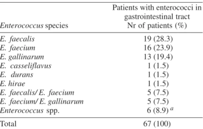
-E. gallinarum (7.5%). Six patients harbored other asso-ciation of enterococci species in gastrointestinal tract as indicated in Table II.
The vancomycin resistance genotype in E. gallinarum
TABLE I
Sequences of the species-specific primers used in this study
Species Gene Product (bp) Primers (5’ – 3’)
Enterococcus faecalis ddlE. faecalis 941 ATCAAGTACAGTTAGTCTT ACGATTCAAAGCTAACCTG Enterococcus faecium ddlE. faecium 550 GCAAGGCTTCTTAGAGA
CATCGTGTAAGCTAACTTC
Enterococcus gallinarum vanC-1 822 GGTATCAAGGAAACCTC
CTTCCGCCATCATAGCT Enterococcus casseliflavus vanC 2-3 439 CTCCTACGATTCTCTTG CGAGCAAGACCTTTAAG
765 765 765 765 765 Mem Inst O swaldo Cruz, Rio de Janeiro, Vol. 99(7), November 2004
and E. casseliflavus were determined by PCR and all gave positive results for the presence of vanC-1 and vanC-2
genes, respectively.
The distribution of antibacterial resistance according to species is presented in Table III. The results show that only E. gallinarum and E. casseliflavus were resistant to vancomycin. Resistance to penicillin among the E. faecium
and the E. faecalis isolates were 9.1% and 9.4%, respec-tively. In contrast, resistance to chloramphenicol was 33.3% and 40.6%, erythromycin 66.7% and 75%, tetracy-cline 42.4% and 56.2%, and to quinupristin-dalfopristin 3% and 12.5%, respectively. E. faecium and E. faecalis,
with high-level gentamicin resistance, were 18.2% and 28.1%, respectively. High-level streptomycin resistance was detected in 21.2% of E. faecium and 21.9% of E. faecalis isolates. E. avium and E hirae were susceptible to all drugs tested (Table III).
Although the antibiotic resistance mentioned can ex-ist independently, it can be combined in a single strain that could be a multiresistant strain. In this study the
multiresistant strain, defined as that presenting resistance for at least one aminoglycoside (gentamicin and/or strep-tomycin) plus resistance for another three classes of an-tibiotics, was verified in 13 isolates of Enterococcus spp. (Table IV).
Neither isolate of E. faecalis nor E. faecium was a β -lactamase producer.
DISCUSSION
This study investigated the species occurrence and antibacterial resistance patterns of enterococci isolated from rectal swabs of hospitalized patients in clinics that have high-risk for vancomycin-resistant enterococci (VRE) colonization in a university hospital in Brazil. The trans-mission dynamics of VRE and factors contributing to their dissemination are complex. Numerous variables and in-teractions need to be considered, however gastrointesti-nal VRE colonization appears to play a major role in the development of VRE infections (Montecalvo et al. 1995). The isolates obtained in this study were E. faecium
(34%) followed by E. faecalis (33%) and we do not have a clear dominance of E. faecalis as expected. According to date published in other Brazilian study (Mondino et al. 2003), E. faecalis was the prevalent specie (53.6%) among intestinal strains. A diversity in the species distribution might be obtained when enterococci isolated in different geographical regions are involved. The most frequent enterococcal species isolated from clinical specimens are
E. faecalis and E. faecium, which are responsible for 90% of nosocomial infections (Mutnick et al. 2003), however according to Kühn et al. (2003), these two species repre-sented 78% of the enterococci isolated from intestinal tract of hospitalized patients.
Motile enterococci, including E. gallinarum and E. casseliflavus, are rarely encountered in human clinical specimens and are primarily found in the gastrointestinal tract in poultry, in foods, and in domestic fowls. Although these species are infrequently isolated from clinical speci-mens, they have been implicated in a wide variety of inva-sive infections in humans, especially immunocompromised TABLE II
Distribution of enterococci species in the gastrointestinal tract of the patients studied
Patients with enterococci in gastrointestinal tract Enterococcus species Nr of patients (%)
E. faecalis 19 (28.3)
E. faecium 16 (23.9)
E. gallinarum 13 (19.4) E. casseliflavus 1 (1.5)
E. durans 1 (1.5)
E. hirae 1 (1.5)
E. faecalis/ E. faecium 5 (7.5) E. faecium/ E. gallinarum 5 (7.5) Enterococcus spp. 6 (8.9) a
Total 67 (100)
a: patients colonized by other enterococci association (two or more species).
TABLE III
Species distribution of enterococci resistant to antimicrobials
Enterococcus species Antimicrobials - Number of strains (%)
(number tested) VAN PEN CLO ERI TET QUI/DAL GEN a STR a
E. faecalis (32) - 3 (9.4) 13 (40.6) 24 (75) 18 (56.2) 4 (12.5) 9 (28.1) 7 (21.9) E. faecium (33) - 3 (9.1) 11 (33.3) 22 (66.7) 14 (42.4) 1 (3) 6 (18.2) 7 (21.2) E. gallinarum (23) 23 (100) 1 (4.3) 3 (13) 8 (34.8) 7 (30.4) 2 (8.7) 3 (13) 1 (4.3) E. casseliflavus (5) 5 (100) - - 2 (40) 1 (20) - -
-E. durans (2) - - 2 (100) 1 (50) - - -
-E. avium (1) - - -
-E. hirae (1) - - -
-Total (97) 28 (28.9) 7 (7.2) 29 (29.9) 57 (58.7) 40 (41.2) 7 (7.2) 18 (8.5) 15 (15.5)
766 766 766 766
766 Antimicrobial Resistance of Enterococcus sp. • Andresa M aschieto et al.
or chronically ill patients, and sometimes are nosocomially acquired (Reid et al. 2001, Dargere et al. 2002).
In this study the prevalence of vanC phenotype en-terococci was significantly higher (23.7% of E. gallinarum
and 5.2% of E. casseliflavus) than in several studies that reported intestinal colonization by enterococci vanC. In 1997, the prevalence of colonization by these species in another hospital in Brazil was 1.2% (Cereda et al. 1997), Canada presented 5% of these species (Toye et al. 1997), Lebanon 1.4% (Zouain & Araj 2001), and Kuwait 4.5% (Udo et al. 2003). The high prevalence obtained in this study is very important because several studies have re-ported infection or colonization by E. gallinarum carry-ing genes that determine high resistance to vancomycin:
E. gallinarum vanA in Belgium(Dukta-Malen et al. 1994), in Italy (Biavasco et al. 2001), in Japan (Takayama et al. 2003),and in Brazil (Camargo et al. 2004). E. gallinarum
vanB in Switzerland (Liassine et al. 1998) and Australia (Schooneveldt et al. 2000).
Several hospitals located in São Paulo and some other Brazilian cities reported both outbreaks and isolated cases of VRE infection/colonization (Zanella et al. 1999, 2003, Cereda et al. 2001, 2002, Reis et al. 2001), however the enterococci strains obtained in this study were van sus-ceptible, indicating that vancomycin retains its therapeu-tic efficacy against E. faecalis and E. faecium in this hos-pital.
The prevalence of resistance to penicillin remains low in this hospital, indicating that this antimicrobial agent would be a therapeutic option. On other hand, the ab-sence of vancomycin resistance among enterococci strains studied permits the use of vancomycin in combi-nation with aminoglycoside as an alternative to penicillin to treat allergic patients or against β-lactamase producing strains. However, the high rate of aminoglycoside resis-tance obtained in this study could not maintain a thera-peutic efficacy and the synergistic effect when combined
with cell wall active agents such as vancomycin and ampi-cillin in the treatment of enterococcal infections.
The high rates of resistance to chloramphenicol, eryth-romycin and tetracycline observed in this study restricts the use of these drugs at this hospital.
Quinupristin-dalfopristin is almost inactive against E. faecalis. In contrast, most isolates of E. faecium are sus-ceptible to this agent. The mechanism of resistance to dalfopristin in E. faecalis is related with an efflux pump that appears to be intrinsic in this species (Eliopoulos 2003). Date obtained in this study are in concordance with those published because 13% of the E. faecalis were found to be resistant to quinupristin-dalfopristin. However, among E. faecium isolates obtained in this study, 3.8% showed resistance to this agent.
Although the antibiotic resistance mentioned above can exist independently, it can be combined in a single strain resulting in multiresistance. The several multiresis-tant enterococci strains obtained in this study are a cause of concern due to limitations in clinical use, specially by loss of synergistic combinations which are often needed for treatment of enterococcal infections (Schouten 1999). In spite of the conditions that could facilitate VRE colonization, we did not find either vancomycin resistant
E. faecalis or E. faecium in this hospital. However, recom-mendation to prevent the spread of VRE are essential due the resistance to several antimicrobials showed by en-terococcal strains obtained in this study.
REFERENCES
Biavasco F, Paladini C, Vignaroli C, Foglia G, Manso E, Varaldo PE 2001. Recovery from a single blood culture of two En-terococcus gallinarum isolates carrying both vanC-1 and vanA cluster genes and differing in glycopeptide suscepti-bility. Eur J Clin Microbiol Infect Dis 5: 309-314. Camargo ILBC, Barth AL, Pilger K, Seligman BGS, Machado
A, Darini ALC 2004. Enterococcus gallinarum carrying vanA gene cluster. First report in Brazil. Braz J Med Biol TABLE IV
Multiresistance patterns of enterococci strains
Enterococcus MIC (µg/ml) HLAR
strains VAN PEN CLO ERI TET QUI/DAL GEN SM
E. faecalis HC 131 S/A 2(S) 16(R) 128(R) > 256(R) 64(R) 0,5(S) > 500(R) < 2000(S) E. faecium HC 135 S/A 4(S) 4(S) 64(R) > 256(R) 256(R) 1(S) > 500(R) < 2000(S) E. faecalis HC 135 S/B 4(S) 1(S) 64(R) > 256(R) 256(R) 1(S) > 500(R) < 2000(S) E. faecium HC 137 S/A 2(S) 1(S) 64(R) > 256(R) 128(R) 0,5(S) > 500(R) < 2000(S) E. faecalis HC 141 S/A 1(S) 4(S) 64(R) 256(R) 16(R) 0,5(S) > 500(R) < 2000(S) E. faecalis HC 153 S/A 4(S) 1(S) 32(R) 128(R) 32(R) 1(S) < 500(S) > 2000(R) E. faecalis HC 154 S/C 4(S) 0,5(S) 32(R) 128(R) 32(R) 1(S) < 500(S) >2000(R) E. faecium HC 156 S/A 2(S) 16(R) 32(R) > 256(R) 128(R) 1(S) < 500(S) > 2000(R) E. gallinarum HC 185 S/A 8(S) 2(S) 32(R) > 256(R) 128(R) 2(S) > 500(S) > 2000(R) E. faecalis HC 186 S/B 1(S) 16(R) 64(R) 8(R) 0,5(S) 1(S) < 500(S) > 2000(R) E. faecalis HC 221 S/A 2(S) 8(S) 64(R) > 256(R) 0,125(S) 4(R) > 500(R) < 2000(S) E. faecalis BAC 58S/A 2(S) 2(S) 32(R) > 256(R) 32(R) 0,5(S) > 500(R) > 2000(R) E. faecalis BAC 61 S/C 4(S) 4(S) 64(R) > 256(R) 32(R) 4(R) < 500(S) > 2000(R)
767 767 767 767 767 Mem Inst O swaldo Cruz, Rio de Janeiro, Vol. 99(7), November 2004
Res, in press.
Cereda RF, Gales AC, Silbert S, Jones RN, Sader HS 2002. Molecular typing and antimicrobial susceptibility of van-comycin-resistant Enterococcus faecium in Brazil. Infect Control Hosp Epidemiol 23: 19-22.
Cereda RF, Pignatari AC, Hashimoto A, Sader H 1997. In vitro antimicrobial activity against enterococci isolated in a uni-versity hospital in São Paulo, Brazil. Braz J Infect Dis1: 83-90.
Cereda RF, Sader HS, Jones RN, Sejas L, Machado AM, Zanatta YP, Rego STMS, Medeiros EAS 2001. Enterococcus faecalis resistant to vancomycin and teicoplanin (VanA phenotype) isolated from a bone marrow transplanted pa-tient in Brazil. Braz J Infect Dis 5: 40-46.
Dargere S, Vergnaud M, Verdon R, Saloux E, LePagE O, Leclercq R, Bazin C 2002. Enterococcus gallinarum endocarditis occurring on native heart valves. J Clin Microbiol 40: 2308-2310.
Dutka-Malen S, Blaimont B, Wauters G, Courvalin P 1994. Emergence of high-level resistant Enterococcus gallinarum and Enterococcus casseliflavus. Antimicrob Agents Chemother 34: 1675-1677.
Dukta-Malen S, Evers S, Courvalin P 1995. Detection of glyco-peptide resistance genotypes and identification to the spe-cies level of clinically relevant enterococci by PCR. J Clin Microbiol 33: 24-27.
Eliopoulos GM 2003. Quinupristin-Dalfopristin and Linezolid: evidence and opinion. Clin Infect Dis 36: 473-481. Facklam RR, Sahm DF, Teixeira ALM 1999. Enterococcus. In
PR Murray, EJ Baron, MA Pfaller, FC Tenover, RH Yolken (eds), Manual of Clinical Microbiology, 7th ed., American Society for Microbiology, Washington DC, p. 297-315. Kühn I, Iversen A, Burman LG, Olsson-Liljequist B, Franklin
A, Finn M, Aarestrup F, Seyfarth AM, Franklin A, Finn M, Blanch AR, Vilanova X, Taylor H, Caplin J, Moreno MA, Dominguez L, Herrero IA, Möllby R 2003. Comparison of enterococcal populations in animals, humans, and the envi-ronment – A European study. Inter J Food Microbiol 88: 133-145.
Leclercq R 1997. Enterococci acquire new kinds of resistance. Clin Infect Dis24 (Suppl. 1): 80S-84S.
Liassine N, Frei R, Jan I, Auckenthaler R 1998. Characteriza-tion of glycopeptide-resistant enterococci from a Swiss Hos-pital. J Clin Microbiol36: 1853-1858.
MacFaddin JF 2000. Biochemical Tests for Identification of Medical Bacteria, 3rd ed., Lippincot Williams & Wilkins, US, 912 pp.
Mondino SS, Castro AC, Mondino PJ, Carvalho MG, Silva KM, Teixeira LM 2003. Phenotypic and genotypic charac-terization of clinical and intestinal enterococci isolated from inpatients and outpatients in two Brazilian hospitals. Microb Drug Resist 9: 167-174.
Montecalvo MA, Lancestre H, Carraber M, Gedris C, Chung M, VanHorn K, Wormser GP 1995. Natural history of colo-nization with vancomycin resistant Enterococcus faecium. Infect Control Hosp Epidemiol 16: 680-685.
Mutnick AH, Biendenbach DJ, Jones RN 2003. Geographic variations and trends in antimicrobial resistance among En-terococcus faecalis and Enterococcus faecium in the SEN-TRY antimicrobial surveillance program (1997-2000). Diagn Microbiol Infect Dis 46: 63-68.
NCCLS-National Committee for Clinical Laboratory Standards 2000. Methods for Dilution Antimicrobial Susceptibility Tests for Bacteria that Grow Aerobically, 5th ed., Wayne, PA. Reid KC, Cockerill FR, Patel R 2001. Clinical and
epidemio-logical features of Enterococcus casseliflavus/flavescens and Enterococcus gallinarum bacteriemia: a report of 20 cases. Clin Infect Dis 32: 1540-1546.
Reis AO, Cordeiro JC, Machado AM, Sader HS 2001. In vitro antimicrobial activity of Linezolid tested against vancomy-cin-resistant enterococci isolated in Brazilian hospitals. Braz J Infect Dis 5: 243-251.
Ruoff KL, Whiley RA, Beighton D 1999. Streptococcus. In PR Murray, EJ Baron, MA Pfaller, FC Tenover, RH Yolken (eds), Manual of Clinical Microbiology, 7th ed., American Society for Microbiology, Washington DC, p. 283-296. Schooneveldt JM, Marriott RK, Nimmo GR 2000. Detection
of vanB determinant in Enterococcus gallinarum in Austra-lia. J Clin Microbiol38: 3902.
Schouten MA 1999. Vancomycin resistant enterococci. Preva-lence in Europe. Clin Microbiol Infec8 (Supp.1): 136. Takayama Y, Sunakawa K, Akahoshi T 2003. Meningitis caused
by Enterococcus gallinarum in patients with ventri-culoperitoneal shunts. J Infect Chemother9: 348-350. Toye B, Shymanski J, Bobrowska M, Woods W, Ramotar K
1997. Clinical and epidemiologic significance of enterococci intrinsically resistant to vancomycin (possessing the VanC genotype). J Clin Microbiol3: 3166-3170.
Udo EE, Al-Sweih N, Phillips AO, Chugh TD 2003. Species prevalence and antibacterial resistance of enterococci iso-lated in Kuwait hospitals. J Med Microbiol 52: 163-168. Zanella RC, Brandileone MCC, Bokermann S, Almeida SCG,
Valdetaro F, Votório F, Moreira MFA, Villins M, Salomão R, Pignatari AC 2003. Phenotypic and genotypic character-ization of VanA Enterococcus isolated during the first noso-comial outbreak in Brazil. Microb Drug Resist 9: 283-291. Zanella RC, Valdetaro F, Lovgren M, Tyrrel GJ, Bokermann S, Almeida SCG, Vieira VSD, Brandileone MCC 1999. First confirmed case of a vancomycin-resistant Enterococcus faecium with Van A phenotype from Brazil: isolation from a meningitis case in São Paulo. Microb Drug Resist5: 159-162.