INTRODUCTION
1. Departamento de Ciências Biológicas, Universidade Federal do Triângulo Mineiro, Uberaba, MG. 2. Departamento de Clínica Médica, Universidade Federal do Triângulo Mineiro, Uberaba, MG. Address to: Dra. Adriana Gonçalves de Oliveira. DCB/UFTM. Praça Manoel Terra 330, 38015-050 Uberaba, MG, Brasil.
Phone: 55 34 3318-5480; Fax: 55 34 3318-5462 e-mail: [email protected]
Received in 22/10/2010 Accepted in 17/11/2010
Trends in antimicrobial resistance among clinical isolates of
enterococci in a Brazilian tertiary hospital: a 4-year study
Evolução da resistência aos antimicrobianos entre isolados clínicos de enterococos em um
hospital terciário brasileiro: um estudo de 4 anos
Natália Conceição
1, Cristina da Cunha Hueb Barata de Oliveira
2, Paulo Roberto da Silva
1, Bárbara Godoi
Melo Ávila
1and Adriana Gonçalves de Oliveira
1ABSTACT
Introduction: In the past two decades members of the genus Enterococcus have emerged as important nosocomial pathogens worldwide. his study prospectively analyzed the distribution of species and trends in antimicrobial resistance among clinical isolates of enterococci in a Brazilian tertiary hospital from 2006-2009. Methods: Enterococcal species were identiied by conventional biochemical tests. he antimicrobial susceptibility proile was performed by disk difusion in accordance with the Clinical and Laboratory Standards Institute (CLSI). A screening test for vancomycin was also performed. Minimal inhibitory concentration (MIC) for vancomycin was determined using the broth dilution method. Molecular assays were used to conirm speciation and genotype of vancomycin-resistant enterococci (VRE). Results: A total of 324 non-repetitive enterococcal isolates were recovered, of which 87% were E. faecalis
and 10.8% E. faecium.he incidence of E. faecium per 1,000 admissions increased signiicantly (p < 0.001) from 0.3 in 2006 to 2.3 in 2009. he VRE rate also increased over time from 2.5% to 15.5% (p < 0.001). All VRE expressed high-level resistance to vancomycin (MIC >256μg/ mL) and harbored vanA genes. he majority (89.5%) of VRE belonged to E. faecium species, whichwere characteristically resistant to ampicillin and quinolones. Overall, ampicillin resistance rate increased signiicantly from 2.5% to 21.4% from 2006-2009. Resistance rates for gentamicin, chloramphenicol, tetracycline, and erythromycin signiicantly decreased over time, although they remained high. Quinolones resistance rates were high and did not change signiicantly over time. Conclusions: he data obtained show a signiicant increasing trend in the incidence of E. faecium resistant to ampicillin and vancomycin.
Keywords: Antimicrobial resistance proile. Enterococci. Vancomycin-resistant enterococci.
Enterococcus faecium.
RESUMO
Introdução: Nas últimas duas décadas, os enterococos emergiram como importantes patógenos nosocomiais no mundo inteiro. Neste estudo, foi analisada a distribuição das espécies e a evolução da resistência aos antimicrobianos entre isolados clínicos de enterococos obtidos em um hospital terciário, no período de 2006 a 2009. Métodos: As espécies foram identiicadas por testes bioquímicos convencionais e o peril de sensibilidade foi determinado pelo método de disco difusão. A sensibilidade à vancomicina foi também determinada pela triagem em agar e pela concentração inibitória mínima (CIM). Testes moleculares foram utilizados para conirmar as espécies e determinar os genótipos dos enterococos resistentes à vancomicina (VRE). Resultados: Foram analisadas 324 amostras de enterococos, sendo 87% E. faecalis e 10,8%
E. faecium. A incidência de E. faecium por 1.000 pacientes internados aumentou signiicativamente (p < 0,001) de 0,3 em 2006 para 2,3 em 2009. A taxa de VRE também aumentou signiicativamente de 2,5% para 15,5% (p < 0,001). Todos os VRE apresentaram genótipo VanA e CIM >256μg/mL para vancomicina. A maioria (89,5%) dos VRE pertencia à espécie E. faecium e foram resistentes à ampicilina e quinolonas. Foi observado um aumento signiicativo na taxa de resistência à ampicilina, de 2,5% (2006) para 21,4% (2009). As taxas de resistência para gentamicina, cloranfenicol, tetraciclina e eritromicina diminuíram signiicativamente no período do estudo. Para as quinolonas, as taxas de resistência foram elevadas não alteraram signiicativamente, no período do estudo. Conclusões: Os resultados do presente estudo mostram um aumento signiicativo na incidência de E. faecium resistentes à ampicilina e vancomicina.
Palavras-chaves: Peril de resistência a antimicrobianos. Enterococos. Enterococos resistentes à vancomicina. Enterococcus faecium.
Enterococci are widespread in nature and are normal constituents of the human gastrointestinal tract, but nowadays they have been recognized as important pathogens, especially among hospitalized patients. Enterococci may cause a range of diferent disorders, such as urinary tract infections, intraabdominal abscesses, wound infections, endocarditis and bacteraemia1. According to the
SENTRY Antimicrobial Surveillance Program, enterococci are the fourth most common pathogen of bacteremia in North America and the ith in Europe2. In Brazil, they are the eighth agent of
bacteremia overall and the third among the Gram-positive cocci3.
Intrinsic or acquired resistance to various commonly used antimicrobial agents is a remarkable characteristic of enterococci4. Acquired resistance
to glycopeptides (vancomycin and teicoplanin), penicillins and aminoglycosides (high-level resistance) are the most clinically important, because therapeutic options in these cases are limited.
Six types of acquired vancomycin resistance have been reported in enterococci; however, the most prevalent are VanA and VanB, in which the genes encoding resistance are associated with mobile genetic elements that allow resistance to spread clonally and laterally5. he VanC type confers an
intrinsic nontransferable low-level resistance to vancomycin that has been observed primarily in
Enterococcus gallinarum and Enterococcus casselilavus. Clinical vancomycin-resistant enterococci (VRE) isolates were irst recognized in the 1980s in Europe and USA4. Approximately ten years later,
the irst VRE were isolated in Brazil, in the States of Paraná and São Paulo, located in the southern and southeastern regions of the country, respectively6,7.
Although these VRE isolates belong to Enterococcus faecium species, until recently in Brazil, Enterococcus faecalis was the predominant VRE commonly reported in hospitals in the State of São Paulo8-13.
METHODS
RESULTS
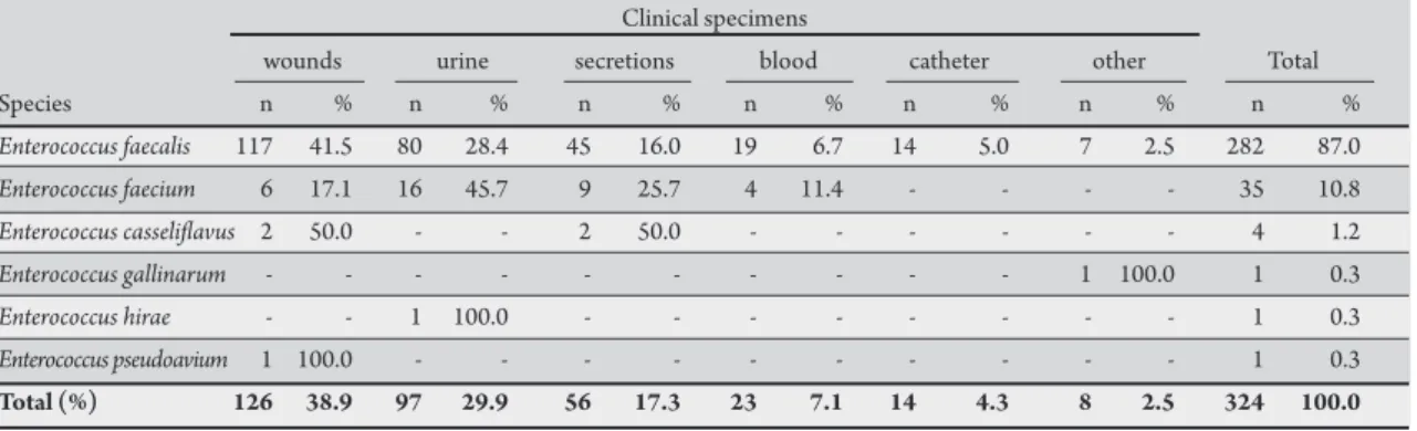
TABLE 1 - Distribution of Enterococcus species isolated from hospitalized patients from 2006 to 2009, according to clinical specimens. Clinical specimens
wounds urine secretions blood catheter other Total
Species n % n % n % n % n % n % n %
Enterococcus faecalis 117 41.5 80 28.4 45 16.0 19 6.7 14 5.0 7 2.5 282 87.0
Enterococcus faecium 6 17.1 16 45.7 9 25.7 4 11.4 - - - - 35 10.8
Enterococcus casselilavus 2 50.0 - - 2 50.0 - - - 4 1.2
Enterococcus gallinarum - - - 1 100.0 1 0.3
Enterococcus hirae - - 1 100.0 - - - 1 0.3
Enterococcus pseudoavium 1 100.0 - - - - - - - 1 0.3
Total (%) 126 38.9 97 29.9 56 17.3 23 7.1 14 4.3 8 2.5 324 100.0
published data on this subject are still sporadic in Brazil. In this study, our group analyzed the distribution of species and trends in antimicrobial resistance among enterococci recovered from clinical specimens in a Brazilian tertiary hospital over a four-year period. he genotypes of VRE isolates were also determined.
Study design
A prospective study was conducted from 2006 to 2009 in the hospital of the Triangulo Mineiro University Hospital of the Federal University (Universidade Federal do Triângulo Mineiro, UFTM). his hospital, located in the State of Minas Gerais in southeastern Brazil, is a 294-bed tertiary-care teaching hospital with a 40-bed intensive care unit (ICU) and a full range of medical specialties. Over the study period, an average of 11,000 patients were admited annually. All enterococci isolates recovered from hospitalized patients were included in the study, but only the irst isolate from each patient was used.
Phenotypic identiication of Enterococcus species
Isolates were identified at genus level by Gram staining, cellular morphology, absence of catalase production, hydrolysis of L-pyrrolidonyl-β-naphthylamide (PYR test), hydrolysis of esculin in presence of bile salts (bile-esculin test) and tolerance to 6.5% NaCl. Species identification was determined based on tests of carbohydrate fermentation, arginine hydrolysis, mobility, yellow pigment production and growth in 0.04% tellurite14.
Susceptibility testing
he antimicrobialsusceptibility proile was performed using the disk difusion method. he antimicrobials agents tested were: vancomycin (30µg), teicoplanin (30µg), ampicillin (10µg), penicillin (10U), streptomycin (300µg), gentamicin (120µg), norloxacin (10µg), ciproloxacin (5µg), chloramphenicol (30µg), tetracycline (30µg), and erythromycin (15µg). Beta-lactamase production was tested with chromogenic nitrocein disk (Becton, Dickinson and Company, CeinaseTM, USA), in accordance with the manufacturer's
instructions. A screening test for vancomycin was performed on BHI agar supplemented with 6μg/mL of this drug for all enterococcal isolates. For isolates resistant according to the screening test, the minimal inhibitory concentration (MIC) for vancomycin was determined using the broth dilution method. All susceptibility tests were performed and interpreted according to guidelines established
25923, S. aureus ATCC 29213, and E. faecalis ATCC 29212 were used for quality control.
Molecular testing
Bacterial DNA was extracted from the enterococcal isolates that were phenotypically resistant to vancomycin using the QIAamp® DNA Mini kit (Qiagen, Hilden, Germany), in accordance with the manufacturer's guidelines. Detectionof vancomycin resistance genes was performed by a multiplex polymerase chain reaction (PCR) assay, in accordance with procedures described16 by Woodford et al. Species
identiication of VRE isolates was conirmed by PCR, as described previously17. PCR products were analyzed by electrophoresis on 1.5%
agarose gels and stained by ethidium bromide.
Statistical analysis
To evaluate the trend in antimicrobial resistance among enterococci over time, the χ2-test for trend was performed using Epi
Info (CDC, Atlanta, GA) statistical sotware (version 3.5.1). he signiicance level was set at p ≤ 0.05.
Ethical considerations
he present study was approved by Research Ethics Commitee of the UFTM.
A total of 324 non-repetitive enterococcal isolates were consecutively recovered during the study period. hese isolates were recovered from diferent clinical specimens, but they were more frequent in wounds (38.9%) and in urine (29.9%). Table 1
shows the distribution of enterococcal species according to clinical specimens. he species identiied were E. faecalis (87%), E. faecium
(10.8%), E. casselilavus (1.2%), E. gallinarum (0.3%), E. hirae (0.3%) and E. pseudoavium (0.3%).
Figure 1 shows the incidence of E. faecalis, E. faecium and other enterococcal species per 1,000 patient admissions from 2006 to 2009. A signiicant increasing trend in the incidence of E. faecium
(p < 0.001) from 0.3 in 2006 to 2.3 in 2009 was observed, but not of E. faecalis or other enterococcal species over time.
he rate of VRE also increased signiicantly over time (p < 0.001), from 2.5% in 2006 to 15.5% in 2009 in our institution, although in 2007 there were no VRE and in 2008 the rate was only 1.4%
DISCUSSION
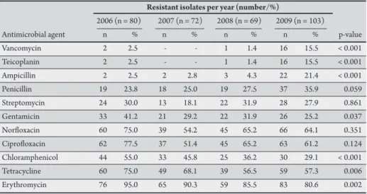
TABLE 2 - Trends in antimicrobial resistance among clinical isolates of enterococci recovered from hospitalized patients from 2006 to 2009.
Resistant isolates per year (number/%)
2006 (n = 80) 2007 (n = 72) 2008 (n = 69) 2009 (n = 103)
Antimicrobial agent n % n % n % n % p-value
Vancomycin 2 2.5 - - 1 1.4 16 15.5 < 0.001
Teicoplanin 2 2.5 - - 1 1.4 16 15.5 < 0.001
Ampicillin 2 2.5 2 2.8 3 4.3 22 21.4 < 0.001
Penicillin 19 23.8 18 25.0 19 27.5 37 35.9 0.059
Streptomycin 24 30.0 13 18.1 22 31.9 28 27.9 0.861
Gentamicin 33 41.2 21 29.2 22 31.9 26 25.2 0.037
Norloxacin 60 75.0 39 54.2 45 65.2 66 64.1 0.351
Ciproloxacin 62 77.5 37 51.4 45 65.2 63 61.2 0.124
Chloramphenicol 44 55.0 33 45.8 25 36.2 30 29.1 < 0.001
Tetracycline 60 75.0 49 68.1 39 56.5 59 57.3 0.006
Erythromycin 76 95.0 65 90.3 59 85.5 83 80.6 0.002
In
cide
n
c
e of
E
n
ter
oc
oc
c
u
s
2006
Year
2007 2008 2009
7.0
6.0
5.0
4.0
3.0
2.0
1.0
0
*
FIGURE 1 - Incidence of Enterococcus faecalis (gray bars), Enterococcus faecium (black bars) and others species of Enterococcus (white bars) per 1,000 patient admissions from 2006 to 2009. *χ2 for trend p < 0.001.
as E. faecium (VREfm). he irst two VREfs were isolated in 2006. hereater, no other VREfs were isolated during the study period. he irst VREfm appeared at the end of 2008, since then an increasing number of VREfm were noted in 2009.
All VRE isolates uniformly harbored vanA genes, as demonstrated by PCR. hey were resistant to teicoplanin and expressed high-level resistance to vancomycin (MIC >256μg/mL). he two VREfs showed the same antimicrobial susceptibility proile characterized
by resistance to norfloxacin, ciprofloxacin, chloramphenicol, tetracycline, and erythromycin, and susceptibility to ampicillin, penicillin, streptomycin, and gentamicin. All VREfm were resistant to ampicillin, penicillin, norloxacin, ciproloxacin, and erythromycin and susceptible to streptomycin, gentamicin, chloramphenicol, and tetracycline.
Enterococcus gallinarum (n = 1) and E. casselilavus (n = 4) isolates were susceptible to vancomycin by the disk difusion method, but grew on agar screening with 6µg/mL of vancomycin, while the MIC observed for this drug was 8µg/mL. hese ive isolates were not considered as VRE in this study.
As demonstrated in Table 2, ampicillin and penicillin resistance rates increased from 2006 to 2009 from 2.5% to 21.4% and 23.8% to 35.9%, respectively, but only the increasing rate of ampicillin was statistically signiicant (p < 0.001). Among the 282 E. faecalis and 35
E. faecium isolates, 4 (1.4%) and 25 (77.4%) of them, respectively, were resistant to ampicillin and to penicillin. However, 63 (22.3%)
E. faecalis isolateswere resistant to penicillin, but remained susceptible to ampicillin. Beta-lactamase producing isolates were not detected. Rates of resistance to gentamicin (41.2% to 25.2%), chloramphenicol (55% to 29.1%), tetracycline (75% to 57.3%), and erythromycin (95% to 80.6%) signiicantly decreased from 2006 to 2009, as demonstrated in Table 2. Regarding streptomycin, norloxacin and ciproloxacin, the resistance rates did not change signiicantly over the four-year study period.
Although there are at least 30 species of the genus Enterococcus, both E. faecalis and E. faecium are the most common species causing human infections1,14. Similarly, in this study, E. faecalis was the most
prevalent species followed by E. faecium, while the other species were rarely recovered from clinical specimens. Nevertheless, a signiicant increasing trend in the incidence of E. faecium per 1,000patient admissions from 0.3 in 2006 to 2.3 in 2009 was veriied, approximately an eight-fold increase. In a study conducted in a hospital in Greece, the authors reported a similar increased incidence of E. faecium
infections (0.7 in 2002 to 2.4 in 2007), although that hospital is larger than ours, with more than 60,000 admissions annually18.
he vancomycin-resistant enterococci rate also increased in our
institution, by almost the same proportion as the incidence of E. faecium
incidence. Most (89.5%) of the VRE isolates were E. faecium exhibiting
the vanA genotype. In a retrospective study conducted at a tertiary
Brazilian hospital located in the State of São Paulo, Furtado et al. also
showed an increase in the rate of VRE over time from 9.5% in 2000 to 14.7% in 2001 and to 15.8% in 2002. However, the authors did not
identify the VRE species or genotypes19. According to more recent data
from the SENTRY Program, the percentage of VRE in Brazil increased from 6.9% in 2003 to 31.1% in 2008 and the majority (68.5%) of
these isolates were E. faecium20. hese VRE rates are much higher than
that observed in other Latin America countries and in our hospital.
In the last two decades, the importance of E. faecium as a
he authors declare that there is no conlict of interest. CONFLICT OF INTEREST
FINANCIAL SUPPORT
REFERENCES spread of a hospital-adapted complex of E. faecium designated as clonal
complex-17 (CC-17), which is associated with the majority of hospitals outbreaks and clinical infections on all continents21,22. his complex
is characterized by ampicillin and quinolones resistance and by the presence of a putative pathogenicity island. Currently, the increase in
E. faecium resistant to ampicillin usually precedes increasing rates of VREfm in various locations around the world, especially in certain European countries, where until recently, VRE rates were low4,23-26. In
the USA, since the 1990s, E. faecium isolates account for more than 95% of all VRE recovered and most of them are also resistant to ampicillin24.
Likewise, our group observed a signiicant increasing trend in resistance to ampicillin among clinical isolates of enterococci due to increased incidence of E. faecium in our institution, since resistance rates to this drug among E. faecium (71.4%) were much higher than among E. faecalis (1.4%) isolates. Of note, all VREfm isolated in this study were resistant to both ampicillin and quinolones, showing the same antimicrobial resistance proile observed for E. faecium of CC-17. Nevertheless, molecular epidemiological studies conducted with Brazilian E. faecium isolates from several hospitals showed that this complex is not common in our country27,28.
In contrast to trends in ampicillin resistance, a signiicant decrease in resistance rates to chloramphenicol and tetracycline occurred from 2006 to 2009 and it is probably related to the increasing incidence of
E. faecium in the present study. Decreasing rates of chloramphenicol resistance among enterococci has previously been observed from 1997 to 1999 in the USA (19% to 12%) and Latin America (34% to 27%), according to the SENTRY Program29. In addition, all VREfm isolates
during this study were uniformly susceptible to both drugs. A similar chloramphenicol resistance rate among VREfm of 0.5% was observed in North America21; however, chloramphenicol use for the treatment of
VRE infections is known to result in the development of resistance30.
Regarding tetracycline, although the enterococci resistance rate decreased over time, it remained high in 2009 (57%), but was slightly lower than that reported by the SENTRY Program for Latin America (67.2%)31 and by other Brazilian studies performed with clinical
enterococci strains isolated in 1996 and 1997 (62%)32 and in 2006
and 2007 (66.5%)8. Likewise, the resistance rates to erythromycin
were very high, despite the signiicant decrease observed (95% to 80.6%), conirming that resistance to this drug is very common among enterococci1,8,31,33.
Overall, moderate rates of high-level resistance to aminoglycosides were observed over the four-year, similar to those observed recently in Brazil of 32.1% and 26.7% and in the USA of 30.6% and 25.2% for streptomycin and gentamicin, respectively3,34. Regarding quinolones,
resistance rates did not change signiicantly during the study period, whereas the overall resistance rates to ciproloxacin were slightly higher compared to Latin America (50%) and the USA (58%)29,31.
In conclusion, the present data show an increasing incidence of E. faecium resistant to ampicillin and vancomycin,corroborating the worldwide trends. Considering that all the VREfm identiied in our institution expressed resistance to ampicillin and quinolone, it is quite probable that they belong to the hospital-adapted CC-17. However, future molecular characterization is necessary to verify this, since there are no studies demonstrating the spread of E. faecium
CC-17 in Brazil to date. herefore, more rigorous strategies should be developed in order to prevent further spread of E. faecium in Brazil.
The authors would like to thank the Clinical Pathology Laboratory staf at the Triangulo Mineiro Federal University (Minas Gerais, Brazil) for providing the enterococci isolates.
Fundação de Amparo à Pesquisa do Estado de Minas Gerais and the Fundação de Ensino e Pesquisa de Uberaba.
1. Murray BE. The life and times of the Enterococcus. Clin Microbiol Rev 1990; 3:46-65.
2. Biedenbach DJ, Moet GJ, Jones RN. Occurrence and antimicrobial resistance patern comparisons among bloodstream infection isolates from the SENTRY Antimicrobial Surveillance Program (1997-2002). Diagn Microbiol Infect Dis 2004; 50:56-69.
3. Gales AC, Sader HS, Ribeiro J, Zoccoli C, Barth A, Pignatari AC. Antimicrobial susceptibility of gram-positive bacteria isolated in Brazilian hospitals participating in the SENTRY Program (2005-2008). Braz J Infect Dis 2009;13:90-98. 4. Murray BE. Vancomycin-resistant enterococcal infections. N Engl J Med
2000;342:710-721.
5. Courvalin P. Genetics of glycopeptide resistance in gram-positive pathogens. Int J Med Microbiol 2005; 294:479-486.
6. Dalla-Costa LM, Souza DC, Martins LTF, Zanella RC, Brandileone MC, Bokermann S, et al. Vancomycin-resistant Enterococcus faecium: irst case in Brazil. Braz J Infect Dis 1998;2:160-163.
7. Zanella RC, Valdetaro F, Lovgren M, Tyrrel GJ, Bokermann S, Almeida SC, et al. First conirmed case of a vancomycin-resistant Enterococcus faecium with vanA phenotype from Brazil: isolation from a meningitis case in São Paulo. Microb Drug Resist 1999; 5:159-162.
8. Bender EA, Freitas ALP, Reiter KC, Lutz L, Barth AL. Identiication, antimicrobial resistance and genotypic characterization of Enterococcus spp. isolated in Porto Alegre, Brazil. Braz J Microbiol 2009; 40:693-700.
9. Caiaffa Filho HH, Almeida GD, Oliveira GA, Sarahyba L, Mamizuka EM, Buratini MN. Molecular characterization of van genes found in vancomycin resistant Enterococcus spp. isolated from Hospital das Clínicas, FMUSP, São Paulo, Brazil. Braz J Infect Dis 2003; 7:173-174.
10. Cereda RF, Sader HS, Jones RN, Sejas L, Machado AM, Zanata YP, Rego STMS, Medeiros EAS. Enterococcus faecalis resistant to vancomycin and teicoplanin (VanA phenotype) isolated from a bone marrow transplanted patient in Brazil. Braz J Infect Dis 2001; 5:40-46.
11. Cordeiro JC, Silbert S, Reis AO, Sader HS. Inter-hospital dissemination of glycopeptides resistant Enterococcus faecalis in Brazil. Clin Microbiol Infect 2004; 3:260-262.
12. Ribas RM, Darini ALC, Moreira TA, Freitas C, Gontijo Filho PP. Vancomycin-resistant vanA phenotype Enterococcus faecalis: irst case in Minas Gerais state and epidemiological considerations. Braz J Infec Dis 2007; 11:439-440. 13. Zanella RC, Brandileone MCC, Bokermann S, Almeida SCG, Valdetaro F, Vitório F,
et al. Phenotypic and genotypic characterization of vanA Enterococcus isolated during the irst nosocomial outbreak in Brazil. Microb Drug Resist 2003; 9:283-291. 14. Teixeira LM, Carvalho MGS, Facklam RR. Enterococcus. In:Murray BE, Baron EJ,
Jorgensen JH, Landry ML, Pfaller MA, editors. Manual of Clinical Microbiology. Washington (DC): American Society for Microbiology; 2007. p. 430-442. 15. Clinical and Laboratory Standards Institute. Performance standards for
16. Woodford N, Morrison D, Johnson AP, Briant V, George RC, Cookson B. Application of DNA probes for rRNA and vanA genes to investigation of a nosocomial cluster of vancomycin-resistant enterococci. J Clin Microbiol 1993; 31:653-658.
17. Dutka-malen S, Evers S, Courvalin P. Detection of glicopeptide resistance genotypes and identiication to the species level of clinically relevant enterococci by PCR. J Clin Microbiol 1995; 33:24-27.
18. Protonotariou E, Dimitroulia E, Pournaras S, Pitiriga V, Soianou D, Tsakris A. Trends in antimicrobial resistance of clinical isolates of Enterococcus faecalis
and Enterococcus faecium in Greece between 2002 and 2007. J Hosp Infect 2010; 75:225-227.
19. Furtado GHC, Martins ST, Coutinho AN, Soares GMM, Wey SB, Medeiros EAS. Incidence of vancomycin-resistance Enterococcus at a university hospital in Brazil. Rev Saude Publica 2005; 39:41-46.
20. Sader HS, Moet GJ, Jones RN. Antimicrobial resistance among gram-positive bacteria isolated in Latin American hospitals. J Chemother 2009; 21:611-620. 21. Deshpande LM, Fritsche TR, Moet GJ, Biendenbach DJ, Jones RN. Antimicrobial
resistance and molecular epidemiology of vancomycin-resistant enterococci from North America and Europe: a report from the SENTRY Antimicrobial Surveillance Program. Diagn Microbiol Infect Dis 2007; 58:163-170. 22. Leavis HL, Bonten MJM, Willems RJL. Identiication of high-risk enterococcal
clonal complexes: global dispersion and antibiotic resistance. Curr Opin Microbiol 2006; 9:454-460.
23. Oteo J, Cuevas O, Navarro C, Aracil B, Campos J. Trends in antimicrobial resistance in 3469 enterococci isolated from blood (EARSS experience 2001-06, Spain): increasing ampicillin resistance in Enterococcus faecium. J Antimicrob Chemother 2007; 59:1044-1045.
24. Shepard BD, Gilmore MS. Antibiotic-resistant enterococci: the mechanisms and dynamics of drug introduction and resistance. Microbes Infect 2002; 4:215-224.
25. Treitman AN, Yarnold PR, Warren J, Noskin GA. Emerging incidence of
Enterococcus faecium among hospital isolates (1993 to 2002). J Clin Microbiol 2005; 43:462-463.
26. Werner G, Coque TM, Hammerum AM, Hope R, Hryniewicz W, Johnson A, et al. Emergence and spread of vancomycin resistance among enterococci in Europe. Euro Surveill 2008; 13:1-11.
27. Camargo ILBC, Gilmore MS, Darini ALC. Multilocus sequence typing and analysis of putative virulence factors in resistant and vancomycin-sensitive Enterococcus faecium isolates from Brazil. Clin Microbiol Infect 2006; 12:1123-1130.
28. Titze-de-Almeida R, Van Belkum A, Felipe MSS, Zanella RC, Top J, Willems RJL. Multilocus sequence typing of hospital associated Enterococcus faecium from Brazil reveals their unique evolutionary history. Microb Drug Res 2006; 12:121-125. 29. Low DE, Keller N, Barth A, Jones RN. Clinical prevalence, antimicrobial
susceptibility, and geographic resistance paterns of Enterococci: results from the SENTRY Antimicrobial Surveillance Program, 1997-1999. Clin Infect Dis 2001; 32:S133-145.
30. Lautenbach E, Gould CV, LaRosa LA, Marr AM, Nachamkin I, Bilker WB, et al. Emergence of resistance to chloramphenicol among vancomycin-resistant enterococcal (VRE) bloodstream isolates. Intern J Antimicrob Agents 2004; 23:200-203.
31. Sader HS, Jones RN, Gales AC, Silva JB, Pignatari AC. SENTRY antimicrobial surveillance program report: Latin American and Brazilian result for 1997 through 2001. Braz J Infect Dis 2004; 8:25-79.
32. d’Azevedo PA, Dias CAG, Lemos SK, Bitencourt JAF, Teixeira LM. Antimicrobial susceptibility among Enterococcus isolates from the city of Porto Alegre, RS, Brazil. Braz J Infect Dis 2004; 35:199-204.
33. Sader HS, Gales AC, Pfaller MA, Mendes RE, Zocolli C, Barth A, et al. Pathogen frequency and resistance paterns in Brazilian hospitals: summary of results from three years of the SENTRY Antimicrobial Surveillance Program. Braz J Infect Dis 2001; 5:200-214.