Streptococcus mutans
-induced secondary
caries adjacent to glass ionomer
cement, composite resin and amalgam
restorations
in vitro
Cárie secundária adjacente a restaurações de
cimento de ionômero de vidro, resina composta
e amálgama induzida por
Streptococcus mutans
in vitro
Abstract: The aim of this study was to deine, in vitro, the potential to inhibit secondary caries of restorative materials currently used in dental practice. Standard cavities were prepared on the buccal and lingual surfaces of ifty extracted human third molars. The teeth were randomly divided into ive groups, each one restored with one of the follow-ing materials: glass ionomer cement (GIC); amalgam; light-cured composite resin; ion-releasing composite; and light-cured, luoride-containing composite resin. The teeth were thermocycled, sterilized with gamma irradiation, exposed to a cariogenic challenge using a bacterial system using Streptococcus mutans, and then prepared for microscopic obser-vation. The following parameters were measured in each lesion formed: extension, depth, and caries inhibition area. The outer lesions developed showed an intact surface layer and had a rectangular shape. Wall lesions were not observed inside the cavities. After Analysis of Variance and Component of Variance Models Analysis, it was observed that the GIC group had the smallest lesions and the greatest number of caries inhibition areas. The le-sions developed around Amalgam and Ariston pHc restorations had an intermediate size and the largest lesions were observed around Z-100 and Heliomolar restorations. It may be concluded that the restorative materials GIC, amalgam and ion-releasing composites may reduce secondary caries formation.
Descriptors: Dental caries; Streptococcus mutans; Dental materials.
Resumo: O objetivo deste estudo foi deinir, in vitro, o potencial de materiais restaurado-res, usados rotineiramente na prática clínica, na inibição da cárie secundária. Cavidades padronizadas foram preparadas nas faces vestibulares e linguais de 50 terceiros molares humanos extraídos. Os dentes foram divididos aleatoriamente em 5 grupos, cada um res-taurado com um dos seguintes materiais: cimento de ionômero de vidro (CIV); amálgama; resina composta fotopolimerizável; compósito que libera íons, e resina composta foto-polimerizável contendo lúor. Os dentes foram termociclados, esterilizados com radiação gama, expostos a um desaio cariogênico utilizando um sistema bacteriano com Strepto-coccus mutans e preparados para observação microscópica. Os parâmetros medidos em cada lesão formada foram: extensão, profundidade e área de inibição de cárie. As lesões externas formadas apresentaram camada supericial intacta e formato retangular. Não foram vistas lesões de parede no interior das cavidades. Após Análise de Variância e Aná-lise de Componentes de Variância, foi observado que o grupo CIV apresentou as menores lesões e o maior número de áreas de inibição de cárie. As lesões formadas ao redor das res-taurações de amálgama e Ariston pHc apresentaram tamanho intermediário e as maiores lesões foram observadas ao redor das restaurações dos grupos Z-100 e Heliomolar. Pode ser concluído que os materiais restauradores CIV, amálgama e compósitos que liberam íons podem reduzir a formação de cáries secundárias.
Descritores: Cáries dentárias; Streptococcus mutans; Materiais dentários. Adriana Gama-Teixeira(a)
Maria Regina Lorenzeti Simionato(b)
Silvia Nagib Elian(c)
Maria Angela Pita Sobral(d)
Maria Aparecida Alves de Cerqueira Luz(d)
(a) MSc; (d)PhDs – Department of Operative
Dentistry, School of Dentistry, University of São Paulo.
(b) PhD, Department of Microbiology, Institute
of Biomedical Sciences, University of São Paulo.
(c) PhD, Department of Statistics, Institute of
Mathematics and Statistics, University of São Paulo.
Corresponding author:
Maria Aparecida Alves de Cerqueira Luz Rua Duarte Azevedo, 284, cj. 22, Santana São Paulo - SP - Brazil
CEP: 02036-021 E-mail: [email protected]
Introduction
Secondary caries has been considered a major reason for the replacement of restorations2,14. This
fact has prompted the development of materials with the promise of having anticariogenic properties like the release of OH−, calcium and luoride which are able to inhibit bacterial growth, decreasing both supericial colonization and acid production by mi-croorganisms. Furthermore, luoride can react with dental enamel, thus enabling remineralization.9
Materials with other ions with cariostatic prop-erties can also reduce the development of caries through inhibition of bacterial activity.9,15
Amal-gam, for instance, contains some of these cariostatic agents, such as Ag, Cu, and Zn ions, which can be released by the restoration. Anticariogenic ion-re-leasing composites with biological principles have been produced, promising to have an anticariogenic behavior through the release of different ions, ac-cording to the speciic oral conditions.19
Because of the large variety of restorative materi-als, some criteria should be established in order to support their choice regarding their anticariogenic potential. The aim of this study was to compare car-ies formation around different restorative materials, using an in vitro bacterial system to induce the de-velopment of lesions.
Material and Methods
Fifty extracted, non-erupted, human third mo-lars without previous lesions or visible enamel de-fects were stored in distilled water at 4°C. Approval by the Ethics Committee, Institute of Biomedical Sciences, University of São Paulo (ICB-USP, proto-col 048/CEP) and by the Ethics Committee, School
of Dentistry, University of São Paulo (FOUSP, pro-tocol 10/00) was obtained.
Standard Class-V cavities, 4 mm-long mesiodis-tally, 2 mm-wide occluso-cervically and 2-mm deep, were prepared in the middle third of the buccal and lingual surfaces of each tooth, with a cylindrical plain cut diamond bur (n. 1090, KG Sorensen, Baru-eri, SP, Brazil) in a high speed air turbine handpiece under water spray.
The teeth were randomly divided into 5 groups (n = 10), each one restored with one of the follow-ing materials on the buccal and lfollow-ingual surfaces: group 1 - Glass Ionomer Cement - GIC (Ketac Fil Plus, ESPE, Medizin, Germany); group 2 - amalgam (Dispersalloy, Caulk Dentsply, Milford, DE, USA); group 3 – light-cured composite resin (Z-100, 3M, St. Pauls, MN, USA); group 4 - anticariogenic ion-releasing composite (Ariston pHc, Vivadent, Schaan, Liechtenstein); group 5 – light-cured luoride-con-taining composite resin (Heliomolar, Vivadent, Schaan, Liechtenstein). Following preparation, all cavities were cleaned with aqueous slurry of pumice and anionic detergent. The restorative techniques are summarized in Table 1.
The restored teeth were thermocycled at 5°C and 55°C distilled water baths for 700 cycles. Steel wires were attached to the roots of each tooth. The dental crowns were painted with 2 coats of acid-resistant nail varnish (Revlon, Oxford, NC, USA), except on the restorations, and on a 2-mm wide border around them. The tooth/wire units were sterilized with gamma irradiation (25 kGy).
The development of carious lesions was induced
in vitro with a bacterial system, following the meth-od used by Dummer et al.3 (1982) and Gilmour et
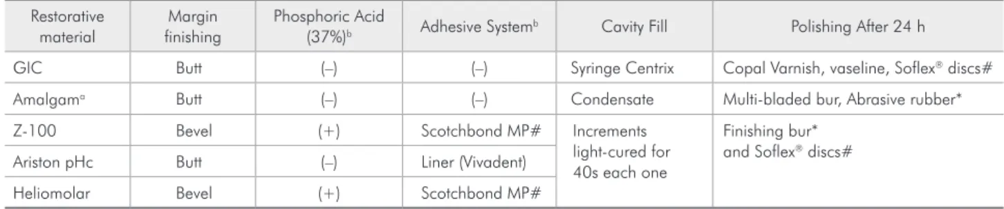
Table 1 - Restorative techniques used.
Restorative material
Margin finishing
Phosphoric Acid
(37%)b Adhesive System
b Cavity Fill Polishing After 24 h
GIC Butt (–) (–) Syringe Centrix Copal Varnish, vaseline, Soflex® discs#
Amalgama Butt (–) (–) Condensate Multi-bladed bur, Abrasive rubber*
Z-100 Bevel (+) Scotchbond MP# Increments light-cured for 40s each one
Finishing bur* and Soflex® discs#
Ariston pHc Butt (–) Liner (Vivadent) Heliomolar Bevel (+) Scotchbond MP#
al.5 (1990), that was modiied in the incubation
pe-riod, which was enlarged, and in the culture medi-um, which was changed. The microorganism used was Streptococcus mutans (ATCC 25175), which was incubated in TSB (Difco-Becton, Dickinson and Company - Sparks, MD, USA) with 5% sucrose (37°C for 24 h) in order to induce bacterial growth. The bacteria were plated in a solid culture medium (TSA, Difco-Becton, Dickinson and Company - Sparks, MD, USA) to obtain isolated colonies and incubated again (37°C for 24 h). Then, the grown colonies were transferred to tubes containing TSB and 5% sucrose, starting a preconditioning of the microorganism with sucrose for 6 days. This way, the tube containing the inoculum broth was ob-tained with approximately 1.84 × 10⁶
colony-form-ing units (CFU)/ml. Each tooth was immersed in a tube containing TSB + 5% sucrose and 200 µl of the inoculum broth. The teeth were maintained in this bacterial system for 30 days, being transferred to a fresh tube every 48 h. During the incubation period, tests were performed to check for contaminants. Af-ter 30 days, the teeth were removed from the bacte-rial system and the steel wires were removed.
The teeth were sectioned with double face dia-mond discs (Discolex, KG Sorensen, Barueri, SP, Brazil) to separate the buccal from the lingual sur-faces. Each aspect was embedded in epoxy resin (Resigel - Redlease®, Varzea Paulista, SP, Brazil),
and sectioned with a Labcut Machine® (Extec 1010,
Excel Technologies Inc., Enield, CT, USA) in a buc-colingual direction, resulting in specimens with 500 µm in thickness. Two of these sections were randomly chosen and ground by hand to a thickness around 100 µm using sandpaper numbers 150, 220, 400, and 600 (Norton Abrasivos Brasil - Igarassu, PE, Brazil).
Each chosen section was examined under a light microscope (Citoval 2 with CarlZeiss lens - Laboral 4, Zeiss, Thornwood, NY, USA) coupled to a soft-ware (DIRACOM 3, Imagelab 2000, São Paulo, SP Brazil) to capture and process the images (LIDO, University of São Paulo) in order to do the following measurements (variables) in the occlusal and cervical outer lesion: extension (E) - from one lesion margin up to the opposite margin; depth (D) - at the deepest
point of the lesion; inhibition area (IA) - distance between lesion and restoration margin. So, four positions of each tooth (buccal/occlusal, lingual/oc-clusal, buccal/cervical and lingual/cervical) were analyzed following those criteria due to anatomical and histological differences of the crowns, and the data were subjected to statistical analyses. For each variable, Analysis of Variance was used to test the equality of means between the groups. Because of the dependence of the four measurements in each tooth, Component Variance Models were also used in the analysis.13
The same sections were also examined using po-larized light microscopy, embedding in water and quinoline, in order to visualize the lesions.
Results
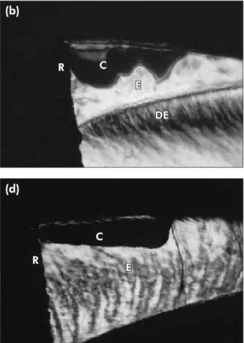
The enamel surface subjected to the cariogenic challenge presented a white and opaque appearance. Microscopically, outer lesions with or without caries inhibition zones were observed (Figure 1), whereas wall lesions were not seen in the cavities in any of the specimens. Polarized-light examination showed an intact surface layer above the lesion body, which had a rectangular shape and positive and negative birefringence when embedded in water and quino-line, respectively. When quinoline was used, we could observe, in some sections, the dark zone and the translucent zone under it.
The samples’ means were grouped when the val-ues for the material groups and for the dental sites were similar. Tables 2, 3 and 4 show the statistically different groups and sites.
(p = 0.0275). Tables 2 and 3 show the estimates and the conidence intervals for the means of these vari-ables.
The GIC group showed the highest number and size (0.05 mm ± 0.01) of inhibition areas (IA), sta-tistically different from those of the other groups. Differences among these other groups were not sta-tistically detected (p > 0.05). The equality of means
was observed for all dental positions (p = 0.1693). These data are shown in Table 4.
Discussion
The bacterial method for in vitro caries forma-tion has been used because it allows not only the demineralization of dental tissue, but it also favors the investigation of different anticariogenic
proper-(a)
C
E
IC
R
(b)
C
E
DE R
(d)
E C
R
(c)
R C
E
(e)
DE
C
E
R
ties of restorative materials or microleakage around restorations. Because of that, the bacterial method is considered to reproduce very closely the in vivo
conditions.3,5
It has been reported that restorative materials can induce a variable amount of bacterial retention, and that is an important parameter in the etiology of sec-ondary caries formation. However, restorative materi-als can inhibit bacterial growth and favor the reminer-alization through ion release and, if the material allows a good marginal seal, it can be considered one of the protecting mechanisms against secondary caries.11
Good marginal seal also depends on the site of the crown where the cavosurface margin is located. Some sites make the marginal seal more dificult than others
due to anatomical landmarks, and this is the reason for the model of statistical analyses adopted in this study considering the four sites of the crown: occlusal and cervical of the buccal and lingual portions of the restorations. These are important factors, especially in the treatment of patients with high caries-risk.
In the current study, the microleakage necessary for the development of wall lesions did not occur, or it was too small, probably due to the restorative techniques and thermocycling method used. All le-sions clearly extended straight into the cavity wall at a 90° angle or curved upward. Probably, a method that induces a gap between the dental wall and the restoration would induce enough microleakage to allow the development of wall lesions.5,19
The conditions of our study and the absence of wall lesions allowed us to ignore microleakage events, despite the high complexity of the environ-ment at the tooth-restoration interface where the bacterial media was used for the cariogenic chal-lenge.6 We can assume that the different responses
observed were promoted by the materials used, as the in vitro restorations were easily accessed and clearly visualized favoring the marginal seal.
Although the possibility of outer lesions of sec-ondary caries is poorly affected by the anticario-Table 2 - Means, standard deviation (SD) and confidence intervals (CI) of lesion extension (mm).
Restorative Material* Occlusal sites* Cervical sites*
Mean SD CI Mean SD CI
Ketac Fil 1.52 0.07 (1.38, 1.66) 1.37 0.07 (1.23, 1.51) Amalgam and Ariston pHc 1.63 0.05 (1.53, 1.73) 1.48 0.05 (1.38, 1.58) Z-100 and Heliomolar 1.81 0.05 (1.71, 1.91) 1.66 0.05 (1.56, 1.76)
*Statistically similar groups and sites are grouped. Confidence coefficients are 0.95.
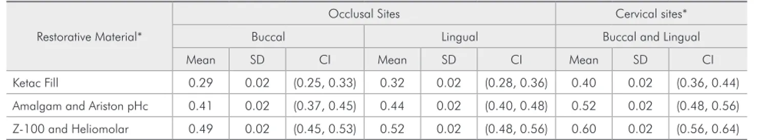
Table 3 - Means, standard deviation (SD) and confidence intervals (CI) of lesion depth (mm).
Restorative Material*
Occlusal Sites Cervical sites* Buccal Lingual Buccal and Lingual Mean SD CI Mean SD CI Mean SD CI Ketac Fill 0.29 0.02 (0.25, 0.33) 0.32 0.02 (0.28, 0.36) 0.40 0.02 (0.36, 0.44) Amalgam and Ariston pHc 0.41 0.02 (0.37, 0.45) 0.44 0.02 (0.40, 0.48) 0.52 0.02 (0.48, 0.56) Z-100 and Heliomolar 0.49 0.02 (0.45, 0.53) 0.52 0.02 (0.48, 0.56) 0.60 0.02 (0.56, 0.64)
*Statistically similar groups and sites are grouped. Confidence coefficients are 0.95.
Table 4 - Means, standard deviation (SD) and confidence intervals (CI) of inhibition area (mm).
Restorative Material* All dental positions* Mean SD CI Ketac Fil 0.05 0.01 (0.03, 0.07) Ariston pHc, Amalgam,
Z-100 and Heliomolar 0.01 0.00 (0.00, 0.02)
genic properties of restorative materials,6 it has been
observed that the depth of these lesions is mainly altered by the concentration of cariostatic agents such as mercury, silver, zinc, copper and luoride on the dental surface.9 Thus, the capacity of
restor-ative materials to release such agents as well as the ability of their incorporation by the adjacent dental structure should be considered in tooth protection against secondary caries. The smallest lesions close to the GIC restorations probably occurred due to the high concentration and release of luoride from GIC,16 and it has been already shown that this
situ-ation can decrease signiicantly S. mutans levels in a cariogenic challenge in situ.1 Our results are also in
agreement with those of other studies.8,9
The anticariogenic action of metallic elements released by amalgam restorations can explain the small lesions observed in this group, suggesting lower secondary caries risk and slower progres-sion of a leprogres-sion around amalgam restorations than around resin ones.12,20 The small lesions also found
with Ariston pHc may be related to the release of calcium, luoride and hydroxyl and this inding is in agreement with the results of another study10 that
also showed that Ariston pHc hampers demineral-ization next to the restoration. In contrast, the Z-100 and Heliomolar composite resin groups devel-oped large lesions according to all parameters and positions studied, in agreement with the results of another study that also observed wider outer lesions using composite resin8 probably due to the lack of
luoride release and antibacterial properties of the composite resin restorations.17,18
Composite resins, even those containing luoride, do not show any anticariogenic action,7 and have no
other carious inhibitory substance.12 In contrast, it
was demonstrated in vitro that resinous materials can favor S. mutans growth4 and the deposit of
bac-terial colonies on the restoration,11,20 probably due
to its organic constitution. Furthermore, the resin-ous matrix may prevent the contact of luoride with water, resulting in a low release of this ion. On the other hand, composite resins adhering to the dental structure through adhesives improve the marginal seal, which probably inhibited wall lesions.
Besides the ability to inhibit caries development, other factors should be evaluated when choosing a restorative material. Adhesion, marginal seal, and the mechanical and aesthetic properties are also rel-evant characteristics that should be taken into ac-count. Other experimental designs should also be tested to further evaluate microleakage in secondary caries development.
Conclusions
GIC was the material that statistically promoted the best protection against secondary caries devel-opment when used as a restorative material in vi-tro, followed by amalgam and Ariston pHc. The composite resins used did not seem to promote any protection of the dental structure against secondary outer lesions.
Acknowledgments
We wish to thank Prof. Moacyr Domingos No-velli for his assistance with the Imagelab software, and Prof. Patricia Gama for her English language revision. This study was Supported by The State of São Paulo Research Foundation (FAPESP, grant n. 99/12518-5).
References
1. Benelli EM, Serra MC, Rodrigues Jr AL, Cury JA. In situ
anticariogenic potential of glass ionomer cement. Caries Res. 1993;27(4):280-4.
2. Burke FIT, Wilson NHF, Cheung SW, Mjor IA. Influence of patient factors on age of restorations - a failure and reasons for their placement and replacement. J Dent. 2001;29(5):317-24.
3. Dummer PMH, Edmunds DH, Green RM. Demineralization of human enamel by Streptococcus mutans NCTC 10832
using a sequential batch culture technique. Caries Res. 1982;16(2):193-6.
4. Friedl KH, Schmalz G, Hiller KA. Flüssigklitskulturen zur prüfung der wirkung zahänarztlischer werkstoffe auf des Bak-terienwachstun. Dtsch Zahnarzte Z. 1992;47(12):2826-31. 5. Gilmour SM, Edmunds DH, Dummer PM. The production
6. Grossman ES, Matejka JM. Reliability of outer lesion sec-ondary caries as a predictor of wall lesions. Am J Dent. 1999;12(1):31-6.
7. Hara AT, Turssi CP, Serra MC, Nogueira MC. Extent of the cariostatic effect on root dentin provided by fluoride-contain-ing restorative materials. Oper Dent. 2002;27(5):480-7. 8. Hattab FN, Mok NY, Agnew EC. Artificially formed caries
like lesions around restorative materials. J Am Dent Assoc. 1989;118(2):193-7.
9. Hsu CYS, Donly KJ, Drake DR, Ewfel JS. Effects of aged fluoride-containing restorative materials on recurrent root caries. J Dent Res. 1998;77(2):418-25.
10. Kielbassa AM, Schulte-Monting J, Garcia-Godoy F, Meyer-Lueckel H. Initial in situ secondary caries formation: effect of various fluoride-containing restorative materials. Oper Dent. 2003;28(6):765-72.
11. Krasse BG. Prediction and prevention of recurrent caries based on microbiological assays. In: Anusavice KJ. Quality evalu-ation of dental restorevalu-ations. Chicago: Quintessence; 1989. p. 199-209.
12. Leinfelder KF. Criteria for clinical evaluation of composite resin restorations. In:Anusavice KJ. Quality evaluation of dental restorations. Chicago: Quintessence; 1989. p. 139-49.
13. Lindsey JK. Models for repeated measurement. Oxford: Clar-endon; 1993.
14. Mjor I A, Shen C, Eliasson ST, Richter S. Placement and re-placement of restorations in general dental practice in Iceland. Oper Dent. 2002;27(2):117-23.
15. Orstavik D. Antibacterial properties of and element re-lease from some dental amalgams. Acta Odontol Scand. 1985;43(4):231-9.
16. Preston AJ, Mair LH, Agalamanyi EA, Higham SM. Fluor-ide release from aesthetic dental materials. J Oral Rehabil. 1999;26(2):123-9.
17. Sarret DC. Clinical challenges and the relevance of materi-als testing for posterior composite restorations. Dent Mater. 2005;21(1):9-20.
18. Savarino L, Breschi L, Tedaldi M, Ciapetti G, Tarabusi C, Greco M et al. Ability of restorative and fluoride releasing materials to prevent marginal dentine demineralization. Bio-materials. 2004;25(6):1011-7.
19. Schiffner U. Inhibition of enamel and root dentin demineral-ization by Ariston pHc: an artificial mouth study. Am J Dent. 1999;12 Spec No:S10-2.