Annicele da Silva Andrade(a) Maria Beatriz Duarte Gavião(b) Gustavo Hauber Gameiro(c) Moara De Rossi(a)
(a) Graduate Student; (b)Professor – Department of Pediatric Dentistry, Piracicaba Dental School, University of Campinas, Piracicaba, São Paulo, Brazil. (c) Professor, Department of Physiology,
Universidade Federal do Rio Grande do Sul – UFRGS, Porto Alegre, Rio Grande do Sul, Brazil.
Corresponding author: Maria Beatriz Duarte Gavião Av. Limeira, 901
Piracicaba - SP - Brazil CEP: 13414-903
E-mail: [email protected]
Received for publication on Mar 25, 2010 Accepted for publication on Apr 29, 2010
Characteristics of masticatory
muscles in children with unilateral
posterior crossbite
Abstract: The aim of this study was to detect possible differences in the EMG (electromiography) activity, chewing rate (CR), cycle dura-tion (CD) and preferred chewing side (PCS) between children with and without unilateral posterior crossbite. Thirty-seven children aged from 7 to 10 years were selected from the clinic of the Department of Pedi-atric Dentistry, Piracicaba Dental School, Brazil, and divided into two groups: unilateral posterior crossbite (UPCB group, n = 17), and normal occlusion (NOccl group, n = 20). The PCS was determined using a visual spot-checking method. The EMG activity was recorded during mastica-tion, and two chewing sequences of 20 s were evaluated to establish each subject’s CR (cycles/sec) and CD. UPCB and NOccl groups did not have a PCS. The EMG activity and the cycle characteristics did not differ be-tween the groups. The correlations bebe-tween CD, CR and EMG activ-ity were statistically signiicant for the masseter and anterior temporalis muscles only in the NOccl group, in which there was also a signiicant correlation between the EMG activity of masseter and anterior tempo-ralis. In conclusion, these indings suggest that although children with and without UPCB presented a bilateral masticatory pattern with similar CR and CD, balanced EMG activity of masseter and anterior temporalis muscles was observed only in the NOccl group. These results indicate that in children, UPCB can alter the coordination of masticatory muscles during mastication.
Descriptors: Malocclusion; Electromyography; Mastication.
Introduction
Posterior crossbite is deined as a malocclusion in the canine, molar and/or premolar regions, characterized by the buccal cusps of the maxil-lary teeth occluding lingually against the buccal cusps of the correspond-ing mandibular teeth.1 It may develop during eruption of the primary
dentition and can involve the permanent dentition at a later stage of de-velopment. It may originate from a skeletal or dental malrelationship, or both, and may lead to a mandibular displacement.2
Some studies in children and adolescents have shown that posterior crossbite has been associated with asymmetrical function of the masti-catory muscles,3-5 signs and symptoms of temporomandibular disorders
(TMD), such as pain, headache, and muscle tenderness,6,7 which may
been used to monitor mastication.9 Among these
techniques, electromyography (EMG), the measure-ment of the electrical activity of muscles, has found wide application.10 EMG has been used to identify
differences in chewing patterns between individu-als, and to classify them into groups according to their chewing eficiency.11
According to Planas12 (1997), the crossbite side
presents a greater number of occlusal contacts in function, being the preferred chewing side. Howev-er, these characteristics were not conirmed by other studies.9,13 Chewing side preference was deined by
Christensen and Radue14 (1985) as: “when
mastica-tion is performed consistently or predominantly on the right or left side of the dentition”. Considering that masticatory function during growth has a bio-logical impact on the growing structures, a unilat-eral mastication may lead to asymmetric anatomical structures (bones, temporomandibular joint, mus-cles, and teeth) on completion of growth.12,15 Thus,
understanding whether and how posterior crossbite malocclusion inluences the preferred chewing side and EMG activity of masticatory muscles would be particularly appropriate.
This study was designed to detect whether there is any difference in the EMG activity during mas-tication, chewing rate, cycle duration and preferred chewing side between children with and without unilateral posterior crossbite.
Material and Methods
A cross-sectional study design was used with subjects recruited as a convenience sample of 37 children aged 7-10 years, who were to start dental treatment at the Department of Pediatric Dentistry, Piracicaba Dental School, University of Campinas, Piracicaba, SP, Brazil. The Research Ethics Com-mittee of the Dental School approved the project (Protocol Nos. 020/2006 and 023/2006).
The children were divided into two groups: children with unilateral posterior crossbite (UPCB group,) consisting of 17 subjects aged from 7 to 10 years (8.65 ± 1.23); from these, 12 subjects had uni-lateral crossbite on the left side, ive on the right side. The control group consisted of 20 children with nor-mal occlusion (NOccl group) (mean age 8.64 ± 1.15
years). The children and their parents or guardians received an oral and written explanation about our aims and research methodology. After obtaining the consent of term, we conducted an anamnesis to ver-ify their medical and dental history and oral habits. The exclusion criteria for both groups were the pres-ence of symptoms of craniomandibular dysfunction, major dental reconstructions, previous or current orthodontic treatment, caries, and/or severe gingi-vitis or missing teeth. Only subjects with unilateral presentation of posterior crossbite with a functional shift of the mandible toward the crossbite side were selected. Children with crossbite resulting from den-tal inclination were not considered.
Preferred chewing side
The preferred chewing side was determined using a modiication of the visual spot-checking method described by Christensen and Radue16 (1985).
Sub-jects were given a 1.7 g piece of Trident (Cadbury, Bauru, São Paulo, Brazil) (Sugar Free chewing gum) and were instructed to chew in their habitual man-ner. After 15 s, they were asked to stop chewing, clench the gum between their teeth and grin broadly so that the position of the chewing gum could be observed and recorded as right or left. This proce-dure was carried out seven times consecutively. The term “observed preferred chewing side” (OPCS) was used when the child chewed 5/7, 6/7 or 7/7 times on the same side.
Chewing characteristics
Two chewing sequences of 20 seconds were re-corded, as described above. The average of the number of cycles in these two sequences was then divided by 20 s to establish each subject’s automatic habitual chewing rate (cycle/s). Furthermore, the mean duration of each cycle during the 20 seconds was calculated.
Electromyographic evaluation
and a subsequent ampliication of 50 times with a common mode rejection ratio of 130 dB to 60 Hz. The data were sent to a 14-bit A/D converter and sampled at 2,000 Hz. A differential double elec-trode was used, with 100 times pre-ampliication and two contacts measuring 10.0 x 1.0 mm, with a distance of 10.0 mm between them, impedance up-wards of 10 GΩ and a common rejection value of 130 dB to 60 Hz, crafted in silver and ixed in a res-in capsule measurres-ing 40 x 20 x 5 mm. Durres-ing the experiment, the child remained comfortably seated, with a straight back and head oriented in the Frank-fort plane parallel to the loor. Both the skin and the electrodes were cleaned with 70 percent GL ethyl alcohol in order to eliminate any residues of grease or pollution, and for the anterior temporalis muscle, the skin was shaved, when necessary.
The electrodes were placed on the masseter and anterior temporalis in the following orientations: masseter - level halfway between the zygomatic arch and the gonial angle, close to the level of the occlu-sal plane; anterior portion of the temporalis muscle - in front of the anterior border of the hairline. A ground electrode was also used on the right hand to reduce electromagnetic interferences and other acquisition noise. The muscle activity was recorded during two chewing sequences (chewing gum) of 20 seconds and the means of root mean square (RMS) were used. The mean of two measurements was con-sidered. The methodology for signal treatment was in accordance with Merletti17 (1999). The children
were instructed to chew in their habitual manner.
Statistical analysis
Comparisons between sides (crossbite side vs. non-crossbite side/UPCB group or right side vs. left side/NOccl group) were performed by the paired t-test. The chewing rate and EMG activity were
compared between groups using the unpaired t-test. Fisher’s exact test was used to associate the preferred chewing side in the two groups. The rela-tionship between chewing characteristics and EMG activity was evaluated by the appropriate Pearson’s and Spearman coeficient tests. The level of signii-cance was set at α = 0.05.
Results
Table 1 shows the sample distribution, the total crossbite group and control group in relation to pre-ferred chewing side. There was no statistical differ-ence between groups.
Graph 1 shows the comparison between sides in the UPCB and NOccl groups. There were no signii-cant differences between the crossbite side and the normal one in the UPCB group, neither between the right and left sides in the NOccl group. (p > 0.05).
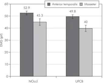
Since there were no differences between sides, the mean EMG values of the right and left masseter and anterior temporalis were considered for comparisons between groups. Graph 2 shows the respective val-ues. There were no signiicant differences between groups (p > 0.05).
Graph 3 shows the chewing cycle characteristics for UPCB and NOccl groups, in which no signii-cant differences could be observed (p > 0.05).
Table 1 - Sample distribution in relation to preferred chew-ing side in children with UPCB and NOccl groups.
UPCB group
12 left side/5 right side NOccl group
Left 2/0 4
Right 3/1 7
Bilateral 7/4 9
Total 17 20
(Fisher’s exact test)
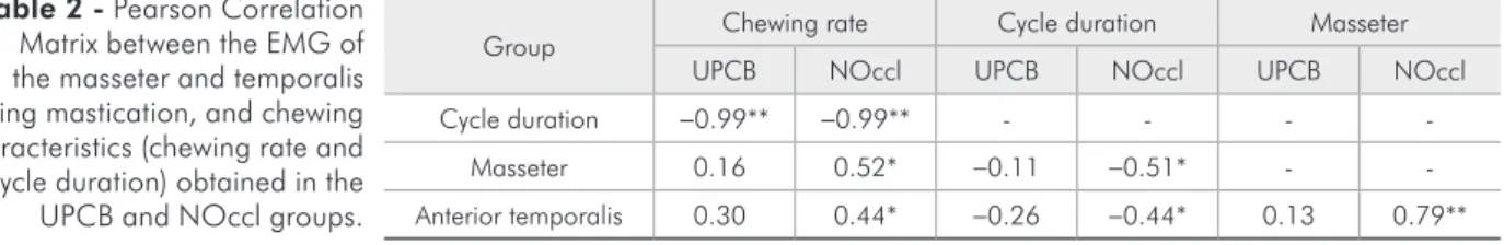
Group Chewing rate Cycle duration Masseter
UPCB NOccl UPCB NOccl UPCB NOccl
Cycle duration -0.99** -0.99** - - -
-Masseter 0.16 0.52* -0.11 -0.51* -
-Anterior temporalis 0.30 0.44* -0.26 -0.44* 0.13 0.79**
*p < 0.05; **p < 0.001.
Table 2 reports the correlation between chewing characteristics and EMG activity during chewing in the UPCB group and in the NOccl group. A signii -cant correlation was observed between chewing rate and cycle duration, in both the UPCB and NOccl groups. For the masseter, the correlation between cycle duration and EMG activity in the NOccl group was negative, moderate and signii cant. Con-versely, the correlation between chewing rate and EMG activity of masseter was positive, moderate and signii cant. As regards the anterior temporalis
muscle, the correlations exhibited the same pattern observed in the masseter. Therefore, the EMG ac-tivity of the masticatory muscles showed signii cant correlations with chewing characteristics only in the NOccl group. Moreover, the correlation between the EMG activities of masseter and anterior tempo-ralis was strong and statistically signii cant only in the NOccl group.
Discussion
In the present study, using the visual
spot-check-0 10 20 30 40 50
E
MG
(
µ
V
)
60 Anterior temporalis Masseter
NOccl UPCB
52.9
45.3
49.8
40
Graph 2 - EMG data (µV) of chewing during 20 s. Data are
plotted as mean ± SEM (p > 0.05, unpaired t-tests).
Graph 3 - Chewing cycle characteristics for UPCB and NOccl groups. (A) Chewing rate (cycles/s); (B) cycle duration (s). Data
are plotted as mean ± S.E.M (p > 0.05, unpaired t-tests).
0 0.5 1
C
ycles
/s
ec
1.5
NOccl UPCB
1.37 1.42
0 0.3
0.2
0.1 0.4 0.5 0.6 0.7 0.8 0.9 1
2
0
s
ec/
cy
cle
NOccl UPCB
0.74 0.72
A B
0 10 20 30 40 50
E
MG
(
µ
V
)
Crossbite side
Normal side
Right side
Left side 60
70
4
9
.1
3
8
.8 41
.6
4
3
.3
6
0
.2
4
8
.6
4
5
.5 51
.0
Anterior temporalis Masseter
NOccl
UPCB
Graph 1 - EMG data (µV) from each masticatory muscle during chewing gum (20 s) in children with and without
pos-terior crossbite. Data are plotted as mean ± SEM (p > 0.05,
ing method, it was observed that most of the chil-dren with unilateral posterior crossbite showed a bilateral pattern of mastication, without a preferred side, in agreement with previous indings.10,14,18,19
There were no signiicant differences regarding the preferred chewing side between UPCB and NOccl groups (Table 1). These indings could be justiied by the fact that chewing is considered an event deter-mined at two levels, an individual central chewing pattern generator, and peripheral events inducing chewing adaptations.20 Establishment of the
indi-vidual central chewing pattern begins with tooth eruption, and is well established in a child with a complete deciduous dentition.21 Once the central
chewing pattern has been established, it appears to be relatively resistant to change.20 Therefore, since
the children were evaluated during mixed dentition, they could perhaps persist with the chewing pattern they acquired early.
The above-mentioned comments can be sup-ported by the absence of signiicant differences in the electrical activity of masseter and anterior tem-poralis muscles observed between sides in both groups, as well as in inter-groups comparisons (Graphs 1 and 2). These results are in agreement with those obtained by Alarcón et al.13 (2000) and
Andrade et al.22 (2009), in which no signiicant
dif-ferences were found between sides in both cross-bite and normocclusive subjects. This could mean that during chewing there is a symmetric function of the masticatory muscles, and therefore chewing can be predominantly bilateral, despite the presence of UPCB, as was also previously considered.9,13,18,22
Conversely, Egermark-Eriksson et al.6 (1990) found
that crossbite subjects preferred to chew unilateral-ly, whereas Ingervall and Thilander18 (1975) showed
that patients with lateral shift had a lower activ-ity of the anterior and posterior temporalis muscles than normocclusive subjects, but the activity of the masseter muscles was similar in both groups. The discrepancies among the results in different studies may be due to differences between the samples, the location of the measuring points, and the use of dif-ferent EMG techniques.
Comparisons between the chewing character-istics in children with and without UPCB showed
a similar rhythm and cycle duration during masti-cation (Graph 3), showing that those with UPCB have probably generated adaptive responses to the morphologic alterations, allowing similar masti-catory function. Throckmorton et al.20 (2001)
re-ported that patients with unilateral posterior cross-bite chewed more slowly than did the controls, and after treatment their cycle duration was shortened to equal those of control values. This controver-sial inding could be due to different methodolo-gies, since they recorded chewing using an intraoral splint attached to each subject’s lower teeth, and in the present study the mean EMG activity during chewing was recorded without any intraoral appli-ance. It is well known that masticatory rhythm is sensitive to sensory input. This peripheral level re-sponds to interferences by slowing or even stopping the chewing cycle, and by lowering the amount of occlusal force used during mastication.23,24
Howev-er, the chewing cycle duration is much more sensi-tive to the central pattern generator located in the brain stem,25 and the chewing rate is relatively
re-sistant to change. Therefore, the results of the pres-ent study suggest that the occlusal interferences as-sociated with crossbite are insuficient to alter the output of the central pattern generator as regards the chewing rate.
Some studies showed that children with a uni-lateral posterior crossbite exhibit unusual chewing patterns when chewing on the affected side and this is characterized by an increased frequency of re-verse sequencing.3,20 Ben-Bassat et al.26 (1993) and
Throckmorton et al.20 (2001) reported that
success-ful treatment of a unilateral crossbite with palatal expansion did not eliminate the reverse-sequencing chewing cycles. However, there is evidence that the treatment of posterior crossbite with an orthodon-tic functional appliance (Function Generating Bite) was able to reduce the prevalence of reverse se-quencing chewing cycles in children with this mal-occlusion.27
cor-relations between these muscles were found in the UPCB (Table 2). These results suggest that poste-rior crossbite negatively affects the performance of masticatory muscles during mastication, leading to a poor muscular coordination in children with this malocclusion. Although the cycle shape has not been evaluated in the present study, the unbalanced muscular activity observed in UPCB could be an im-portant inding, indicating some dysfunction in the mastication process. If the asymmetric masticatory function during growth has a biological impact on the growing structures, which may lead to physical and functional problems,5 the present results
rein-force the importance of the fact that such asymme-tries may be prevented by orthodontic therapy at an early stage in development.28-30
Conclusion
The present investigation demonstrated that both groups, with and without UPCB did not pres-ent a preferred chewing side. The results obtained by EMG evaluation during chewing indicate a bi-lateral masticatory pattern in both groups, in which the chewing rate and cycle duration were also simi-lar. However, children with UPCB presented poor muscular coordination during mastication.
Acknowledgements
We are grateful to The National Council for Scientiic and Technological Development (CNPq, BR) for the scholarship received by the irst author and The State of São Paulo Research Foundation (FAPESP, SP, Brazil) (Process 05/03472-4) for inan-cial support.
References
1. Bjork A, Krebs A, Solow B. A method for epidemiologic registration of malocclusion. Acta Odontol Scand. 1964 Feb;22(1):27-41.
2. Daskalogiannakis J. Glossary of orthodontic terms. Berlin: Quintessence Publishing Group; 2009. 297 p.
3. Pinto AS, Buschang PH, Throckmorton GS, Chen P. Mor-phological and positional asymmetries of young children with functional unilateral posterior crossbite. Am J Orthod Dentofacial Orthop. 200 Nov;120(5):513-20.
4. Sonnesen L, Bakke M, Solow B. Bite force in pre-orthodon-tic children with unilateral crossbite. Eur J Orthod. 2001 Dec;23(6):741-9.
5. Andrade Ada S, Gameiro GH, Derossi M, Gavião MB. Poste-rior Crossbite and Functional Changes. Angle Orthod. 2009 Mar;79(2):380-6.
6. Egermark-Eriksson I, Carlsson GE, Magnusson T, Thilander B. A longitudinal study of malocclusion in relation to signs and symptoms of craniomandibular disorders in children and adolescents. Eur J Orthod. 1990 Nov;12(4):399–407. 7. Vanderas AP, Papagiannoulis L. Multifactorial analysis of the
aetiology of craniomandibular dysfunction in children. Int J Paediatr Dent. 2002 Sep;12(5):336-46.
8. Sonnesen L, Bakke M, Solow B. Malocclusion traits and symp-toms and signs of temporomandibular disorders in children with severe malocclusion. Eur J Orthod. 1998 Oct;20(5):543– 59.
9. Salioni MA, Pellizoni SE, Guimarães AS, Juliano Y, Alonso LG. Functional unilateral posterior crossbite effects on mas-tication movements using axiography. Angle Orthod. 2005 May;75(3):362-7.
10. Wendy E, Brown. Method to investigate differences in chewing behaviour in humans. J Texture Stud. 1994;25(1):1-16. 11. Braxton D, Dauchel C, Brown WE. Association between
chewing efficiency and mastication patterns for meat, and in-fluence on tenderness perception. Food Qual Prefer. 1996 Jul-Oct;7(3-4):217–23.
12. Planas P. Reabilitação Neuroclusal, 2nd Ed. Rio de Janeiro:
Medsi; 1997. 355 p.
13. Alarcón JA, Martín C, Palma JC. Effect of unilateral pos-terior crossbite on the electromyographic activity of human masticatory muscles. Am J Orthod Dentofacial Orthop. 2000 Sep;118(3):328-41.
14. Christensen LV, Radue JT. Lateral preference in mastication. A feasibility study. J Oral Rehabil. 1985 Sep;12(5):421-7. 15. Liu C, Kaneko S, Soma K. Effects of a mandibular lateral shift
on the condyle and mandibular bone in growing rat’s. Angle Orthod. 2007 Sep;77(5):787-93.
16. Christensen LV, Radue JT. Lateral preference in mastica-tion: an electromyographic study. J Oral Rehabil. 1985 Fall;12(5):429-34.
17. Merletti R. Standards for reporting EMG data. J Electromyogr Kinesiol. 1999 Feb;9 (1): III-IV.
18. Ingervall B, Thilander B. Activity of temporal and masseter muscles in children with a lateral forced bite. Angle Orthod. 1975 Oct;45(4):249–58.
19. Martín C, Alarcón JA, Palma JC. Kinesiographic study of the mandible in young patients with unilateral posterior crossbite. Am J Orthod Dentofacial Orthop. 2000 Nov;118(5):554-8. 20. Throckmorton GS, Buschang PH, Hayasaki H, Pinto AS.
poste-rior unilateral crossbite in children. Am J Orthod Dentofacial Orthop. 2001Nov;120(5):5521–9.
21. Wickwire NA, Gibbs CH, Jacobson P, Lundeen HC. Chewing patterns in normal children. Angle Orthod. 1981 Jan;51(1):148–60.
22. Andrade AS, Gavião MB, Derossi M, Gameiro GH. Electro-myographic activity and thickness of masticatory muscles in children with unilateral posterior crossbite. Clin Anat. 2009 Mar;22(2):200-6.
23. Ingervall B, Carlsson GE. Masticatory muscle activity before and after elimination of balancing side occlusal interference. J Oral Rehab. 1982 May;9(3):183-92.
24. Bakke M, Michler L, Moller E. Occlusal control of mandibu-lar elevator muscles. Scand J Dent Res. 1992 Oct;100(5):284-91.
25. Thexton AJ. Mastication and swallowing: an overview. Br Dent J. 1992 Oct 10;173(6):197-206.
26. Ben-Bassat Y, Yaffe A, Brin I, Freeman J, Ehrlich Y. Func-tional and morphological-occlusal aspects in children
treat-ed for unilateral posterior cross-bite. Eur J Orthod. 1993 Feb;15(1):57-63.
27. Piancino MG, Talpone F, Dalmasso P, Debernardi C, Lewin A, Bracco P. Reverse-sequencing chewing patterns before and after treatment of children with a unilateral posterior cross-bite. Eur J Orthod. 2006 Oct;28(5):480-4.
28. Castelo PM, Bonjardim LR, Pereira LJ, Gavião MB. Facial dimensions, bite force and masticatory muscle thickness in preschool children with functional posterior crossbite. Braz Oral Res. 2008 Jan-Mar;22(1):48-54.
29. Lam PH, Sadowsky C, Omerza F. Mandibular asymmetry and condylar position in children with unilateral posterior cross-bite. Am J Orthod Dentofacial Orthop. 1999 May;115(5):569-75.