Luiz Fernando Machado Silveira(a) Carina Folgearini Silveira(b) Luis Antônio Suita de Castro(c) João Batista César Neto(d) Josué Martos(e)
(a) PhD, Associate Professor, Department of Clinics; (b)Graduate Student; (e)PhD, Adjunct Professor, Department of Clinics – School of Dentistry, Federal University of Pelotas, Pelotas, RS, Brazil.
(c) MSc, EMBRAPA-CPACT; Laboratory of Immunology and Microscopy, Pelotas, RS, Brazil.
(d) PhD, Professor, Department of Stomatology, School of Dentistry, University of São Paulo (USP), São Paulo, SP, Brazil.
Corresponding author: Luiz Fernando Machado Silveira Rua Santa Cruz, 2434/602 Pelotas - RS - Brazil CEP: 96015-710
E-mail: lfms1960@gmail.com
Received for publication on Jun 23, 2009 Accepted for publication on Dec 17, 2009
Crown-down preflaring in the
determination of the first apical file
Abstract: In this study, scanning electron microscopy (SEM) was used to evaluate the adaptation of the irst apical ile after prelaring in mesio-buccal (MB) and mesiolingual (ML) canals of mandibular molars con-sidering the tactile sensibility as a reference. The mesial canals (n = 22) of human mandibular molar teeth were used, and the irst instrument to bind to the working length was determined after prelaring and crown-down shaping. Digital images of the root apex were acquired and a single examiner determined the contact of the ile with the walls using Image J software. The results showed that the ile was in contact in 47.83% and 31.71% in the MB and ML canals, respectively. When the apexes are fused, the average was 40.03%. A descriptive analysis showed that the irst apical ile did not touch all dentin walls in any of the samples.
Descriptors: Molar / anatomy & histology; Root canal preparation; Root canal therapy; Microscopy, electron, scanning; Endodontics.
Introduction
The success of endodontic treatment demands knowledge of the root canal system. However, bidimensional radiographic images do not repre-sent anatomical conditions. Most canals do not communicate with peri-odontal tissue precisely at the root apex, and the apical foramen often does not have a round shape.1 Morphologic analyses have shown that
canals are oval-shaped or irregular in the apical third and present their largest diameter at the buccolingual aspect.2,3,4
This concept is conirmed by the fact that the irst ile that clinically binds to the apex is smaller than the anatomical diameter of the apical foramen.5 Weine6 (1989) proposed that the apical enlargement must be
three sizes larger than the irst ile that bound at the working length.6
However, if the apical diameter corresponds to 0.25 mm in its smallest extension and 0.50 mm in its largest extension, binding will occur with instrument #25, and enlargement will be achieved with instrument #40 without touching the whole perimeter of the apical foramen. Baugh and Wallace7 (2005) concluded that the apical preparation should be larger
than that previously proposed by Kerekes and Tronstad5 (1977).5,7
How-ever, to achieve a more accurate estimate of the apical diameter, removal of the interferences along the canal is necessary.8 If the canals are narrow
diameter.8
In cases of pulp necrosis, in which the bacteri-al presence is inherent, disinfection of the canbacteri-al as proposed by Weine6 (1989) allows for a higher
re-duction in the bacterial population.6,9,10,11 A deeper
penetration of the irrigation with a larger volume may facilitate this disinfection.9,12
With regard to illings, Wu et al.2 (2000) and
Weiger et al.13 (2006) conirmed that many of the
canal areas (especially in the apical region) are not touched during canal preparation due to its oval or irregular conigurations.
The aim of the present study was to determine the adaptation of the irst apical ile after prelar-ing in the mesiobuccal (MB) and mesiolprelar-ingual (ML) canals of mandibular molars considering the tactile sensibility as a reference, using scanning electron microscopy.
Material and Methods
Twelve human mandibular molars, partially intact with no determined age and complete root formation, were selected for this study. All teeth presented moderate root canal curvature (> 35 de-grees), according to the Schneider method. The teeth had been previously cleaned with periodontal curettes (Golgran, São Caetano do Sul, SP, Brazil) sterilized in an autoclave (Dabi Atlante; Ribeirão Preto, SP, Brazil) and stored in saline solution. Only the mesial roots, comprising a total of 22 canals (12 mesiobuccal and 10 mesiolingual) were used in this study.
The caries were removed with round diamond burs and the access to the root canals was performed using round steel burs # 4 and # 6 (KG Sorensen, São Paulo, Brazil) and the conic steel bur endo-Z (Dentsply Maillefer, Ballaigues, Switzerland) at high speed under water spray. After completing the access to the canals, canals were repeatedly irrigated and aspirated with 1% sodium hypochlorite for debris removal. The full working length was established by deducting 1 mm from the actual canal length, which had been determined by inserting a size 06 K-ile (Dentsply Maillefer, Ballaigues, Switzerland) into the canal until the tip of the ile was just visible at the apical foramen.
The cervical thirds of the canals were then lared with batt burs #2 and #4 (Dentsply Maillefer, Bal-laigues, Switzerland) and Gates-Glidden #2 and #3 (Dentsply Maillefer, Ballaigues, Switzerland), under irrigation with 1% sodium hypochlorite. After pre-laring, the canals were then crown-down prepared according to the following protocol: the #45 (Dent-sply Maillefer, Ballaigues, Switzerland) ile was used to initiate the preparation (in all cases the #45 ile did not reach the working length). The #45 ile (Dentsply Maillefer, Ballaigues, Switzerland) was inserted to the point of the irst resistance, and four balanced-force movements were performed without apical pressure. The #45 ile (Dentsply Maillefer, Ballaigues, Switzerland) was removed, and the canal was irrigated with 1% sodium hypochlorite. Next, the immediately smaller ile (#40, Dentsply Maille-fer, Ballaigues, Switzerland)) was used to prepare the canal walls using the same technique. It was successively repeated until the working length was reached, such that the ile that bound at the work-ing length was the smallest one. The ile diameter was recorded for both canals and the diameter of the irst apical ile was considered.
The irst apical ile was attached to the root with cyanoacrylate (SuperBonder-Loctite, São Paulo, Brazil), and the roots were then sectioned perpen-dicularly 1 mm from the apex with a 0.30-mm-thick diamond disk (KG Sorensen, São Paulo, SP, Brazil). The working length region was evaluated using a scanning electron microscope (Zeiss DSM 940A, Oberkochen, Germany) at 200 X magniica-tion. The images were recorded digitally and stored as TIFF (tagged image ile format) iles. Subsequent-ly, the percentage of contact of the ile with the ca-nal (PCFC) at the apex region was obtained using Image J software (http://rsbweb.nih.gov/ij, National Institutes of Health, Bethesda, MD, USA). The soft-ware was calibrated according to the reference bar provided by scanning electronic microscopy. Finally, the canal perimeter was measured with the analyze tool. The same tool was used to obtain the percent-age of ile-to-canal contact.
Results
The relationship between the perimeter of con-tact of the instrument bound at the apical region and the perimeter of the canal in the mesiobuccal and mesiolingual canals is expressed in Graph 1 and Tables 1 and 2. When the canals merged, the perim-eter obtained was expressed as the sum of the areas of contact of the two iles (40.03%).
Discussion
This study was conducted on the mesial root of mandibular molars, where the canals have very small diameters.5,14 Tactile sensibility is an
impor-tant parameter for the execution of several dental procedures; although subjective, tactile sensitivity is frequently used by experienced clinicians for the de-termination of the apical diameter in endodontics.
During root canal shaping, the anatomical diame-ter of the apical area is dediame-termined through the iden-tiication of the irst ile that, in the working length, inds resistance in the dentinal walls, and binds to them.15,16 Both the detection of apical constriction
and the determination of instrument size are accom-plished through the clinician’s tactile sensibility. This approach is based on the supposition that the root ca-nal is narrow at the apical third and that the ile can pass without interference until the apical region.
The magnitude of apical shaping depends on the determination of the irst ile size that binds at the apical constriction, while the detection of this constriction is provided by tactile sensitivity.
How-ever, tactile sensitivity cannot be used to determine whether the instrument is bound at the constriction zone or at any other interference along the root ca-nal. The tactile sensation of the apical constriction
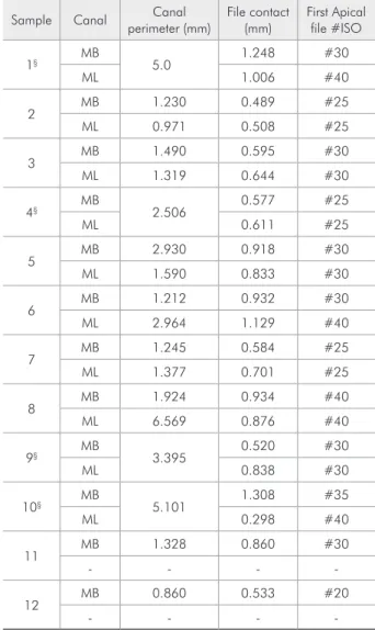
Table 1 - Perimeter of the canal at the working length re-gion (mm) and contact of the first apical file to the canal walls (mm), of each sample.
Sample Canal Canal perimeter (mm)
File contact (mm)
First Apical file #ISO
1§ MB 5.0 1.248 #30
ML 1.006 #40
2 MB 1.230 0.489 #25
ML 0.971 0.508 #25
3 MB 1.490 0.595 #30
ML 1.319 0.644 #30
4§ MB 2.506 0.577 #25
ML 0.611 #25
5 MB 2.930 0.918 #30
ML 1.590 0.833 #30
6 MB 1.212 0.932 #30
ML 2.964 1.129 #40
7 MB 1.245 0.584 #25
ML 1.377 0.701 #25
8 MB 1.924 0.934 #40
ML 6.569 0.876 #40
9§ MB 3.395 0.520 #30
ML 0.838 #30
10§ MB 5.101 1.308 #35
ML 0.298 #40
11 MB 1.328 0.860 #30
- - -
-12 MB 0.860 0.533 #20
- - -
-§Fused canals; - Absent canal; MB=mesiobuccal; ML=mesiolingual.
Table 2 - Contact (%) of the first file to the canal walls at the apex (MB and ML canals).
MB canal ML/MB canal ML canal
Individual foramen
47.83%
foramen fused
40.03%*
Individual foramen
31.71%
* In cases of fused canals/foramens two files were used to determine the percentage of contact. The sum of the result (%) for each file was considered the total contact in such cases.
0
12.2
5.8
16.00
6.41
milimete
r
(mm)
Mesiobuccal Mesiolingual MB/ML Fused 10
12 16 18
14
4
2 6 8
Root canal perimeter File contact
14.79
4.69
may be improved in cases where early prelaring has been performed.14,17,18
Silveira et al.8 (2008) veriied that the removal
of anatomic interferences along the root canal made the estimate of the irst ile that binds at the apical region more precise. The diameter of the irst apical ile used following this procedure presented an in-crease of two diameters of instruments, when com-pared to samples without interference removal.
Tactile sensitivity determines the number of in-struments necessary for the enlargement of the api-cal area. Therefore, the determination of the apiapi-cal diameter, which according to current endodontic concepts has the same biological importance as the working length, is dependent on a procedure that is not objective and does not present scientiic evidence to orient its clinical use. The present data reinforce that the clinical determination of the irst apical ile by tactile sensitivity, performed after the removal of interferences along the canal, promotes a contact area considered satisfactory for the objectives of endodon-tic therapy. The dificulties in adapting the iles at the apical area are predominantly related to the anatomi-cal form and are particularly related to the fusion of the mesial canals of the lower molar where the neigh-boring canal constitutes one of the canal walls.
The current indings indicate that the changes in root canal geometry after preparation are more de-pendent on the type of canal than on the technique or instruments used to shape the canals.19 If the apical
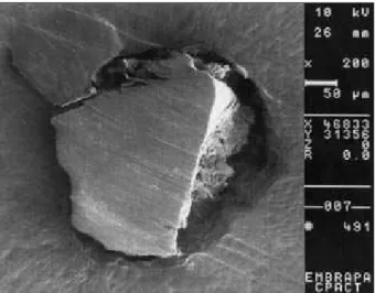
region is round, the irst ile that binds in the work-ing length will most likely wear the entire canal pe-rimeter with three more ile diameters. When this re-gion is oval, however, the ile will not simultaneously touch the whole diameter (Figures 1-3).
Consequent-Figure 1 - Example of file adaptation on the dentin walls at 1 mm from the anatomic apex (original magnification 200 X).
Figure 2 - Sample showing the incomplete contact of the apical file with the dentin walls of the anatomic apex (origi-nal magnification 200 X).
ly, the objective of removing the infected dentin layer and preventing the development or persistence of api-cal periodontitis might not be accomplished.20,21
The removal of interference from the root canal, as in this study, resulted in a level of contact with the walls of the canal in the apical third insuficient for the goals of endodontics. However, contact was achieved at around 40% of the apical areas. It is noteworthy that the apical anatomy interferes in a signiicant way with the goal of achieving contact with all walls.
New techniques and technologies should be de-veloped with the objective of predicting in a more accurate manner the apical diameter for shaping. Such developments are fundamental for cleaning, disinfecting and illing the root canal and, conse-quently, for success in endodontics.
Conclusion
The technique employed in the present study did not allow the irst apical ile to touch all dentin walls of the apical region.
References
1. Grande NM, Plotino G, Pecci R, Bedini R, Pameijer CH, Somma F. Micro-computerized tomographic analysis of ra-dicular and canal morphology of premolars with long oval canals. Oral Surg Oral Med Oral Pathol Oral Radiol Endod. 2008 Sep;106(3):e70-6.
2. Wu MK, R’oris A, Barkis D, Wesselink PR. Prevalence and extent of long oval canals in the apical third. Oral Surg Oral Med Oral Pathol Oral Radiol Endod. 2000 Jun;89(6):739-43.
3. Wu MK, Wesselink PR. A primary observation on the prep-aration and obturation of oval canals. Int Endod J. 2001 Mar;34(2):137-41.
4. Martos J, Ferrer-Luque CM, González-Rodriguez MP, Castro LAS. Topographical evaluation of the major apical foramen in permanent human teeth. Int Endod J. 2009 Apr;42(4):329-34. 5. Kerekes K, Tronstad L. Morphometric observations on the root
canals of human molars. J Endod. 1977 Mar;3(3):114-8. 6. Weine FS. Endodontic Therapy. St. Louis: Mosby; 1989.
In-tracanal treatment procedures, basic and advanced topics. p.256-369.
7. Baugh D, Wallace J. The role of apical instrumentation in root canal treatment: a review of the literature. J Endod. 2005 May;31(5):333-40.
8. Silveira LF, Martos J, Pintado LS, Teixeira RA, César Neto JB. Early flaring and crown-down shaping influences the first file bind to the canal apical third. Oral Surg Oral Med Oral Pathol Oral Radiol Endod. 2008 Aug;106(2):e99-101. 9. Siqueira JF Jr, Rôças IN, Santos SR, Lima KC, Magalhães
FA, de Uzeda M. Efficacy of instrumentation techniques and irrigation regimens in reducing the bacterial population within root canals. J Endod. 2002 Mar; 28(3):181-4.
10. Card SJ, Sigurdsson A, Orstavik D, Trope M. The effective-ness of increased apical enlargement in reducing intracanal bacteria. J Endod. 2002 Nov;28(11):779-83.
11. Mickel A, Chogle S, Liddle J, Huffaker K, Jones JJ. The role of apical size determination and enlargement in the reduction of intracanal bacteria. J Endod. 2007 Jan;33(1):21-3.
12. Vianna ME, Horz HP, Gomes BP, Conrads G. In vivo evalua-tion of microbial reducevalua-tion after chemo-mechanical prepara-tion of human root canals containing necrotic pulp tissue. Int Endod J. 2006 Jun; 39(6):484-92.
13. Weiger R, Bartha T, Kalwitzki M, Löst C. A clinical meth-od to determine the optimal apical preparation size. Part I. Oral Surg Oral Med Oral Pathol Oral Radiol Endod. 2006 Nov;102(5):686-91.
14. Contreras MAL, Zinman EH, Kaplan SK. Comparison of the first file that fits at the apex, before and after early flaring. J Endod. 2001 Feb;27(2):113-6.
15. Grossman LI, Oliet S, Del Río CE. Endodontic Practice. 11th
ed. Philadelphia: Lea & Febiger; 1988. Preparation of the root canal: equipment and technique for cleaning, shaping and irrigation. p.179-227.
16. Walton RE, Rivera EM. Cleaning and shaping. In: Walton RE, editor. Principles and Practice of Endodontics. Philadelphia: Saunders; 1996. p.201-233.
17. Seidberg BH, Alibrandi BV, Fine H, Logne B. Clinical inves-tigation of measuring working lengths of root canals with an electronic device and with digital tactile sense. J Am Dent Assoc. 1975 Feb;90(2):379-87.
18. Stabholz A, Rotstein I, Torabinejad M. Effect of preflaring on tactile detection of the apical constriction. J Endod. 1995 Feb;21(2):92-4.
19. Peters OA, Laib A, Göhring TN Barbakow F. Changes in root canal geometry after preparation assessed by high-resolution computed tomography. J Endod. 2001 Jan;27(1):1-6. 20. Iqbal MK, Ku J. Instrumentation and obturation of the
api-cal third of root canals: addressing the forgotten dimension. Compend Contin Educ Dent. 2007 Jun;28(6):314-20. 21. Oliveira LD, Carvalho CAT, Valera MC, Koga-Ito CY, Jorge