Ali Nazemi grew up in Miayneh, Iran. He obtained his BSc in Chemistry from K. N. Toosi University of Technology, Tehran, Iran in 2005. He then moved to Canada and completed his MSc in the area of Inorganic Chemistry at the University of Toronto under the supervision of Dr. Datong Song in 2009. He then decided to continue his graduate studies in the area of polymer and macromolecular c h e m i s t r y. H e i s c u r r e n t l y a P h D candidate at The University of Western Ontario under the supervision of Dr. Elizabeth R. Gillies. His research focuses on surface functionalization of polymersomes with dendrons for a wide range of biomedical applications and development of photodegradable dendrimers and dendrimer-based assemblies.
Elizabeth Gillies was raised in Ontario, Canada and obtained her BSc degree in Chemistry from Queen’s University, Kingston, Canada in 2000. She then moved to the University of California, Berkeley where she completed her PhD degree in 2004 under the supervision of Jean Fréchet. Her research involved the development of dendrimer-based anti-cancer drug delivery vehicles as well as pH-sensitive linkages for drug delivery applications. She then moved to Bordeaux, France where she performed postdoctoral work in the group of Ivan Huc, working on water-soluble helical aromatic oligoamide foldamers for biological applications. In 2006, she began as an Assistant Professor at the University of Western Ontario in London, Canada and was promoted to Associate Professor in 2006. Her research group works on the development of biodegradable polymers, polymer assemblies, and dendrimers for tissue engineering and drug delivery applications.
*Correspondence: E. R. Gillies. Department of Chemistry, The University of Western Ontario. 1151 Richmond St., Lon-don, Canada N6A 5B7. E-mail: [email protected]
Department of Chemistry, The University of Western Ontario, London, Canada, 2Department of Chemical and Biochemical Engineering, The
University of Western Ontario, London, Canada
A wide variety of nanomaterials have demonstrated promise in medical applications such as drug delivery and imaging. In these applications, the surface chemistry of the materials is critical as it plays an important role in determining the toxicity and biodistribution behavior of the material. We review here the functionalization of nanomaterials with
dendrons as an eficient method to alter the surface chemistry of the materials, introducing
new properties and functions. Described here is the functionalization of superparamagnetic iron oxide nanoparticles (SPIO) with dendritic guanidines to enhance their transport into cells for magnetic resonance imaging applications. The introduction of dendrons bearing peripheral hydroxyls, amines, guanidines, carbohydrates and Gd(III) chelates to polymer vesicles (polymersomes) is also described. These dendritic moieties allow for modulation
of toxicity, cell uptake, protein binding, and contrast agent eficiency, while at the same
time allowing the stabilities of the polymersomes to be maintained. Thus, this approach holds promise for the development of a wide range of multifunctional materials for pharmaceutical applications.
Uniterms: Dendrimer. Superparamagnetic Iron Oxide Nanoparticles. Polymersome. Pharmaceutical applications.
Uma grande variedade de nanomateriais tem demonstrado aplicações médicas promissoras, tais como liberação de fármacos e em imagens. Nestas aplicações, a superfície química dos materiais é crítica, uma vez que exerce papel importante na determinação da toxicidade e comportamento de biodistribuição do material. Aqui, nós revisamos a funcionalização de nanomateriais, como dendrons, como método eiciente de alterar a superfície química destes compostos, introduzindo novas propriedades e funções. Descritos aqui estão nanopartículas superparamagnéticas de óxido de ferro (do inglês, SPIO), com guanidinas dendríticas para aumentar seu transporte para o interior das células, úteis em imagens de ressonância magnética. A introdução de dendrons contendo hidroxilas, aminas, guanidinas, carboidratos e quelatos de Gd(III) periféricos em vesículas poliméricas (polymersomes) também está descrita. Esses grupos dendríticos permitem a modulação de toxicidade,
captura celular, ligação à proteína e eiciência como agente de contraste, enquanto que, ao
mesmo tempo, permitem a manutenção da estabilidade das vesículas poliméricas. Assim, essa abordagem é promissora para o desenvolvimento de grande variedade de materiais multifuncionais para aplicações farmacêuticas.
INTRODUCTION
In recent years, a diverse array of nanosized materi-als has been investigated for biomedical applications. For example, polymer assemblies such as micelles and vesicles have been widely investigated as carriers that can encapsu-late drugs and release them in a controlled manner, while at the same time prolonging their circulation time and protecting them from premature degradation (Choudhury, He, 2012; Ohya et al., 2012; Tian et al., 2012). Various nanogels, microparticles and nanoparticles composed of biodegradable polymers have also been investigated with similar goals (Balmert, Little, 2012; Balmert, Little, 2012; Beck-Broichsitter et al., 2012; Patel et al., 2012). In medical imaging applications, superparamagnetic iron oxide nanoparticles (SPIO) have been demonstrated to provide contrast in a variety of applications owing to their
ability to perturb local magnetic ields, leading to regions
of hypointensity, or signal voids in images (Corot et al., 2006; Laurent et al., 2008; Villaraza et al., 2010). Semi-conductor nanocrystals, termed quantum dots, have been shown to provide enhanced optical properties relative to
conventional luorophores, leading to their use in vivo in optical imaging and cell tracking, high resolution cellular imaging, and the study of various cellular processes at the molecular level (Cinteza, 2010). Gold nanoparticles of various shapes and sizes are one of the most widely used classes of bionanomaterials with applications in imaging, diagnostics, photothermal therapy, drug and gene delivery and other areas (Dykman, Khlebtsov, 2012).
While the bulk composition of a bionanomaterial is important for its function and long-term biocompatibility, the functionalities present at its surface are also critical. It
is the surface of a material that will irst comes into contact
with the biological system and as such will play a major role in its toxicity and biodistribution behavior (Yang et al., 2010). The surface can also provide sites for the in-troduction of drugs and moieties to target the material to
speciic sites in vivo (Pang et al., 2010; Pittet et al., 2006). Furthermore, the high levels of multivalency available at the surface of a nanomaterial can provide therapeutic properties by inhibiting undesirable multivalent interac-tions between host cells and pathogens including bacteria and viruses using ligands such as carbohydrates (Mammen
et al., 1998; Rai et al., 2006).
Dendrimers are a class of macromolecules at the interface of polymer and synthetic organic chemistry. In contrast to hyperbranched polymers, which are prepared from branched (multifunctional) monomer units in a single polymerization reaction, resulting in incomplete branching and polydisperse materials, dendrimers are synthesized
from branched monomer units in a stepwise manner, lead-ing to monodisperse or very low polydispersity products (Tomalia et al., 1985; Frechet, Tomalia, 2001; Newkome
et al., 2001). In addition, it is possible to achieve precise control over their molecular shape, dimensions, density,
polarity, lexibility and solubility by choosing different
building/branching units and surface functional groups (Tomalia et al., 1990; Caminade et al., 2005). Because of their monodispersities, high loading capacities, large-scale production, and bioconjugation capabilities, dendritic structures have been recognized to be ideal building blocks for biomedical applications (Gillies, Fréchet, 2005; Medi-na, El-Sayed, 2009; Rolland et al., 2009; Jain et al., 2010). Generally, dendrimers are globe- or ellipsoid-shaped, and they consist of three distinct regions: a central core, repeated branches, and surface functional groups (Aulenta et al., 2003). The central core should be a molecule with at least two reactive functional groups. The repeated branches are organized in a series of radi-ally concentric layers called “generations’’ (Caminade
et al., 2005). When iterative growth is conducted from
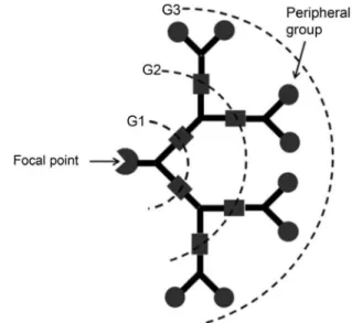
focal point or core molecule containing one reactive group, wedge-shaped structures, commonly referred to as “dendrons” result (Figure 1). In addition to the interest in dendrimers and dendrons for biomedical applications, our group has also been interested in the use of dendrons for
the surface modiication of bionanomaterials. It was antici -pated that their high multivalency would be advantageous in terms of displaying ligands or other functionalities at the surfaces of materials, while at the same time their bulky
FIGURE 1 - Schematic representation of a dendron showing the focal point, generations (G1, G2, and G3 represent the
irst, second and third generations respectively), and peripheral
processes, diagnosis of disease and the study of disease progression. MRI can be performed without contrast agents, but in many cases, the use of contrast agents can provide increased signal contrast between normal and diseased tissues, or aid in the understanding of biological phenomena. In addition to low molecular weight com-plexes based on Gd(III), (Caravan et al., 1999; Merbach, Toth, 2001; Terreno et al., 2010) which are commonly used clinically, the development of new agents based on
SPIO has been of signiicant interest over the last couple
of decades (Corot et al., 2006; Laurent et al., 2008; Vil-laraza et al., 2010). These nanoparticles induce local
inho-mogeneities in the magnetic ield, resulting in regions of
hypointensity in T2-weighted MR images. There are two primary means of administering SPIO. In one approach, SPIO is injected intravenously and arrives at the target site passively or through the conjugation of active targeting groups to the nanoparticle surface (Tsourkas et al., 2005; Lee et al., 2007). In another approach, in particular for the study of biological processes such as cancer metastasis and stem cell transplantation, cells are labeled with SPIO, implanted in vivo and then tracked by MRI (Bulte et al., 2001; Pittet et al., 2006; Foster et al., 2008).
It has been found that even single SPIO labeled cells can be detected in vivo by MRI, demonstrating the im-mense promise of cell tracking by MRI (Heyn et al., 2006). However, the ability to detect these labeled cells by MRI depends on the quantity of SPIO in the cells. While phago-cytic cells such as macrophages can be readily labeled with
SPIO, this labeling is more dificult for non-phagocytic
cells such as cancer cells. For biomedical applications, SPIO is often coated with water-soluble polymers such as dextran (Weissleder et al., 1995) or poly (ethylene ox-ide) (PEO), (Li et al., 2005) which impart solubility and biocompatibility, but do not enhance the cellular uptake of SPIO. Various transfection agents have been used to enhance the uptake of SPIO, but the complexes formed with these agents are often polydisperse and challenging to control (Frank et al., 2003). Alternatively, cell-penetrating
of chromophores into cells in a manner similar to HIV-Tat and protamine, which contain high levels of the amino acid arginine. Thus, the concept of dendritic cell-penetrating agents seemed promising, but these agents had not previ-ously been demonstrated to deliver large payloads such as nanoparticles into cells. A polyester dendron based on 2,2-bis(hydroxymethyl)propionic acid was selected as the dendritic scaffold due to its ease of synthesis, biode-gradability and biocompatibility (Gillies et al., 2005). A dendron having a focal point alkyne and eight peripheral guanidines was prepared by divergent growth of the 3rd
generation dendron 1 from propargyl alcohol (Wu et al., 2005), installation of peripheral amines using a protected b-alanine anhydride, deprotection to the amine terminated dendron 2 (Li et al., 2007), coupling of a protected
guani-dine derivative, and inally deprotection to provide den
-dron 3 (Figure 2) (Martin et al., 2008). Using a luorescein derivative of 3, it was demonstrated by low cytometry
studies with GL261 mouse glioma cells that the eficacy of the luorescein labelled dendritic guanidine transporter
was comparable to that of a fluorescein labelled
HIV-Tat47-57 oligopeptide (FITC-LC-YGRKKRRQRRR-NH2)
(Martin et al., 2008).
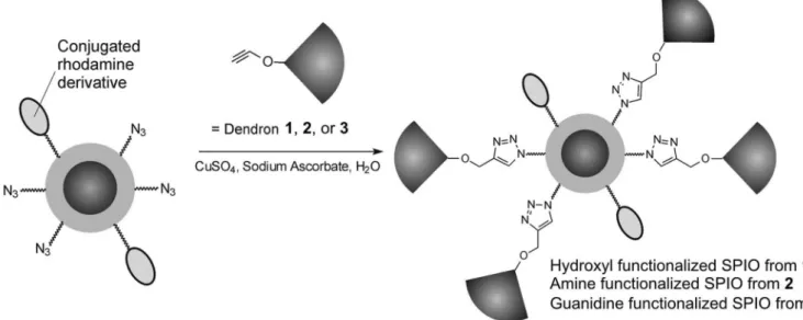
SPIO coated with dextran and functionalized with azides, as well as a rhodamine dye was prepared with the aim of performing the conjugation reaction between the dendrons and the SPIO by highly chemoselective and functional group tolerant Cu(I) catalyzed “click” dition reactions (Lutz, Zarafshani, 2008). These cycload-dition reactions have been found to be very high yielding, even in water and in the presence of significant steric hindrance, and were therefore ideal for the conjugation of macromolecules such as dendrons to nanoparticles (Helms
click reaction proceeded to completion with complete dis-appearance of the peak at 2100 cm-1 corresponding to the
azide group. Dynamic light scattering (DLS) showed that the dendron-functionalized nanoparticles remained similar in size to the initial SPIO, with only a small increase in size and very minimal aggregation. In addition, transmis-sion electron microscopy (TEM) was used to verify that the nanoparticle core was not affected. It was necessary to remove excess copper salts from the reaction mixture by dialysis against ethylenediaminetetraacetic acid(EDTA), followed by pure water.
The cell uptake of the SPIO into GL261 cells was
investigated by low cytometry based on the luorescence
of the rhodamine dye that was conjugated at the same density of each set of nanoparticles (Martin et al., 2008). After time points of 30 min and 2 h (Figure 4a), only the cells incubated with the dendritic guanidine functionalized
particles exhibited signiicantly increased luorescence. These results were conirmed by confocal microscopy,
which demonstrated that the SPIO was indeed taken up by cells and not simply adsorbed on the cell surface. It was also demonstrated that this enhanced uptake led to an enhanced MRI response (Figure 4b). GL261 cells were labeled with the dendritic guanidine functionalized SPIO, and then washed and pelleted. T2 measurements of unlabeled and labeled cell pellets demonstrated a 10-fold
reduction in T2 value for the labeled cells. This signii -cant difference in T2 values illustrates the utility of these nanoparticles for labeling cells for detection by MRI. The toxicities of the various nanoparticles were also investi-gated in GL261 cells using an MTT assay (Figure 4c). (Freshney, 2000) Consistent with previous experiments demonstrating the high biocompatibilities of dendrimers based on 2,2-bis(hydroxymethyl)propionic acid (Gillies et al., 2005), the dendritic hydroxyl and the dendritic amine functionalized SPIO had a very similar toxicity to the dextran coated particles that are widely used for cell label-ing applications. The dendritic guanidine functionalized particles exhibited somewhat increased toxicity at higher concentrations, but they remained relatively non-toxic at the low concentrations used for cell labeling. It is possible that their toxicity at higher concentrations arises from their ability to penetrate and create defects in cell membranes, as has been observed for various polycationic nanopar-ticles and transporters (Leroueil et al., 2008).
Overall, this study demonstrated several key ind
-ings. First, it represented the irst successful example of the
transport of a biologically relevant nanoparticle cargo by a dendritic guanidine transporter. The resulting system can provide improved labeling of cells with SPIO for enhanced sensitivity in cellular MRI. In addition, it demonstrated FIGURE 2 - Synthesis of hydroxyl 1, amine 2, and guanidine 3
that dendrons are an effective means of presenting biologi-cally relevant functionality on a nanoparticle surface and a synthetic means for expanding this dendritic functional-ization approach to a diverse array of azide functionalized nanomaterials.
DENDRITIC FUNCTIONALIZATION OF
POLYMERSOMES
Over the past few decades, amphiphilic copolymers have been demonstrated to assemble into a vast array of morphologies ranging from spherical micelles (Alexan-dridis, Lindman, 2000) to cylindrical micelles (Cai et al., 2007), helical rods (Cornelissen et al., 1998), toroids
(Pochan et al., 2004), vesicles (Discher, Eisenberg, 2002; Zhang, Eisenberg, 1995), tubes (Yan et al., 2004), and multicompartment cylinders (Cui et al., 2007). Among these, polymer vesicles in particular, commonly referred to as polymersomes have received increasing attention. Interest in polymersomes can likely be attributed in part to their structural similarity to phospholipid vesicles (li-posomes), which are important components of biological systems where they serve as cell membranes, and they have also been widely investigated in areas such as drug and gene delivery (Torchilin, Weissig, 2003). Like phos-pholipid vesicles, polymersomes possess a hydrophobic membrane as well as an aqueous core, allowing for the incorporation of both hydrophilic and hydrophobic mol-FIGURE 3 - “Click” conjugation of dendrons 1, 2, and 3 to azide functionalized SPIO to provide dendritic hydroxyl, amine, and guanidine functionalized SPIO respectively (Martin et al., 2008).
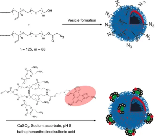
FIGURE 5 - Schematic showing the functionalization of polymersomes bearing peripheral azide groups with dendrons having focal point alkynes.
ecules, and thus potentially multifunctional properties. In comparison with liposomes, polymersomes have been shown to exhibit properties such as increased strength and decreased permeability, which may be advantageous in their application as synthetic biomaterials (Discher et al., 1999; Discher, Eisenberg, 2002). Indeed, over the past decade there have been many reports emerging on their use as carriers of proteins (Ranquin et al., 2005; Liu et al., 2010), hydrophilic drugs (Ahmed et al., 2006; Yang et al., 2010), and imaging agents (Ghoroghchian et al., 2005).
As described above, the surface chemistry of polymersomes is critical in terms of controlling their interactions with biological systems. The introduction of dendritic groups via “click” [3+2] cyloaddition reactions of azide functionalized polymersomes with dendrons hav-ing focal point alkynes provides the opportunity to alter the surface chemistry in a single step without changing the block copolymers comprising the polymersome mem-branes. This provides a unique opportunity to impart new biological properties and functions.
In our initial work, as shown in Figure 5, poly-mersomes composed of the amphiphilic linear diblock copolymer poly(butadiene-b-ethylene oxide) (PBD-PEO)
were used (Li et al., 2007). An azide was introduced to the polymer terminus and polymersomes were prepared con-taining varying ratios of the azide and hydroxyl terminated polymers. Dendron 2 having an alkyne focal point and peripheral amine groups was used and ~1 rhodamine dye per dendron was introduced to the dendron’s periphery in order to track and quantify the conjugation reaction. This dendron was reacted with the polymersomes under stan-dard click conditions involving CuSO4, sodium ascorbate,
and the ligand bathophenanthrolinedisulfonic acid. It was found that the use of this ligand prevented the adsorption of copper ions to the dendritic amines. The conjugation
yields for the various polymersomes were quantiied based
on the UV-visible absorbance of the rhodamine dye on the dendrons.
surface during the 24 h reaction. At higher azide content, the conjugation yields dropped off dramatically. This was attributed to steric hindrance at the polymersome surface due to the bulky nature of the dendrons. As these poly-mersomes were micron-sized, they were also observed by
luorescence confocal microscopy. That the polymersomes were luorescent was further conirmation of the successful
conjugation of the rhodamine-labeled dendrons (Figure 6b). In addition, well-dispersed polymersomes were ob-served at low azide content. On the other hand, at higher
azide content, signiicant aggregation was observed which
may be attributed to either a disruption of the hydrophilic/ hydrophobic balance within the polymersome membrane upon dendron conjugation, or to interactions between the dendrons on different polymersomes. However, at azide content £ 20% this method was highly promising for the surface functionalization of polymersomes.
In a subsequent study, the surface functionaliza-tion of PBD-PEO polymersomes with dendritic versus non-dendritic ligands was studied (Martin et al., 2009). Mannose was selected as the ligand as its multivalent binding to targets such as Concanavalin A (Con A) has been extensively investigated and a number of assays have been developed to evaluate this binding (Bittiger, 1976; Roy, 1996; Kanai et al., 1997; Gestwicki et al., 2002). Mannose functionalized dendron 4 (Figure 7), and a small molecule mannose derivative with an azide functionality were prepared (Martin et al., 2009). A dye-labeled ver-sion of dendron 4 was used to quantify the conjugation to polymersomes and similar results to those described above for dendron 2 were obtained. As shown in Figure 7a dendritic mannose polymersomes were prepared by the “click” conjugation of dendron 4 to polymersomes
con-taining 5% azide functionalized PBD-PEO. Non-dendritic
mannose polymersomes were prepared by irst conjugating
the azide functionalized mannose to an alkyne-terminated PBD-PEO, and then assembly of polymersomes from a 50:50 mixture of mannose and hydroxyl-terminated polymers (Figure 7b). These quantities were selected in order to provide the same overall mannose content in the dendritic and non-dendritic polymersomes, but displayed in a different manner.
The dendritic and non-dendritic polymersomes were compared using a hemagglutination assay. In the presence of the protein Con A, red blood cells cluster due to interactions of cell surface carbohydrates with the
protein. When mannose is present at a suficiently high
concentration, Con A binds preferentially to mannose and the clustering of the blood cells is inhibited. Comparisons of the minimum mannose concentration required to inhibit
the agglutination can provide relative binding afinities
of mannose-based ligands, which was the main point of interest in this study. The known small molecule ligand
α-D-methyl mannopyranoside was used as a point of
reference in this study. Despite their multivalency, the non-dendritic polymersomes provided only a very mod-est 3.7-fold enhancement in affinity on a per mannose basis. In contrast, the dendritic mannose functionalized polymersomes provided a much greater 42-fold increase
in relative binding afinity.
This enhancement relative to the non-dendritic system was attributed to several factors including the relatively rigid display of ligands on the dendritic scaf-fold resulting in an entropic advantage (Mammen et al., 1998), the ability of the dendritic scaffold to overcome steric inhibition of binding by the PEO layer (Lin et al., FIGURE 6 - a) Yield for the “click” conjugation between the dendron and polymersomes as a function of polymersome PBD-PEO-N3
2004), and an enhanced “proximity” effect resulting from the clustered display of ligands on the dendron (Lundquist, Toone, 2002). Through the preparation of the analogous dendritic and non-dendritic systems on dextran coated SPIO, it was demonstrated that these enhancements were generalizable to other nanoparticles and other polymer coatings in addition to PEO (Martin et al., 2009). Thus, this study revealed that it is important to consider carefully not only the choice of biological ligand, but also the mode in which it is conjugated to the surface in order to exploit
the beneits of nanomaterials. Furthermore, it showed that
dendritic scaffolds are an effective means of displaying biological ligands on surfaces.
As described above, the dendritic surface function-alization approach provides a means of easily altering the polymersome surface chemistry in a single step. This provided the opportunity to explore the effect of the den-dritic functionalization with different surface moieties on the physical and biological properties of polymersomes
(Amos et al., 2012). In this case, in contrast to the microm-eter-sized polymersomes described above, the PBD-PEO polymersomes were extruded through a 100 nm membrane to produce polymersomes with diameters of 100-200 nm that would be more suitable for biological applications. The azide and alkyne [3+2] cycloaddition was used to conjugate dendrons 1, 2, and 3 to polymersomes composed of 20% azide termined PBD-PEO (Figure 8). Because of the aggregation that had been previously observed upon conjugation of dendrons to vesicles with higher azide content (Li et al., 2007), we were interested in more subtle effects of the dendritic surface functionalization on the polymersome stability at lower azide content. This was
probed irst by measuring the release rate of an encapsu
FIGURE 8 - “Click” conjugation of dendrons 1, 2, and 3 to azide functionalized PBD-PEO polymersomes to provide dendritic hydroxyl, amine, and guanidine functionalized polymersomes respectively (Amos et al., 2012).
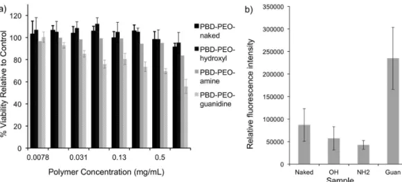
FIGURE 9 - a) Toxicity of naked and dendron functionalized PBD-PEO polymersomes as measured by an MTT assay following 48 hour of incubation with HeLa cells; b) Quantitative comparison of the uptake of naked and dendritic hydroxyl, amine, and guanidine
functionalized polymersomes in HeLa cells following a 1 h incubation at 37 °C, as measured by luorescence confocal microscopy.
ity. No signiicant differences in the release rates of the
dye from naked or the different dendritic functionalized polymersomes were observed, and in each case complete release was observed over a period of 10-15 h. The release of a rhodamine-labeled bovine serum albumin was also investigated. Because of its large size and high polarity/ charge, this protein was not expected to diffuse across the intact polymersome membrane and thus disruption of the membrane would be required for release. Again it
was found that there were no signiicant differences in the
release rates among the naked or different dendritic poly-mersomes, and as expected the release was much slower for the protein, requiring 1-2 weeks to reach completion. Overall, these results demonstrated that the dendritic functionalization approach did not impart any inherent instability to the polymersomes.
The effects of the dendritic groups on the toxicities and cell uptakes of the polymersomes were also
investi-gated (Amos et al., 2012). In an MTT assay, as shown in Figure 9a, it was found that up to 1 mg/mL of polymer,
the highest concentration evaluated, no signiicant toxic
effects were observed for any of the samples except the dendritic guanidine functionalized polymersomes. This was not surprising in light of the previous results with the dendritic guanidine functionalized SPIO and can likely be attributed to the same causes (Martin et al., 2008). How-ever, it was possible to administer these polymersomes
without signiicant toxicity at lower concentrations. The
cell uptake of the polymersomes was of particular interest due to the possibility of using polymersomes to encapsu-late and deliver molecules such as DNA/RNA (Lomas et al., 2007; Christian et al., 2009; Cheng et al., 2011) and proteins, (Demirgoz et al., 2009; Cheng et al., 2011) which cannot normally diffuse through cell membranes. In this case, the uptake was studied using rhodamine-labeled
microscopy (Amos et al., 2012). It was found that the den-dritic guanidine functionalized polymersomes exhibited a
statistically signiicant increase in uptake relative to all of
the other polymersome samples, showing that the dendritic guanidine is a versatile tool for the delivery of nanomateri-als into cells (Figure 9b). The mechanism of cell uptake was investigated through temperature-dependent studies and by examining the colocalization of vesicles with Lyso-Tracker ™ Blue. These studies suggested that endocytosis pathways likely play a major role in the internalization of these systems, though direct translocation may also play a small role.
While the above work demonstrated several impor-tant properties and functions that can be imparted through dendritic surface functionalization of polymersomes, a limitation was the use of PBD-PEO. This copolymer is not biodegradable and while PEO is well established for bio-medical applications, the PBD block presented concerns with respect to the long term behavior in vivo. To address this limitation, a system based on polycaprolactone (PCL)-PEO block copolymers was developed. PCL is a well known biodegradable polymer that is currently approved for uses in tissue engineering (Jenkins et al., 2006; Lam
et al., 2007) and drug delivery (Chandra, Rustgi, 1998;
Sinha et al., 2004). A synthetic method was developed for the preparation of azide terminated (5) and hydroxyl terminated (6) PCL-PEO and these block copolymers
were assembled into polymersomes by a nanoprecipita-tion method involving the dissolunanoprecipita-tion of the copolymer in tetrahydrofuran (THF), followed by a gradual addition of
water, and inally dialysis against water to remove the THF
(Figure 10a) (Nazemi et al., 2011). This led reproducibly to polymersomes with diameters of ~140 nm. It was pos-sible to extrude these polymersomes to reduce their sizes to ~65 nm in diameter, an ideal size for in vivo circulation. By reducing the PCL content in the block copolymers, it was also possible to prepare micelles with diameters of ~ 25 nm using a similar protocol (Figure 10b) (Nazemi et al., 2011).
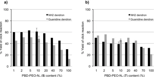
The click functionalizations of the polymersomes and micelles with dendrons 2 and 3 were studied as de-scribed above for the PBD-PEO polymersomes (Nazemi
et al., 2011). With rhodamine-labelled version of dendron
2, which had previous been studied quantitatively with PBD-PEO polymersomes, similar conjugation yields to the PCL-PEO polymersomes were obtained, as measured by UV-visible spectroscopy (Figure 11a). The yields were somewhat lower for rhodamine-labelled version of dendron 3, which was expected based on its larger size. For the micelles, it was expected that at low azide load-ing the conjugation yield should approach 100% as all of the azides should be on the micelle surface. However, as shown in Figure 11b, this was not the case and the yields were similar to those obtained for the polymersomes. The
FIGURE 11 - Yields for the “click” conjugation between the rhodamine-labelled versions of dendrons 2 or 3 and PCL-PEO a) vesicles, and b) micelles as a function of PCL-PEO-N3 content (remainder is PCL-PEO-OMe).
reasons for these lower yields are still unclear at this time, but can perhaps be related to the large size of the dendrons relative to the micelles.
TEM was used to conirm that both the micelles
and polymersomes remained intact following the dendron conjugation. However, upon removal of the
excess dendron by dialysis, signiicant aggregation was
observed for the polymersomes, even at relatively low azide content, unlike for the PBD-PEO polymersomes. Thus, the PEO-PCL polymersomes appear to be more sensitive to aggregation. The reasons for the differences between the two different polymersomes is not clear, but aggregation is believed to result from the cationic charges on dendrons 2 and 3 as studies with hydroxyl terminated dendrons, even of higher generation did not result in aggregation. The removal of the excess dendron by dialysis at a variety of pHs and ionic strengths was investigated with the aim of preventing aggregation was investigated, but unfortunately the aggregation could not be prevented. The micelle systems proved much less sus-ceptible to aggregation, a result likely attributable to their different morphologies. Thus, this work demonstrated that the dendritic functionalization approach could be extended to biodegradable PCL-PEO polymersomes and also to micelles formed from these polymers. Aggrega-tion was problematic for polymersomes funcAggrega-tionalized with dendrons bearing cationic peripheral groups, but not for those with neutral hydroxyl groups, and not for the micelles, even with cationic dendrons.
The above work lay the groundwork for the use
of PCL-PEO polymersomes in speciic biomedical ap
-plications. In a irst demonstration of their utility,
PCL-PEO polymersomes were used to prepare an enhanced Gd(III)-based MRI contrast agent (Nazemi et al., 2012).
As described above in the context of SPIO, MRI is an important noninvasive imaging modality. Most clinically used contrast agents for MRI are based on small molecule chelates of Gd(III) (Caravan et al., 1999; Merbach, Toth, 2001; Terreno et al., 2010). While the availability of these
agents has enabled signiicant developments in MRI, they do suffer from some signiicant limitations including their
low longitudinal relaxivities (r1), typically in the range
of 3 - 5 mM-1s-1, and their short circulation half lives
(Caravan, 2006). These limitations result in the require-ment for large doses and also limits their applicability in molecular imaging (Prince et al., 2009). To address these limitations, Gd(III) complexes have been conjugated to a wide variety of macromolecular scaffolds including den-drimers (Gang et al., 2010; Kobayashi, Brechbiel, 2005), linear polymers (Allen et al., 2006), proteins (Caravan et al., 2002), viral particles (Hooker et al., 2007), micelles (Accardo et al., 2004), liposomes (Schühle et al., 2010), and polymersomes (Cheng, Tsourkas, 2008; Grüll et al., 2010). This can result in improvements in r1 values due
to the slower tumbling rates of macromolecules and the resulting increases in the rotational correlation times of the Gd(III) (Caravan, 2006; Villaraza et al., 2010). In addi-tion, macromolecular systems can exhibit prolonged blood circulation times (Gillies et al., 2005). Polymersomes are a particularly promising scaffold for the development of enhanced MRI contrast agents due to their inherently multifunctional character as different functions such as multimodal imaging agents and drug molecules can be incorporated onto their surface, inside the membrane, or inside the aqueous core.
im-The relaxivities of agents 11, 12, and 15 were
mea-sured at various magnetic ield strengths using a ield cy -cling relaxometer (Nazemi et al., 2012). On a per Gd(III) basis, dendron 11 and polymersomes 12 and 15 exhibited
r1 values of 12.1 ± 0.3, 26.1 ± 1.2, and 10.6 ± 0.4 mM-1s-1,
respectively (20 MHz, 298 K). In comparison with the clinical agent Magnevist® (Gd(III)-DTPA) which has a
relaxivity of 4.6 mM-1s-1 under the same conditions, this
corresponds to 2.6-, 5.7-, and 2.3-fold increases in r1 for
dendron 11, and polymersomes 12 and 15 respectively. The result for the dendron was in the expected range based on results for previous dendritic Gd(III) chelates of similar dendrimer generation (Kobayashi, Brechbiel, 2005; Gang et al., 2010). The enhancement in relaxiv-ity relative to the small molecule was attributed to the crowded nature of the dendron periphery, which inhibits free rotation of the Gd(III) complexes, thus increasing the rotational correlation time. The enhancement for the mobilization on the polymersome surface should provide
further enhancements in relaxivity, while opening pos-sibilities for the multifunctional opportunities described above (Nazemi et al., 2012). To evaluate this hypothesis, three different contrast agents were prepared and studied. As shown in Figure 12a, an isothiocyanate derivative 9
of the clinically used diethylenetriaminepentaacetic acid (DTPA) chelate was reacted with dendron 2 to provide the dendritic chelate 10 in which Gd(III) was coordinated to provide the dendritic Gd(III) complex 11. Dendron 11 was coupled to PCL-PEO polymersomes having a diameter of 140 nm to provide the dendritic polymersome contrast agent 12. On the other hand, as shown in Figure 12b, 9 was irst reacted with propargyl amine to obtain the click -able DTPA derivative 13. This molecule was then used to chelate Gd(III) to provide compound 14 which was then coupled to polymersomes to provide the non-dendritic polymersome contrast agent 15.
Thus, this study demonstrated that different nanoscale components can be readily combined to provide additive effects on relaxivity, while at the same time providing opportunities to exploit the multifunctional potential of polymersomes.
CONCLUDING REMARKS AND FUTURE
DI-RECTIONS
A method has been developed for the conjugation of dendrons to the surfaces of nanomaterials. Thus far, the nanomaterials investigated have been dextran coated SPIO, PBD-PEO polymersomes and PCL-PEO polymer-somes and micelles, while the dendrons can have periph-eral hydroxys, amines, guanidines, carbohydrates or metal chelates. The only requirement is the presence of azide moieties on the surface of the nanomaterial and an alkyne at the focal point of the dendron, so it is a highly versatile strategy. In general, it has been found that the yields of the azide + alkyne [3+2] cycloaddition reactions are high when the densities of azides on the nanomaterial surface FIGURE 13 - Longitudinal relaxivity (r1) of dendron 11,
polymersome 12, and polymersome 15 in phosphate buffer (0.1
M, pH 7.4) as a function of ield strength at 298 K.
used to study the effects of surface chemistry on the properties of materials. In vitro studies have shown that release kinetics of small and large molecules, toxicity, and cell uptake are unaffected by changing the surface groups of polymersomes with the exception of dendritic guanidines, which imparted cell penetrating properties. Though these dendritic guanidines do impart moderate levels of toxicity at higher concentrations, they have proved to be a versatile approach for enhancing the delivery of SPIO, vesicles, and micelles into cells. This can provide important functions such as enhancing the sensitivity of MRI and improving the delivery of encap-sulated drug molecules into cells. Another important function that has been demonstrated is the enhancement in binding of dendritic ligands to protein targets, which has implications for both targeted delivery and
multiva-lent therapeutics. In addition, a signiicant enhancement
in the relaxivity of an MRI contrast agent was achieved using a dendritic vesicle system.
Future work will include the continued application of the dendritic functionalization approach to the devel-opment of new functional materials. For example, while the enhancement in protein binding of dendritic ligands was demonstrated with mannose, there are numerous other ligands such as carbohydrates and peptides that
can provide enhanced therapeutic and targeting eficacy
when presented in a multivalent manner. Furthermore, it will be important to exploit the multifunctionality of materials such as vesicles where in addition to the surface functionalization, therapeutics and/or imaging agents can be incorporated into the membrane or core.
ACKNOWLEDGEMENTS
REFERENCES
ACCARDO, A.; TESAURO, D.; ROSCIGNO, P.; GIANOLIO, E.; PADUANO, L.; D’ERRICO, G.; PEDONE, C.; MORELLI, G. Physicochemical properties of mixed micellar aggregates containing CCK peptides and Gd
complexes designed as tumor speciic contrast agents in
MRI. J. Am. Chem. Soc.,v.126, n.10, p.3097-3107, 2004.
AHMED, F.; PAKUNLU, R.I.; SRINIVAS, G.; BRANNAN, A.; BATES, F.; KLEIN, M.L.; MINKO, T.; DISCHER, D.E. Shrinkage of a rapidly growing tumor by drug-loaded polymersomes: pH-triggered release through copolymer degradation. Mol. Pharmaceutics,v.3, n.3, p.340-350, 2006.
ALEXANDRIDIS, P.; LINDMAN, B. Amphiphilic block copolymers: self-assembly and applications. Amsterdam, New York: Elsevier, 2000. 448 p.
ALLEN, M.J.; RAINES, R.T.; KIESSLING, L.L. Contrast agents for magnetic resonance imaging synthesized with ring-opening metathesis polymerization. J. Am. Chem. Soc., v.128, n.20, p.6534-6535, 2006.
AMOS, R.C.; NAZEMI, A.; BONDUELLE, C.V.; GILLIES, E.R. Tuning polymersome surfaces: functionalization with dendritic groups. Soft Matter.,v.8, n.21, p.5947-5958, 2012.
AULENTA, F.; HAYES, W.; RANNARD, S. Dendrimers: a new class of nanoscopic containers and delivery devices. Eur. Polym. J.,v.39, n.9, p.1741-1771, 2003.
BALMERT, S.C.; LITTLE, S.R. Biomimetic delivery with micro- and nanoparticles. Adv. Mater.,v.24, n.28, p.3757-3778, 2012.
BECK-BROICHSITTER, M.; MERKEL, O.M.; KISSEL, T. Controlled pulmonary drug and gene delivery using polymeric nano-carriers. J. Controlled Release,v.161, n.2, p.214-224, 2012.
BITTIGER, H.; SCHNEBLI, H.P. Concanavalin A as a tool. London, New York: John Wiley and Sons, Ltd., 1976. 639 p.
BULTE, J.W.M.; DOUGLAS, T.; WITWER, B.; ZHANG, S.C.; STRABLE, E.; LEWIS, B.K.; ZYWICKE, H.; MILLER, B.; VAN GELDEREN, P.; MOSKOWITZ, B.M.; DUNCAN, I.D.; FRANK, J.A. Magnetodendrimers allow endosomal magnetic labeling and in vivo tracking of stem cells. Nat. Biotechnol.,v.19, n.12, p.1141-1147, 2001.
CAI, S.; VIJAYAN, K.; CHENG, D.; LIMA, E.M.; DISCHER, D.E. Micelles of different morphologies - advantages of
work-like ilomicelles of PEO-PCL in paclitaxel delivery.
Pharm. Res.,v.24, n.11, p.2099-2109, 2007.
C A M I N A D E , A . ; L A U R E N T, R . ; M A J O R A L , J . Characterization of dendrimers. Adv. Drug Delivery Rev., v.57, n.15, p.2130-2146, 2005.
CARAVAN, P. Strategies for increasing the sensitivity of gadolinium based MRI contrast agents. Chem. Soc. Rev., v.35, n.6, p.512-523, 2006.
CARAVAN, P.; CLOUTIER, N.J.; GREENFIELD, M.T.; MCDERMID, S.A.; DUNHAM, S.U.; BULTE, J.W.M.; AMEDIO JR., J.C.; LOOBY, R.J.; SUPKOWSKI, R.M.; HORROCKS JR., W.D.; MCMURRY, T.J.; LAUFFER, R.B. The interaction of MS-325 with human serum albumin and its effect on proton relaxation rate. J. Am. Chem. Soc., v.124, n.12, p.3152-3162, 2002.
CARAVAN, P.; ELLISON, J.J.; MCMURRY, T.J.; LAUFFER, R.B. Gadolinium (III) chelates as MRI contrast agents: structure, dynamics, and applications. Chem. Rev.,v.99, n.9, p.2293-2352, 1999.
CHANDRA, R.; RUSTGI, R. Biodegradable polymers. Prog. Polym. Sci.,v.23, n.7, p.1273-1335, 1998.
CHENG, R.; FENG, F.; MENG, F.; DENG, C.; FEIJEN, J.; ZHONG, Z. Glutathione-responsive nano-vehicles as a promising platform for targeted intracellular drug and gene delivery. J. Control.Release,v.152, n.1, p.2-12, 2011.
CHENG, R.; MENG, F.; MA, S.; XU, H.; LIU, H.; JING, X.; ZHONG, Z. Reduction and temperature dual-responsive crosslinked polymersomes for targeted intracellular protein delivery. J. Mater. Chem.,v.21, n.47, p.19013-19020, 2011.
C H E N G , Z . ; T S O U R K A S , A . P a r a m a g n e t i c p o r o u s polymersomes. Langmuir,v.24, n.15, p.8169-8173, 2008.
CHOUDHURY, N.N.; HE, H. Nanocarriers for the simultaneous co-delivery of therapeutic genes and anticancer drugs. Curr. Pharm. Biotechnol.,v.13, n.7, p.1317-1331, 2012.
copolymers. Science,v.280, n.5368, p.1427-1430, 1998.
COROT, C.; ROBERT, P.; IDEE, J.M.; PORT, M. Recent advances in iron oxide nanocrystal technology for medical imaging. Adv. Drug Delivery Rev.,v.58, n.14, p.1471-1504, 2006.
CUI, H.; CHEN, Z.; ZHONG, S.; WOOLEY, K.L.; POCHAN, D.J. Block copolymer assembly via kinetic control. Science, v.317, n.5838, p.647-650, 2007.
DEMIRGOZ, D.; PANGBURN, T.O.; DAVIS, K.P.; LEE, S.; BATES, F.S.; KOKKOLI, E. PR_b-targeted delivery of tumor necrosis factor-alpha by polymersomes for the treatment of prostate cancer. Soft Matter,v.5, n.10, p.2011-2019, 2009.
DISCHER, B.M.; WON, Y.Y.; EGE, D.S.; LEE, J.C.M.; BATES, F.S.; DISCHER, D.E.; HAMMER, D.A. Polymersomes: tough vesicles made from diblock copolymers. Science, v.284, n.5417, p.1143-1145, 1999.
DISCHER, D.E.; EISENBERG, A. Polymer vesicles. Science, v.297, n.5583, p.967-973, 2002.
DYKMAN, L.; KHLEBTSOV, N. Gold nanoparticles in biomedical applications: recent advances and perspectives. Chem. Soc. Rev.,v.41, n.6, p.2256-2282, 2012.
FOSTER, P.J.; DUNN, E.A.; KARL, K.E.; SNIR, J.A.; NYCZ, C.M.; HARVEY, A.J.; PETTIS, R.J. Cellular magnetic resonance imaging: in vivo imaging of melanoma cells in lymph nodes of mice. Neoplasia,v.10, n.3, p.207-216, 2008.
FRANK, J.A.; MILLER, B.R.; ARBAB, A.S.; ZYWICKE, H.A.; JORDAN, E.K.; LEWIS, B.K.; BRYANT JR, L.H.; BULTE, J.W.M. Clinically applicable labeling of mammalian and stem cells by combining superparamagnetic iron oxides and transfection agents. Radiology,v.228, n.2, p.480-487, 2003.
GESTWICKI, J.E.; CAIRO, C.W.; STRONG, L.E.; OETJEN, K.A.; KIESSLING, L.L. Influencing receptor-ligand binding mechanisms with multivalent ligand architecture. J. Am. Chem. Soc.,v.124, n.50, p.14922-14933, 2002.
GHOROGHCHIAN, P.P.; FRAIL, P.R.; SUSUMU, K.; BLESSINGTON, D.; BRANNAN, A.K.; BATES, F.S.; CHANCE, B.; HAMMER, D.A.; THERIEN, M.J. Near-infrared-emissive polymersomes: self-assembled soft matter for in vivo optical imaging. Proc. Natl. Acad. Sci. U.S.A.,v.102, n.8, p.2922-2927, 2005.
GILLIES, E.R.; DY, E.; FRÉCHET, J.M.J.; SZOKA, F.C. Biological evaluation of polyester dendrimer-poly(ethylene oxide) “bow-tie” hybrids with tunable molecular weight and architecture. Mol. Pharmaceutics,v.2, n.2, p.129-138, 2005.
GILLIES, E.R.; FRÉCHET, J.M.J. Dendrimers and dendritic polymers in drug delivery. Drug Discov.Today,v.10, n.1, p.35-43, 2005.
GRÜLL, H.; LANGEREIS, S.; MESSAGER, L.; CASTELLI, D.D.; SANINO, A.; TORRES, E.; TERRENO, E.; AIME, S. Block copolymer vesicles containing paramagnetic lanthanide complexes: a novel class of T(1)- and CEST MRI contrast agents. Soft Matter,v.6, n.19, p.4847-4850, 2010.
HELMS, B.; MYNAR, J.L.; HAWKER, C.J.; FRÉCHET, J.M.J. Dendronized linear polymers via “click chemistry”. J. Am. Chem. Soc.,v.126, n.46, p.15020-15021, 2004.
HOOKER, J.M.; DATTA, A.; BOTTA, M.; RAYMOND, K.N.; FRANCIS, M.B. Magnetic resonance contrast agents from viral capsid shells: a comparison of exterior and interior cargo strategies. Nano Lett.,v.7, n.8, p.2207-2210, 2007.
HUANG, K.; VOSS, B.; KUMAR, D.; HAMM, H.E.; HARTH, E. Dendritic molecular transporters provide control of delivery to intracellular compartments. Bioconjugate Chem.,v.18, n.2, p.403-409, 2007.
JAIN, K.; KESHARWANI, P.; GUPTA, U.; JAIN, N.K. Dendrimer toxicity: let’s meet the challenge. Int. J. Pharm., v.394, n.1-2, p.122-142, 2010.
JENKINS, M.J.; HARRISON, K.L.; SILVA, M.; WHITAKER, M . J . ; S H A K E S H E F F, K . M . ; H O W D L E , S . M . Characterisation of microcellular foams produced from semi-crystalline PCL using supercritical carbon dioxide. Eur. Polym. J.,v.42, n.11, p.3145-3151, 2006.
JOSEPHSON, L.; TUNG, C.; MOORE, A.; WEISSLEDER, R.
High-eficiency intracellular magnetic labeling with novel
superparamagnetic-Tat peptide conjugates. Bioconjugate Chem.,v.10, n.2, p.186-191, 1999.
KANAI, M.; MORTELL, K.H.; KIESSLING, L.L. Varying the size of multivalent ligands: the dependence of concanavalin A binding on neoglycopolymer length. Biochemistry,v.119, n.41, p.9931-9932, 1997.
KOBAYASHI, H.; BRECHBIEL, M.W. Nano-sized MRI contrast agents with dendrimer cores. Adv. Drug Delivery Rev.,v.57, n.15, p.2271-2286, 2005.
L A M , C . X . F. ; T E O H , S . H . ; H U T M A C H E R , D . W. Comparison of the degradation of polycaprolactone and polycaprolactone-(beta-tricalcium phosphate) scaffolds in alkaline medium. Polym. Int.,v.56, n.6, p.718-728, 2007.
LAURENT, S.; FORGE, D.; PORT, M.; ROCH, A.; ROBIC, C.; VANDER ELST, L.; MULLER, R.N. Magnetic iron oxide nanoparticles: synthesis, stabilization, vectorization, physicochemical characterizations, and biological applications. Chem. Rev.,v.108, n.6, p.2064-2110., 2008.
LEE, J.H.; HUH, Y.M.; JUN, Y.W.; SEO, J.W.; JANG, J.T.; SONG, H.T.; KIM, S.; CHO, E.J.; YOON, H.G.; SUH, J.S.;
CHEON, J. Artiicially engineered magnetic nanoparticles
for ultra-sensitive molecular imaging. Nat. Med.,v.13, n.1, p.95-99, 2007.
LEROUEIL, P.R.; BERRY, S.A.; DUTHIE, K.; HAN, G.; ROTELLO, V.M.; MCNERNY, D.Q.; BAKER JR, J.R.; ORR, B.G. Wide varieties of cationic nanoparticles induce defects in supported lipid bilayers. Nano Lett.,v.8, n.2, p.420-424, 2008.
LEWIN, M.; CARLESSO, N.; TUNG, C.H.; TANG, X.W.; CORY, D.; SCADDEN, D.T.; WEISSLEDER, R. Tat peptide-derivatized magnetic nanoparticles allow in vivo tracking and recovery of progenitor cells. Nat. Biotechnol., v.18, n.4, p.410-414, 2000.
LI, B.; MARTIN, A.; GILLIES, E.R. Multivalent polymer vesicles via surface functionalization. Chem. Commun., v.48, p.5217-5219, 2007.
LI, Z.; WEI, L.; GAO, M.; LEI, H. One-pot reaction to synthesize biocompatible magnetite nanoparticles. Adv. Mater.,v.17, n.8, p.1001-1005, 2005.
LIN, J.J.; SILAS, J.A.; BERMUDEZ, H.; MILAM, V.T.; BATES, F.S.; HAMMER, D.A. The effect of polymer chain length and surface density on the adhesiveness of functionalized polymersomes. Langmuir,v.20, n.13, p.5493-5500, 2004.
LIU, G.J.; MA, S.B.; LI, S.K.; CHENG, R.; MENG, F.H.;
LIU, H.Y.; ZHONG, Z.Y. The highly eficient delivery of
exogenous proteins into cells mediated by biodegradable chimaeric polymersomes. Biomaterials,v.31, n.29, p.7575-7585, 2010.
LOMAS, H.; CANTON, I.; MACNEIL, S.; DU, J.; ARMES, S.P.; RYAN, A.J.; LEWIS, A.L.; BATTAGLIA, G.
Biomimetic pH sensitive polymersomes for eficient DNA
encapsulation and delivery. Pharmaceuticals,v.19, n.23, p.4238-4243, 2007.
LUNDQUIST, J.J.; TOONE, E.J. The cluster glycoside effect. Chem. Rev.,v.102, n.2, p.555-578, 2002.
LUTZ, J.-F.; ZARAFSHANI, Z. Efficient construction of therapeutics, bioconjugates, biomaterials and bioactive surfaces using azide-alkyne “click” chemistry. Adv. Drug Delivery Rev.,v.60, n.9, p.958-970, 2008.
for delivery of chemotherapeutic agents. Chem. Rev.,v.109, n.7, p.2859-3208, 2009.
MERBACH, A.E.; TOTH, E. The chemistry of contrast agents in medical magnetic resonance imaging. Chichester: John Wiley and Sons, 2001. ??? p.
NAZEMI, A.; AMOS, R.C.; BONDUELLE, C.V.; GILLIES, E.R. Dendritic surface functionalization of biodegradable polymer assemblies. J. Polym. Sci. Part A: Polym. Chem., v.49, n.12, p.2546-2559, 2011.
NAZEMI, A.; MARTINEZ, F.; SCHOLL, T.J.; GILLIES, E.R. Biodegradable dendritic polymersomes as modular, high-relaxivity MRI contrast agents. RSC Adv.,v.2, n.21, p.7971-7973, 2012.
NEWKOME, G.R.; MOOREFIELD, C.N.; VOGTLE, F. Dendrimers and dendrons: concepts, syntheses, applications. Weinheim: Wiley-VCH, 2001. 623 p.
OHYA, Y.; TAKAHASHI, A.; NAGAHAMA, K. Biodegradable polymeric assemblies for biomedical materials. Adv. Polym. Sci.,v.247, p.65-114, 2012.
PANG, Z.; FENG, L.; HUA, R.; CHEN, J.; GAO, H.; PAN, S.; JIANG, X.; ZHANG, P. Lactoferrin-conjugated biodegradable polymersome holding doxorubicin and tetrandrine for chemotherapy of glioma rats. Mol. Pharmaceutics,v.7, n.6, p.1995-2005, 2010.
PATEL, T.; ZHOU, J.; PIEPMEIER, J.M.; SALTZMAN, W.M. Polymeric nanoparticles for drug delivery to the central nervous system. Adv. Drug Delivery Rev.,v.64, n.7, p.701-705, 2012.
2009.
RAI, P.; PADALA, C.; POON, V.; SARAPH, A.; BASHA, S.; KATE, S.; TAO, K.; MOGRIDGE, J.; KANE, R.S. Statistical pattern matching facilitates the design of polyvalent inhibitors of anthrax and cholera toxins. Toxicology,v.24, n.5, p.582-586, 2006.
RANQUIN, A.; VERSÉES, W.; MEIER, W.; STEYAERT, J.; VAN GELDER, P. Therapeutic nanoreactors: combining chemistry and biology in a novel triblock copolymer drug delivery system. Nano Lett.,v.5, n.11, p.2220-2224, 2005.
ROLLAND, O.; TURRIN, C.-O.; CAMINADE, A.-M.; MAJORAL, J.-P. Dendrimers and nanomedicine: multivalency in action. New J. Chem.,v.33, n.9, p.1809-1824, 2009.
ROY, R. Syntheses and some applications of chemically deined
multivalent glycoconjugates. Curr. Opin. Struct. Biol.,v.6, n.5, p.692-702, 1996.
SCHÜHLE, D.T.; VAN RIJN, P.; LAURENT, S.; VANDER ELST, L.; MULLER, R.N.; STUART, M.C.A.; SCHATZ, J.; PETERS, J.A. Liposomes with conjugates of a calix[4] arene and a Gd-DOTA derivative on the outside surface; an
eficient potential contrast agent for MRI. Chem. Commun.,
v.46, n.24, p.4399-4401, 2010.
SINHA, V.R.; BANSAL, K.; KAUSHIK, R.; KUMRIA, R.; TREHAN, A. Poly-epsilon-caprolactone microspheres and nanospheres: an overview. Int. J. Pharm.,v.278, n.1, p.1-23, 2004.
TIAN, B.; TAO, X.; REN, T.; WENG, Y.; LIN, X.; ZHANG, Y.; TANG, X. Polypeptide-based vesicles: formation, properties and application for drug delivery. J. Mater. Chem.,v.22, n.34, p.17404-17414, 2012.
TOMALIA, D.A.; BAKER, H.; DEWALD, J.; HALL, M.; KALLOS, G.; MARTIN, S.; ROECK, J.; RYDER, J.; SMITH, P. A new class of polymers: starburst-dendritic macromolecules. Eur.Polym. J.,v.17, n.1, p.117-132, 1985.
TOMALIA, D.A.; NAYLOR, A.M.; GODDARD, W.A. Starburst dendrimers: molecular-level control of size, shape, surface chemistry, topology, and flexibility from atoms to macroscopic matter. Angew. Chem. Int. Ed.,v.29, n.2, p.138-175, 1990.
TORCHILIN, V.P.; WEISSIG, V. Liposomes. Oxford: Oxford University Press, 2003. 420 p.
TSOURKAS, A.; SHINDE-PATIL, V.R.; KELLY, K.A.; PATEL, P.; WOLLEY, A.; ALLPORT, J.R.; WEISSLEDER, R. In vivo imaging of activated endothelium using an anti-VCAM-1 magnetooptical probe. Bioconjugate Chem.,v.16, n.3, p.576-581, 2005.
VILLARAZA, A.J.L.; BUMB, A.; BRECHBIEL, M.W. Macromolecules, dendrimers, and nanomaterials in magnetic resonance imaging: the interplay between size, function, and pharmacokinetics. Chem. Rev.,v.110, n.5, p.2921-2959, 2010.
WEISSLEDER, R.; BOGDANOC, A.; NEUWELT, E.A.; PAPISOV, M. Long-circulating iron oxides for MR imaging. Adv. Drug Delivery Rev.,v.16, n.2-3, p.321-334, 1995.
WENDER, P.A.; KREIDER, E.; PELKEY, E.T.; ROTHBARD, J.; VANDEUSEN, C.L. Dendrimeric molecular transporters: synthesis and evaluation of tunable polyguanidino dendrimers that facilitate cellular uptake. Org. Lett.,v.7, n.22, p.4815-4818, 2005.
WU, P.; MALKOCH, M.; HUNT, J.N.; VESTBERG, R.; KALTGRAD, E.; FINN, M.G.; FOKIN, V.V.; SHARPLESS, K.B.; HAWKER, C.J. Multivalent, bifunctional dendrimers prepared by click chemistry. Chem. Commun., n.46, p.5775-5777, 2005.
YAN, D.; ZHOU, Y.; HOU, J. Supramolecular self-assembly of macroscopic tubes. Science,v.303, n.5654, p.65-67, 2004.
YANG, X.Q.; GRAILER, J.J.; ROWLAND, I.J.; JAVADI, A.; HURLEY, S.A.; MATSON, V.Z.; STEEBER, D.A.; GONG, S.Q. Multifunctional stable and pH-responsive polymer vesicles formed by heterofunctional triblock copolymer for targeted anticancer drug delivery and ultrasensitive MR imaging. ACS Nano,v.4, n.11, p.6805-6817, 2010.