PB 271
Dentine bond strength of a composite resin polymerized with
conventional light and argon laser
Resistência de união à dentina de resina composta polimerizada
com luz halógena e laser de argônio
Patricia Ramos Lloret* Kátia Martins Rode* Míriam Lacalle Turbino**
ABSTRACT: The use of argon laser (488 nm) has been suggested as a new alternative for polymerizing adhesive materials. This study aimed to evaluate the tensile bond strength of a microfilled composite (A110, 3M) inserted by incremental technique (3 increments of 1 mm) and by single increment (3 mm) polymerized by argon laser for 10, 20 and 30 seconds and halogen light for 40 seconds. Eighty (8 groups of 10 teeth) freshly extracted bovine teeth were stored in a freezer in distilled water for one week. The crowns were cross-sectioned from the roots. Pulpectomy was performed and the pulp chambers were sealed with wax. The buccal surfaces of the teeth were ground with wet sandpaper (grains: 120, 400, and 600) to expose the surface dentin, and the teeth were then included in acrylic resin. A metal device was used to fix each sample and a black propylene matrix25 (3 mm high with an internal
mil-limetric delimitation) was used to insert the material according to the groups studied. The polymerization intervals were of 10, 20 and 30 seconds for the laser polymerization and 40 seconds for the conventional polymerization. Tensile tests were performed by a Universal Testing Machine 4442 (Instron) at a speed of 0.5 mm/min and 500 N load. According to the methodology used, the incremental technique increased bond strength values. There was no difference between the studied polymerization techniques when resin was filled in 3 increments.
DESCRIPTORS: Lasers; argon; Composite resins; Dentin; Tooth.
RESUMO: O uso do laser de argônio (488 nm) tem sido sugerido como uma nova alternativa para polimerização de materiais adesivos. Este estudo tem o objetivo de avaliar a resistência adesiva de uma resina composta micro-particulada (A110, 3M) inserida pela técnica incremental (3 incrementos de 1 mm) e de incremento único (3 mm) polimerizada com laser de argônio por 10, 20 e 30 segundos e com luz halógena por 40 segundos. Oitenta (8 gru-pos com 10 dentes) dentes bovinos recém-extraídos foram armazenados em geladeira, em água destilada, por uma semana. As coroas foram separadas das raízes. Foi realizada a pulpectomia, e as coroas foram seladas com cera. As faces vestibulares foram desgastadas com seqüência de lixas (120, 400 e 600) para expor a dentina superficial, e os dentes foram incluídos em resina acrílica. Foi utilizada uma mesa metálica para fixar cada espécime com a matriz de polipropileno preta (3 mm de altura com delimitação interna milimetrada) e inserir a resina de acordo com os grupos estudados. Os tempos de polimerização foram de 10, 20 e 30 segundos para a polimerização com laser e de 40 segundos para a polimerização convencional. Os testes de resistência adesiva foram realizados com a máquina universal de ensaios 4442 (Instron) com velocidade de 0,5 mm/min e carga de 500 N. De acordo com a metodologia usada, a técnica incremental aumentou os valores de resistência adesiva. Não houve diferença entre as técnicas de polimerização usadas quando a resina foi inserida em 3 incrementos.
DESCRITORES: Lasers; argônio; Resinas compostas; Dentina; Dente.
INTRODUCTION
Patients’ demand for aesthetic restorations stimulates many investigations in order to improve composite resin restoration techniques.
Restorative dentistry is undergoing constant and fast progress in material improvement on ad-hesive capacity and durability. Furthermore, while searching for the improvement of resin
compo-sition, another aspect that has interested many investigators was how adhesive materials have been light-cured and the light quality used for this procedure.
Current photoactivated dental resins use a diketone initiator (camphorquinone) and a reduc-ing agent (tertiary amine) to initiate polymerization.
272 273
272 273
This photoinitiator system is highly sensitive to the blue region of the visible light spectrum, with activ-ity peak around 480 nm. As halogen light sources emit a large variety of wavelengths, spectrum filters to strike out the wavelengths that are inactive for camphorquinone are necessary. The useful wave band for light-cured composite polymerization of this type of equipment is narrow. Due to this un-favorable factor of conventional apparatuses, stud-ies on the polymerization capacity of argon laser (488 nm) have been developed to search for better results for composite resin restorations.
This study aimed to evaluate the tensile bond strength of a microfilled composite inserted by the incremental technique and by a single increment technique polymerized by argon laser for 10, 20 and 30 seconds and halogen light, for 40 seconds.
MATERIALS AND METHODS
Eighty freshly extracted bovine teeth were stored in distilled water in a freezer for one week26.
The crowns were cross-sectioned from the roots. Pulpectomy was performed and pulp chambers were sealed with wax (Horus Dentsply, Petrópo-lis, RJ, Brazil). Buccal surfaces of the teeth were ground with wet sandpaper (grains: 120, 400, and 600, Buehler Ltd., Lake Bluff, IL, USA) to expose the surface dentin. Teeth were included in acrylic resin and then stored in distilled water at 37°C.
The Scotchbond Multi Purpose (3M, St. Paul, USA imported by 3M do Brasil Ltda., Dental Prod-ucts, Sumaré, SP, Brazil) adhesive system was applied following the manufacturer's directions. Dentin was etched with 37% phosphoric acid (3M, St. Paul, USA imported by 3M do Brasil Ltda., Dental Products, Sumaré, SP, Brazil) for 15 sec-onds, washed with water and dried with absorbent filter paper (Mellitta, RS, Brazil) to prevent dentin dehydration. Primer (3M, St. Paul, USA imported by 3M do Brasil Ltda., Dental Products, Sumaré, SP, Brazil) was actively applied to dentin surface and dried for 5 seconds followed by the application of the adhesive system, which was polymerized by argon laser (Accucure 3000, LaserMed, Salt Lake City, UT, USA at 200 mW power set for 5 seconds according to the manufacturer's directions or by halogen light (Degulux Soft-Start, Degussa-Hulls, Hanau, Germany) at a power density of 550 mW/ cm² for 10 seconds.
A metal device (Houston Biomaterials Re-search Center, Dental Branch, Houston, Univer-sity of Texas, USA) was used to fix each sample
and a black propylene matrix25 (University of São
Paulo, SP, Brazil, 3 mm high and internal milli-metric delimitation) was used to insert the material according to the groups studied.
The intervals used for laser polymerization were of 10, 20 and 30 seconds and, for conven-tional polymerization, 40 seconds.
The samples were stored for one week13,23 in
distilled water (100% relative humidity) in a black container (protected from external light) at 37°C.
The tensile tests were performed by a Univer-sal Testing Machine 4442 (Instron, Canton, MA, USA) at a speed of 0.5 mm/min and 500 N load.
Table 1 shows all parameters used in this study.
RESULTS
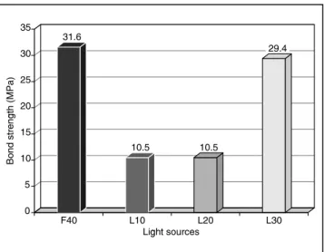
Comparison between polymerization sources with composite resin filled in a single increment
The Kruskal-Wallis test (H = 33.64) revealed statistical difference between the studied groups. Halogen light and the 30-second laser polymer-ization showed the highest bond strength values and there was no statistical difference between these groups. Laser polymerizations for 10 and 20 seconds were unable to achieve acceptable adhe-sion. Graph 1 and Table 2 illustrate the differences detected.
Comparison between polymerization sources with composite resin filled with the incremental technique (3 increments of 1 mm)
Results obtained with the Kruskal-Wallis test (H = 6.22) demonstrate no statistical difference between the polymerization sources studied in this comparison.
TABLE 1 - Parameters used in this study. Laser
10 s Laser 20 s Laser 30 s Photo 40 s
A1
10
P (mW) 200 200 200 275
PD (mW/cm²) 714.28 714.28 714.28 550
E (J) 2 4 6 11
ED (J/cm²) 7.14 14.28 21.42 22
272 273
272 273
Comparison between the filling techniques (single increment versus 3 increments)
The influence of the filling technique on com-posite resin (A110, 3M) bond strength was tested. The Mann-Whitney statistical test was chosen to compare these two groups.
Polymerization by halogen light for 40 sec-onds (z = 1.2851) showed the following values: U1 = 33 (one increment) and U2 = 67 (3 increments
of 1 mm), resulting in no statistical difference be-tween these groups.
argon laser polymerization for 10 seconds (z = −3.7796) revealed statistical difference (5%) between the filling techniques. The incremental technique showed the highest bond strength. Ad-hesion was not observed in the single increment groups polymerized for 10 and 20 seconds with the argon laser. However, it was observed in the group polymerized by laser for 10 seconds using the incremental technique.
There was no statistical difference between the filling techniques in the 30-second laser polymer-ization groups (z = 0.1512).
DISCUSSION
In spite of being almost impossible to repro-duce the clinical situations in vitro, laboratory tensile bond strength tests are commonly used to evaluate the efficacy of restorative systems and also to predict their clinical behavior17. Ferracane,
Greener7 (1986) asserted that it was possible to
correlate mechanical properties with composite resin conversion degree and presume these conver-sion degrees from tests that evaluated composite mechanical properties15.
There are many variables to be considered when bond strength between restorative materials and dental structure is studied.
With regard to the substratum used for ad-hesion tests, many authors show that there is no statistical difference between bond strength in hu-man dentin and in bovine surface dentin14,22.
The standardization of dentin depth is another important factor to be considered. Different den-tin regions present morphological and structural variations that may determine distinct adhesion mechanisms16. McCabe, Rusby13 (1992) concluded
that surface dentin produces higher bond strength values, probably because it is difficult for fluid resin to penetrate in deep dentin since it has more humidity.
The dentin depth was standardized according to Al-Salehi, Burke1 (1997). The study conducted
by Silva et al.23 (1996) demonstrates no statistical
difference in bond strength for periods of up to one week.
This study investigates the variation of polym-erization sources. The type of the polympolym-erization used may directly determine success of a restora-tion21.
According to Kelsey et al.10 (1989), due to the
previously mentioned peculiar characteristics of laser light, there is an optimization of the argon laser beam used in polymerization. Energy loss is reduced when compared with the halogen lamp, reducing the curing time and improving physi-cal properties of composites, such as compressive resistance, diametral bond strength and flexural resistance. Indeed, most investigators are unani-mous in affirming that argon laser improves the physical properties of the tested materials2,3,5,8.
The composite conversion degree is another characteristic that may be improved.
TABLE 2 - Post averages.
Sample Post sum Average
F40 316 31.6
L10 105 10.5
L20 105 10.5
L30 294 29.4
F40: halogen light for 40s; L10: argon laser for 10s; L20: argon laser for 20s; L30: argon laser for 30s.
31.6
10.5 10.5
29.4
0 5 10 15 20 25 30 35
Light sources
Bo
nd
st
re
ng
th
(MPa
)
F40 L10 L20 L30
GRAPH 1 - Composite resin (A110) filled in one
274 275
274 275
This study was performed using the polymer-ization technique with surface contact. Although other investigations demonstrate no energy loss in laser-curing within a distance between light source and resin of up to 6 mm9,11,19, in conventional
light-curing, the greater the distance between light source and resin, the smaller the density power that reaches the composite surface6,18.
According to the results obtained from the comparison between the polymerization sources with the single increment of 3 mm, in the 10- and 20-second laser polymerization groups composite resin did not adhere to dentin. We could suppose that using a power set of higher intensity or in-creasing polymerization time could improve the cure of a microfilled composite. This type of mate-rial has lower light penetration power than hybrid composites because of light dispersion through the organic matrix24. Therefore, changing the
param-eters used could be more advantageous in view of the possibility of filling the resin in a single incre-ment. Moreover, microfilled composite resins have a large amount of monomer that may be converted into polymer, consequently requiring curing pa-rameters different from the ones used for hybrid composites polymerization.
The same comparison with the laser polymer-ization for 30 seconds showed the highest values and was not statistically different from the polym-erization with halogen light. However, in a clinical situation, professionals should not use application parameters different from the ones recommended by the manufacturer, because this could cause damage to adjacent tissue4.
Halogen light and the argon laser for 30 sec-onds demonstrated no statistical difference re-garding the filling technique probably because of the greater exposure time for the laser and the greater energy density of the conventional light favoring greater power of light penetration and, consequently, a deeper curing capacity12.
In the other laser groups (10 and 20 seconds), the incremental technique showed to be more ef-fective with regard to bond strength20.
Laser polymerization of the 3 mm increment composite resin did not show any improvement in bond strength when compared to halogen lamp polymerization.
Analyzing all variables reported in this inves-tigation, further research seems to be necessary in order to define an ideal protocol for the use of argon laser for each type of resin, bearing in mind all the advantages that this new technology can offer, as well as its limitations.
CONCLUSIONS
1. The laser polymerization for 10 and 20 sec-onds in a single increment demonstrated a lower tensile bond strength compared to the 40-second polymerization with halogen light and there was no statistical difference between halogen light (40 seconds) and argon laser-curing for the 30-second interval.
2. There was no statistical difference between the curing sources for the incremental tech-nique.
3. Incremental technique showed the highest tensile bond strength values, except for the polymerizations with halogen light and argon laser for 30 seconds, which did not show sta-tistical difference.
ACKNOWLEGMENTS
This research was supported by FAPESP (The State of São Paulo Research Foundation), process-es numbers: 99/08433-4 and 99/11408-1.
REFERENCES
1. Al-Salehi SK, Burke FJT. Methods used in dentin bonding tests: an analysis of 50 investigations on bond strength. Quintessence Int 1997;28:717-23.
2. Blankenau RJ, Kelsey WP, Powell GL, Shearer GO, Bark-meier WW, Cavel T. Degree of composite resin polym-erization with visible light and argon laser. Am J Dent 1991;4:40-2.
3. Blankenau RJ, Powell GL, Kelsey WP, Barkmeier WW. Post polymerization strength values of an argon laser cured resin. Lasers Surg Med 1991;11:471-4.
4. Brenneise C, Blankenau R. Response of associated oral tissues when exposed to argon laser during polymerization of dental resins. Lasers Surg Med 1997;20(4):467-72. 5. Convington JS, Woods MA, Hoobs HD. Secondary
cur-ing of composite resins: comparison of laser treatment to other methods [abstract IADR n. 2303]. J Dent Res 1999;78:393.
274 275
274 275
curing light intensities [abstract]. J Dent Res 2001;80 Suppl 1744:253.
7. Ferracane JL, Greener EH. The effect of resin formulation on the degree of conversion and mechanical properties of dental restorative resins. J Biomed Mater Res 1986;20:121-31.
8. Fleming MG, Maillet WA. Photopolymerization of com-posite resin using the argon laser. J Can Dent Assoc 1999;65(8):447-50.
9. Kanka J 3rd. Resin bonding to wet substrate. 1. Bonding
to dentin. Quintessence Int 1992;23:39-41.
10. Kelsey WP, Blankenau RJ, Powell GL, Barkmeier WW, Cavel WT, Whisenant BK. Enhancement of physical proper-ties of resin restorative material by laser polymerization. Lasers Surg Med 1989;9:623-7.
11. Lloret PR, Rode KM, Turbino ML, Eduardo CP. Com-parison of the tensile bond strength of composite resin cured with an argon laser using two different distances of the handpiece. J Oral Laser Applic 2001;1 Suppl 1:46. 12. Martins F, Delbem ACB, Santos LRA, Soares HLO,
Martins EOB. Microdureza de resinas em função da cor e luz halógena. Pesqui Odontol Bras 2002;16(3):246-50. 13. McCabe JF, Rusby S. Dentine bonding agents –
char-acteristic bond strength as a function of dentine depth. J Dent 1992;20(4):225-30.
14. Nakamichi I, Iwaku M, Fusayama T. Bovine teeth as possible substitutes in the adhesion test. J Dent Res 1983;62:1076-81.
15. Neves AD, Discacciati JAC, Orefice RL, Jansen WC. Correlação entre grau de conversão, microdureza e con-teúdo inorgânico em compósitos. Pesqui Odontol Bras 2002;16(4):349-54.
16. Pashley DH, Sano H, Ciucci B, Yoshiyama M, Carvalho RM. Adhesion testing of dentin bonding agents: a review. Dent Mater 1995;11:117-25.
17. Retief DH. Standardizing laboratory adhesion tests. Am J Dent 1991;4(5):231-6.
18. Rissi RC, Cabral A. Fotopolimerização: principais va-riáveis clínicas que podem interferir no processo. Rev Assoc Paul Cir Dent 2002;56(2):132-8.
19. Rode KM, Lloret PR, Turbino ML, Eduardo CP. De-termination of the optimum curing distance for composite resin using the argon laser. J Oral Laser Applic 2001;1 Suppl 1:47.
20. Santos LA, Turbino ML, Youssef MN, Matson E. Micro-dureza de resina composta: efeito de aparelhos e tempos de polimerização em diferentes profundidades. Pesqui Odontol Bras 2000;14(1):65-70.
21. Santos SR. Aspectos relevantes na degradação das resinas compostas [Dissertação de Mestrado]. São Paulo: Faculdade de Odontologia da USP; 1998.
22. Schilke R, Baub O, Lisson JA, Schckar M, Geurtsen W. Bovine dentine as a substitute for human dentin in shear bond strength measurements. Am J Dent 1999;12(2):92-6.
23. Silva CM, Coradazzi JL, Pereira JC, Francischone CE. Shear bond strength of an adhesive system in hu-man, bovine and swinish teeth [abstract]. J Dent Res 1996;75:393.
24. Tate WH, Porter KH, Dosch RO. Successful photocur-ing: don’t restore without it. Oper Dent 1999;24:109-14. 25. Turbino ML, Santos LA, Matson E. Microdureza de
resina composta fotopolimerizável: a cor da matriz experi-mental pode alterar os resultados dos testes? Pesqui Odon-tol Bras 2000;14(3):232-6.
26. Verheyen P. Photopolymerization with the argon laser. J Oral Laser Applic 2001;1(1):49-54.