Strontium Incorporation on Microspheres of Alginate/
β
-tricalcium
Phosphate as Delivery Matrices
Ana Paula Duarte Moreiraa,b*, Márcia Soares Saderb, Gloria Dulce de Almeida Soaresb,
Maria Helena Miguez Rocha Leãoa
aTechnology Program in Chemical and Biochemical Processes,
Federal University of Rio de Janeiro – UFRJ, CEP 21941-972, Rio de Janeiro, RJ, Brazil
bProgram of the Metallurgical and Materials Engineering,
Alberto Luiz Coimbra Institute for Graduate Studies and Research in Engineering – COPPE, Polytechnic School, Federal University of Rio de Janeiro – UFRJ,
CEP 21941-972, Rio de Janeiro, RJ, Brazil
Received: December 19, 2013; Revised: April 7, 2014
Strontium (Sr) is known to positively affect the mechanism of bone remodelling. Consequently, calcium phosphate bioceramics associated with alginate matrices containing strontium could improve bone regeneration due to gradual strontium release. This work aims to incorporate Sr on microspheres of alginate (ALG)/β-tricalcium phosphate (β-TCP) and evaluates the in vitro release of strontium ions into a buffer solution at pH 4.0 and 7.4. In this study, strontium chloride was employed as a cross-linking agent and Sr source. Energy dispersive spectroscopy (EDS) showed that strontium was incorporated mainly at the surface of the microspheres produced. The in vitro experiments revealed that there is a rapid strontium release up to 24 h at pH 7.4 due to Sr location on microspheres’ surface. At pH 4.0 both calcium and strontium were released due to the β-TCP dissolution.
Keywords: strontium, microspheres, alginate, β-TCP, drug delivery system
1. Introduction
Calcium phosphate ceramics (CaP) have received great attention to repair and reconstruct damaged or diseased parts of the human skeleton hard tissues, such as bones and teeth1. Among the various calcium phosphate ceramics,
β-tricalcium phosphate (β-Ca3(PO4)2, β-TCP) is considered a suitable candidate as a synthetic bone graft and cement, due to the notable biocompatibility and its compositional similarities to human bones2. Besides that, the dissolution rate of β-TCP is faster than other biomedical ceramics3. This feature makes it resorbable in vivo, with new bone growth replacing the implanted material. This favourable solubility of β-TCP in physiological environments also promotes its use as a great material for drug delivery system (DDS)4.
Changes in the structure of a bioceramic have been proposed in order to improve their performance as substituent bone. In particular, the partial replacement of Ca by Sr ions in the calcium phosphate lattice has been of great interest5, due to the fundamental role of strontium (Sr) in the mineralisation of tissues. Strontium is chemically similar to calcium and can be incorporated into the mineral structure of bone, especially in regions of high metabolic turnover6. Due to the beneficial effects of Sr on bone remodelling, Sr-containing drugs have been widely used for the treatment of osteoporosis. Recent researches have indicated the dual effects of strontium ions. They promote osteoblast activity
by stimulating bone formation and reduce the osteoclastic activity, leading to a decrease in bone resorption7.
However, the poor mechanical properties of β-TCP limit its use to non-load bearing applications8. Thus, the combination of inorganic and organic components has been investigated in order to produce composites with physico-chemical characteristics improved when compared to isolated components9. Some polymers, such as alginate, are used as polymeric matrix in DDS. However, a weak interaction between the polymers and the drugs, burst release of the drugs and the fast degradation of the polymer contribute to the development of alginate-containing composites10.
Alginate is a linear copolymer of 1,4-1inked β -D-mannuronic acid (M) and α-L-guluronic acid (G). It is composed of homopolymeric blocks (GG or MM) and blocks with an alternating sequence (GM) along the chain. Alginate forms stable hydrogels in the presence of divalent cations. The gelation process occurs through the ionic interaction between the cation and the carboxyl group of G units in a model called “egg-box”11. Calcium cations are usually the most frequently used for cross-linking processes. However, other cations may also be employed, such as Sr2+, Ba2+and Zn2+.
buffer solutions are employed as soaking media in order to mimic the environment in which the biomaterial will be implanted. These buffers can simulate a neutral (simulating body fluid) or acidic pH12,corresponding to the inflammatory reactions that precede the osseointegration process.
In this study, strontium ions were employed in the production of the microspheres as a cross-linking agent. The microcapsules were exposed to two different solutions for up to 14 days and changes in the physico-chemical properties and strontium release were evaluated.
2. Material and Methods
β-TCP was obtained by heat treatment (900°C for 4h) of commercial calcium-deficient apatite (TCP powder, Merck, Darmstadt, Germany). β-TCP powder was mixed with alginate (ALG, Alginic acid sodium salt from brown algae, Fluka, Steinheim, Norway), keeping the ratio as ALG/β-TCP 1:15. The mixture was dropped from a syringe into a strontium chloride solution (SrCl2.6H2O, 0.15 mol.L–1, Merck, Darmstadt, Germany) under slow magnetic stirring at room temperature. The microcapsules were kept in the solution overnight to ensure complete exchange of Na for Sr. The microspheres were removed from solution, washed in distilled water and dried in an oven at 37°C. The microcapsule diameters used in this study ranged from 355 to 420 µm.
The produced microspheres were characterised by X-ray diffraction (XRD, XRD-6000 Shimadzu diffractometer) with CuKα radiation operating at 30 kV and 30 mA. X-ray diffraction data were collected in a range of 5–80o with a step size of 0.02o and a count time of 1s per step. Fourier transform infrared spectra (FTIR, Spectrum 100 Perkin Elmer spectrometer) were obtained in the range of 4000–550 cm–1, 32 scans and resolution 4 cm–1, using the ATR (attenuated total reflectance) mode.
Morphological investigation of the microspheres was performed by scanning electron microscopy (SEM, JSM6460 JEOL) operating at 15 kV. Energy dispersive spectroscopy (EDS, System Six 200, Thermo Noran) was used to qualitatively identify strontium on the microspheres. The samples were sputter-coated with a thin gold layer (Emitech, K550, USA) to avoid electrical charging before examination.
The in vitro tests were carried out according to ISO 10993-913, by immersing 250 mg of microspheres into 5 mL of buffer solution in a polypropylene tube. The composition of the buffer solution was sodium chloride (NaCl, 10 mmol.L–1), sodium hydrogen carbonate (NaHCO
3, 45 mmol.L–1) and potassium carbonate (K
2CO3,2 mmol.L–1). Lactic acid was used to adjust the pH to 7.4 (simulating a body fluid) and 4.0 (simulating an inflammatory process). The tubes were kept in horizontal position and incubated at 37°C in an orbital shaker (120 rpm, incubator shaker, M.S. Mistura). The microcapsules remained in contact with the solutions for different times (12, 24, 48, 72 hours, 7 and 14 days). The experiments were performed in triplicate. After each time interval, the fluid was filtered and separated, and the solid material was washed with distilled water, dried at 37°C and re-examined using the already mentioned techniques.
Calcium and strontium contents present in the fluid after in vitro tests were determined using atomic absorption spectrophotometer (AAS, AA-6800 Shimadzu) with (k(Ca) = 422.7 nm and k(Sr) = 460.7 nm). The samples were diluted to an appropriate volume with potassium chloride in hydrochloric acid (HCl, 6 mol.L–1), in order to suppress interferences. AAS was used to analyse the biological fluid before and after the assay. The final result is an average of the values.
3. Results and Discussion
Initially, the reproducibility of the employed method was tested, since, according to the literature14, the usual production of microspheres employs a CaCl2 solution. The experimental process of microcapsules production using SrCl2 solution, as described above, was repeated three times under the same conditions, resulting in more than 90% homogeneous ALG/β-TCP/Sr microspheres.
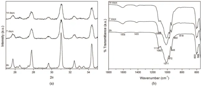
XRD patterns of pure β-TCP and ALG/β-TCP/Sr microspheres are shown in Figure 1. The results showed that there is only one phase present on the microcapsules produced, β-TCP, which is in good agreement with the standard data JCPDS 9-0169. The XRD spectrum of produced microspheres revealed a slight displacement in the diffraction patterns to higher diffraction angles. Furthermore, the high intensity peak (2 1 1) observed in pure β-TCP is less intense in the microcapsules of ALG/β-TCP/Sr. Similar characteristics were described by Rajkumar et al.15 and Son et al.16, showing that a polymeric matrix contraction occurs through chemical bonding between β-TCP and alginate.
Some authors report a shift to lower angles when strontium substitution in the Ca site occurs17. However, it was not observed in the produced microspheres. It seems that Sr is not incorporated in β-TCP lattice in significant amounts and/or that this effect is negligible when compared with the matrix contraction.
FTIR spectra of pure β-TCP, sodium alginate and ALG/
β-TCP/Sr microspheres are shown in Figure 2. The presence
Figure 1. XRD of β-TCP and microspheres of ALG/β-TCP/Sr. Inset
of bands at 1115, 1100, 1080, 1010, 971 and 945 cm–1 can be assigned to vibrational modes of ν3 PO43– groups. The bands at 603 and 590 cm–1 are attributed to ν
4 PO43– groups of β-TCP18. Slight differences in the intensity and in the peaks position of IR bands can be observed comparing the produced microspheres and the pure alginate. The bands at 1594-1604 cm–1 correspond to the asymmetric stretching of the carbonyl C=O group of alginate, and the vibration at 1406-1419 cm–1 is characteristic of the COO– group of
alginate. These bands have low intensity and a small shift compared with the band intensity of the pure alginate. The peak shift observed could be due to a chemical interaction between the mineral phase and the organic matrix. There is a bond formation between calcium atoms of the β-TCP molecule and the oxygen sites of the carboxyl groups in the GG chain of alginate18, but there may also be an interaction between strontium ions and the COO– groups of alginate.
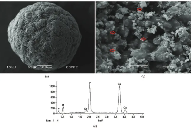
Figure 3 shows the morphological features of the ALG/
β-TCP/Sr microspheres. The microcapsules (Figure 3a) showed a rough surface with rounded particles of the β-TCP well embedded and homogeneously dispersed throughout the porous alginate matrix. Even presenting a rough and irregular surface, with high magnification, cracks were not observed, Figure 3b. The presence of cracks would be undesirable, as the drug should be retained inside the microspheres.
The presence of Sr on microspheres was confirmed by EDS (Figure 3c). However, higher Sr peaks’ intensity were found on microspheres’ surface than inside, when cross-section was analysed (not shown). The formation of beads occurs, instantaneously, when spherical drops come into contact with Sr2+ solution9. In the present case, the cross-linking agent was strontium ions, which have a larger ionic radius than calcium (i.e. 1.13 Å for Sr2+ and 0.99 Å for Ca2+)19. Therefore, during the gelation process, the exchange of large Sr ion probably results in the formation of a crosslink in which the strontium ions are predominant on the surface compared to inside the beads. Moreover, the Figure 2. FTIR spectra of the microspheres of ALG/β-TCP/Sr,
alginate and β-TCP.
gelation process occurs from outside to inside and creates a diffusional barrier for strontium, preventing its migration into the sphere20.
The chemical composition of the starting ALG/β-TCP/ Sr microspheres indicated 0.87 ± 0.08 wt.% of Sr and 34.99 ± 3.56 wt.% of Ca, corresponding to an Sr/Ca molar ratio of 0.025.
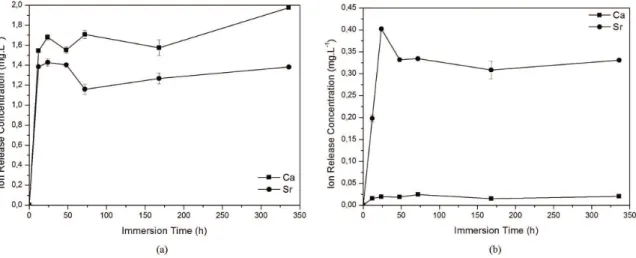
In vitro tests (Figure 4) showed that up to 24h, at both pH levels tested, there is an increase in the calcium and strontium concentration in the solution, denoting a fast release of these ions. Similar results were reported by Suganthi et al.7 who described that the initial burst release could be used for antibiotic delivery to prevent an inflammatory process after surgery. Our study was performed with microspheres of
355-Figure 4. Sr and Ca progressive releases of the ALG/β-TCP/Sr microspheres into (a) pH 4.0 and (b) pH 7.4 buffer with different soaking time (error bars show the standard deviation).
Figure 6. SEM images of the ALG/β-TCP/Sr microspheres after immersion in buffer solution at pH 7.4: (a) 7 days – original magnification = 200 x; (b) 7 days – 5k of magnification; (c) 14 days – original magnification = 200 x and (d) 14 days – 5k of magnification. 420 µm diameters, which are favourable to the dissolution
process due to the high contact surface.
After 24 h soaking, there is a decrease of strontium and calcium concentrations in the medium and, after 72 h soaking, this depletion is more significant at pH 4.0. Based on previous reports21, the β-TCP dissolution initially occurs with Ca2+, Sr2+ and PO
43– ions being released. With increasing soaking time, the ions concentrations rise and the solution becomes super-saturated, resulting in the precipitation of a new calcium phosphate phase. The precipitation process is responsible for decreasing calcium and strontium concentrations after 24h22. This process happens until equilibrium is reached, where, after 7 days, the strontium release is constant.
The strontium and calcium release profiles display a similar trend. Comparing both conditions, the ion relief is more pronounced at pH 4.0 due to the higher dissolution of calcium phosphates in acidic environments23. Thus, this pH provides a fast release of Ca2+, Sr2+ and PO
43– in the first 24 h, as can be seen in Figure 4a.
The Sr/Ca molar ratio remains almost constant and equal to 0.8 during the entire experiment, indicating that the Sr liberation rate is close to Ca. Moreover, as pH 4.0 is favourable to β-TCP dissolution, the release of ions occurs only in the beginning and, after the initial burst, the Sr/Ca molar ratio remains unchanged.
However, at pH 7.4 (Figure 4b) there is a higher liberation of strontium ions when compared with calcium release. These results corroborate with the fact that the strontium ions are found preferentially on the surface instead of inside the microspheres. Therefore, as pH 7.4 is not aggressive and does not promote β-TCP dissolution, the release rate follows all of the previously mentioned stages, being favoured by swelling surface.
Considering the initial Sr content on the microspheres, the Sr maximum amount released during one day was about 66% of the total present in the microcapsules. This corresponds to a low strontium dose per day. Depending on the treatment, it is possible to change the strontium concentration for the best result in order to improve the osteoblast differentiation and reduce osteoclast activity.
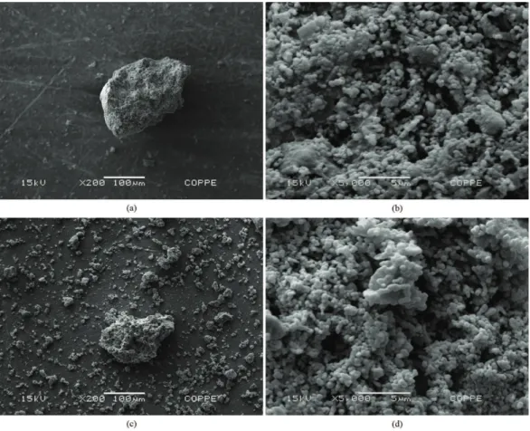
Figure 5 shows the morphological features of the ALG/
β-TCP/Sr microspheres after immersion in buffer solution at pH 4.0 for 7 days (a), and at high magnitude (b), and at the same pH for 14 days (c), and in high magnitude (d). In acidic pH, the carboxylate groups of alginate are protonised and the alginate polymeric chains are more relaxed. Although the protonation hinders water uptake and, hence swelling, the microspheres absorb water at a slow rate20,24. For this reason, the size of microspheres increased, indicating that they are more swelled up to 7 days of immersion. At 14 days, higher β-TCP dissolution occurs, exposing the alginate organic matrix, leading to microcapsules collapse. These results corroborate with data presented by in vitro tests where it was observed that there was a higher Ca and Sr release at acidic pH.
SEM micrographs of the ALG/β-TCP/Sr microspheres after immersion in buffer solution at pH 7.4 for 7 and 14 days are shown in Figure 6. In the body fluid pH, the alginate polymeric chains tend to absorb more water, hence swell quickly and then begin the disintegration and disruption process of egg-box structure. For this reason, after 7 days, the microspheres are shrunken, cracked and β-TCP is more apparent, thus maintaining the microsphere structure (Figure 6a, detail in high magnitude 6b). At 14 days, greater polymeric matrix disintegration occurs, making them unable to maintain the microsphere structure20,24 (Figure 6c, detail in high magnitude 6d).
XRD patterns of the material after dissolution tests at pH 4.0 buffer (Figure 7a) revealed only β-TCP phase. However, the diffraction peaks presented a decrease in the intensity and a width enlargement, characterising a poorly crystalline apatite phase. The dissolution of β-TCP was followed by the
precipitation of a new calcium phosphate phase that was not detectable by XRD. No significant changes were observed in XRD patterns of the samples obtained after pH 7.4 buffer immersion (data not shown).
FTIR spectra of the samples after dissolution tests (Figure 7b) exhibited bands at 1608 and 1422 cm–1 which were more pronounced. These bands correspond to C=O and COO– groups present in the alginate, as well as demonstrating the carbonate presence. These CO32− ions (bands at 1422 and 813 cm–1) are incorporated from CO
2 present in the atmosphere and in the buffer solution. Similar results have been reported by Son et al.16.
4. Conclusions
In this study, ALG/β-TCP/Sr microspheres were prepared through the droplet extrusion process, employing strontium chloride solution as a reticulating agent. Energy dispersive spectroscopy (EDS) showed that strontium was incorporated mainly at the surface of the microspheres produced. The in vitro experiments revealed that there is a rapid strontium release up to 24 h at pH 7.4 due to Sr location on the microspheres’ surface. At pH 4.0, both calcium and strontium were fast released due to β-TCP dissolution. The Sr release up to 24h at pH 4.0 was about 66% of the total present in microspheres. In vitro tests have shown that, at acidic pH, the ions released would be governed by the inorganic phase dissolution, while at neutral pH, organic matrix swelling occurs and, consequently, the ions diffuse into the medium. The use of SrCl2 in the gelation process of the microspheres jointly with an inorganic phase more degradable (β-TCP) allows rapid strontium release in the early stages of implantation, which can be relevant to bone remodelling.
Acknowledgments
The authors would like to thank Faperj, CNPq and CAPES for financial support.
References
1. Dorozhkin SV. Calcium orthophosphates as bioceramics: state of the art. Journal of Functional Biomaterials. 2010;
1(1):22-107. http://dx.doi.org/10.3390/jfb1010022
2. Lin K, Chen L, Qu H, Lu J and Chang J. Improvement of mechanical properties of macroporous β-tricalcium phosphate bioceramic scaffolds with uniform and interconnected pore structures. Ceramics International. 2011; 37(7):2397-2403.
http://dx.doi.org/10.1016/j.ceramint.2011.03.079
3. Samavedi S, Whittington AR and Goldstein AS. Calcium phosphate ceramics in bone tissue engineering: a review of properties and their influence on cell behavior. Acta Biomaterialia. 2013; 9(9):8037-8045. PMid:23791671. http://
dx.doi.org/10.1016/j.actbio.2013.06.014
4. Vahabzadeh S, Edgington J and Bose S. Tricalcium phosphate and tricalcium phosphate/polycaprolactone particulate composite for controlled release of protein. Materials Science and Engineering: C. 2013; 33(7):3576-3582. PMid:23910252. http://dx.doi.org/10.1016/j.msec.2013.04.001
5. Kannan S, Goetz-Neunhoeffer F, Neubauer J, Pina S, Torres PMC and Ferreira JMF. Synthesis and structural characterization of strontium - and magnesium-co-substituted β-tricalcium phosphate. Acta Biomaterialia. 2010; 6(2):571-576. PMid:19679202. http://dx.doi.org/10.1016/j. actbio.2009.08.009
6. Boanini E, Gazzano M and Bigi A. Ionic substitutions in calcium phosphates synthesized at low temperature. Acta Biomaterialia. 2010; 6(6):1882-1894. PMid:20040384. http:// dx.doi.org/10.1016/j.actbio.2009.12.041
7. Suganthi RV, Elayaraja K, Joshy MIA, Chandra VS, Girija EK and Kalkura SN. Fibrous growth of strontium substituted hydroxyapatite and its drug release. Materials Science and Engineering: C. 2011; 31(3):593-599. http://dx.doi. org/10.1016/j.msec.2010.11.025
8. Perera FH, Martínez-Vázquez FJ, Miranda P, Ortiz AL and Pajares A. Clarifying the effect of sintering conditions on the microstructure and mechanical properties of b-tricalcium phosphate. Ceramics International. 2010; 36(6):1929-1935. http://dx.doi.org/10.1016/j.ceramint.2010.03.015
9. Ribeiro CC, Barrias CC and Barbosa MA. Calcium phosphate-alginate microspheres as enzyme delivery matrices.
Biomaterials. 2004; 25(18):4363-4373. Pmid:15046927. http://
dx.doi.org/10.1016/j.biomaterials.2003.11.028
10. Zhang J, Wang Q and Wang A. In situ generation of sodium alginate/hydroxyapatite nanocomposite beads as drug-controlled release matrices. Acta Biomaterialia. 2010; 6(2):445-454. PMid:19596091. http://dx.doi.org/10.1016/j. actbio.2009.07.001
11. Chen CHD, Chen CC, Shie MY, Huang CH and Ding SJ. Controlled release of gentamicin from calcium phosphate/ alginate bone cement. Materials Science and Engineering: C. 2011; 31(2):334-341. http://dx.doi.org/10.1016/j.
msec.2010.10.002
12. Zou C, Cheng K, Weng W, Song C, Du P, Shen G, et al. Characterization and dissolution: eprecipitation behavior of biphasic tricalcium phosphate powders. Journal of Alloys
and Compounds. 2011; 509(24):6852-6858. http://dx.doi.
org/10.1016/j.jallcom.2011.03.158
13. International Organization for Standardization – ISO.
Biological evaluation of medical devices. Part 9: Degradation of materials related to biological testing. ISO; 1994
14. Paula FL, Bareto IC, Rocha-Leão MH, Borojevic R, Rossi AM, Rosa FP, et al. Hydroxyapatite-alginate biocomposite promotes bone mineralization in different length scales in vivo.
Frontiers of Materials Science in China. 2009; 3(2):145-153.
http://dx.doi.org/10.1007/s11706-009-0029-9
15. Rajkumar M, Meenakshisundaram N and Rajendran V. Development of nanocomposites based on hydroxyapatite/ sodium alginate: synthesis and characterization. Materials Characterization. 2011; 62(5):469-479. http://dx.doi.
org/10.1016/j.matchar.2011.02.008
16. Son KD, Yang DJ, Kim MS, Kang IK, Kim SY and Kim YJ. Effect of alginate as polymer matrix on the characteristics of hydroxyapatite nanoparticles. Materials Chemistry and Physics. 2012; 132(2-3):1041-1047. http://dx.doi. org/10.1016/j.matchemphys.2011.12.062
17. Rokidi S and Koutsoukos PG. Crystal growth of calcium phosphates from aqueous solutions in the presence of strontium.
Chemical Engineering Science. 2012; 77:157-164. http://
dx.doi.org/10.1016/j.ces.2012.02.049
18. Xie M, Olderøy MØ, Andreassen JP, Selbach SM, Strand BL and Sikorski P. Alginate-controlled formation of nanoscale calcium carbonate and hydroxyapatite mineral phase within hydrogel networks. Acta Biomaterialia. 2010; 6(9):3665-3675. PMid:20359556. http://dx.doi.org/10.1016/j. actbio.2010.03.034
19. Zhang W, Shen Y, Pan H, Lin K, Liu X, Darvell BW, et al. Effects of strontium in modified biomaterials. Acta Biomaterialia. 2011; 7(2):800-808. PMid:20826233. http:// dx.doi.org/10.1016/j.actbio.2010.08.031
20. Bajpai SK and Sharma S. Investigation of swelling/degradation behaviour of alginate beads crosslinked with Ca2+ and Ba2+ ions.
Reactive & Functional Polymers. 2004; 59(2):129-140. http:// dx.doi.org/10.1016/j.reactfunctpolym.2004.01.002
21. Sánchez-Salcedo S, Balas F, Izquierdo-Barba I and Vallet-Regí M. In vitro structural changes in porous HA/β-TCP scaffolds in simulated body fluid. Acta Biomaterialia. 2009; 5(7):2738-2751. PMid:19394904. http://dx.doi.org/10.1016/j. actbio.2009.03.025
22. Dorozhkin, SV. A review on the dissolution models of calcium apatites. Progress in Crystal Growth and Characterization of Materials. 2002; 44(1):45-61. http://dx.doi.org/10.1016/
S0960-8974(02)00004-9
23. Gentleman E, Fredholm YC, Jell G, Lotfibakhshaiesh N, O’Donnell MD, Hill RG, et al. The effects of strontium-substituted bioactive glasses on osteoblasts and osteoclasts in vitro. Biomaterials. 2010; 31(14):3949-3956. PMid:20170952.
http://dx.doi.org/10.1016/j.biomaterials.2010.01.121 24. Bajpai SK, Saxena SK and Sharma S. Swelling behavior
of barium ions-crosslinked bipolymeric sodium alginate-carboxymethyl guar gum blend beads. Reactive & Functional Polymers. 2006; 66(6):659-666. http://dx.doi.org/10.1016/j.