RBCCV 44205-1547 DOI: 10.5935/1678-9741.20140054
The calcium paradox - What should we have to fear?
Paradoxo do cálcio - o que temos a temer?
Marcos Aurélio Barboza de Oliveira
1, MD; Antônio Carlos Brandi
2, MD; Carlos Alberto dos Santos
2,
MD; Paulo Henrique Husseni Botelho
2, MD; José Luís Lasso Cortez
3, MD; Gilberto Goissis
4,
PhD;
Domingo Marcolino Braile
5,
MD, PhD
1Faculdade de Medicina de São José do Rio Preto (FAMERP), São José
do Rio Preto, SP, Brasil; Centro Universitário de Votuporanga (UNIFEV), Santa Casa Votuporanga, Votuporanga, SP, Brazil.
2Hospital de Base São José do Rio Preto. São José do Rio Preto, SP, Brazil. 3Santa Casa Votuporanga, Votuporanga, SP, Brazil.
4Braile Biomédiaca, Indústria Comércio e Representações, São José so Rio
Preto, SP, Brazil.
5Faculdade de Medicina de São José do Rio Preto (FAMERP), São José do
Rio Preto, SP, Brazil.
Correspondence address:
Marcos Aurélio Barboza de Oliveira
Avenida República do Líbano, 2700, casa 80 - São José do Rio Preto, SP, Brazil - Zip code: 15092-440
E-mail: [email protected]
This study was carried out at São José do Rio Preto Medical School (FAMERP), São José do Rio Preto, SP, Brazil.
No inancial support.
Article received on November 24th, 2013
Article accepted on February 13rd, 2014
Abstract
The calcium paradox was first mentioned in 1966 by Zimmerman et al. Thereafter gained great interest from the scientiic community due to the fact of the absence of calcium ions in heart muscle cells produce damage similar to ischemia-reperfusion. Although not all known mechanisms involved in cellular injury in the calcium paradox intercellular connection maintained only by nexus seems to have a key role in cellular fragmentation. The addition of small concentrations of calcium, calcium channel blockers, and hyponatraemia hypothermia are important to prevent any cellular damage during reperfusion solutions with physiological concentration of calcium.
Descriptors: Heart Arrest, induced. Myocardial Ischemia. Calcium.
Resumo
O paradoxo do cálcio foi pela primeira vez citado em 1966 por Zimmerman et al. A partir daí, ganhou grande interesse por parte da comunidade cientíica internacional devido ao fato da ausência do íon cálcio produzir na célula muscular cardíaca dano semelhante à lesão de isquemia-reperfusão. Apesar de não serem conhecidos todos os mecanismos envolvidos no processo da lesão celular no paradoxo do cálcio, a conexão intercelular mantida somente pelo nexus parece ter papel chave na fragmentação celular. A adição de pequenas concentrações de cálcio, bloqueadores de canal de cálcio, hiponatremia ou hipotermia são importantes para evitar que haja lesão celular no momento da reperfusão com soluções com concentração isiológica de cálcio.
Descritores: Parada Cardíaca Induzida. Isquemia Miocárdica. Cálcio.
Unlike what would be expected, the complete absence of calcium not only caused the cardiac arrest, but also altered the cell membranes of cardiac myocytes, culminating in the reperfusion phase with their necrosis, explaining the term “paradox”[1].
In the following years many researchers have studied possible physiological mechanisms of paradox, culminating INTRODUCTION
In 1960 Zimmerman et al.[1,2] described massive lysis of
Abbreviations, acronyms & symbols
DNP Dinitrophenol ADP Adenosine diphosphate ATP Adenosine triphosphate
LV Left ventricle
with a signiicant amount of studies on the subject, many be -ing presented in 1983 at the IX World Congress of the Soci-ety for Heart Research, held in London[3].
In this event was compiled much of what we knew at the time about the lack of calcium in cardioplegic solution, the extensive myocardial damage that this solution causes and alternative ways for developing a safe hypocalcemic cardio-plegia[4-6].
After nearly 50 years of the discovery of this paradox, this study aims to discuss some harmful effects of calcium paradox in the heart, considering its importance, molecular mechanisms, cellular ultrastructural changes, additive pro-tective or harmful effect when placed in combination with other solutions and some ways to avoid it.
Importance of calcium paradox
In the 1980s, the calcium metabolism in the heart has been extensively studied. At that time, there was consensus on the consequences of the succession of a medium without
calcium followed by another illed of it to heart muscle cells,
which rapidly internalizes this ion, leading it to lysis and heart
failure. This phenomenon is similar to reperfusion injury[7].
Another key aspect is the understanding of mechanisms in-volved, as cardioplegic solutions should not cause cellular damage. Hypocalcemic cardioplegic solutions are effective to induce cardiac arrest[8]. However, substances that interrupt
or mitigate undesirable side effects must be present to pre-vent pre-ventricular dysfunction after cardiopulmonary bypass
[7].
Studies on metabolic pathways that promote or disrupt the process[9,10], as well as their relationship to heart failure[11,12]
have been published, which we will discuss briely below.
Causal mechanism
Several hypotheses were formulated to explain the calci-um paradox as increased permeability of calcicalci-um in the sar-colemma[13], the glycocalyx[14] and separation of intercalated
discs[15,16], but no further clariied the whole mechanism of the
calcium paradox.
It is also possible that intracellular hypercalcemia is not the primary cause of the calcium paradox. Its increase may occur as a result of damage to sarcolemma accompanied by an entry of moderate amount of calcium to structurally altered[17] cells.
Isolated absence of calcium can cause cell damage, but its deleterious effect is potentiated in media with anoxia, caf-feine, 2,4-dinitrophenol (DNF), ventricular balloon
(mechan-ical strain), etc.. All these conditions cause injury to the myo -cardium even in the absence of extracellular calcium[18-22].
Another mechanism accepted is that calcium comes into the cell in a massive way, causing damage and cell death[23].
Structural cellular changes
The irst description of structural changes in myocyte
perfused in calcium-free medium was performed by Muir et al.[16], who observed changes in glycocalyx and intercalated
disks of myocytes in isolated rat hearts. Intercalated disks are
complex structures divided into several regions, the major
one being occupied by adherens fascia. These are the places of greatest tension between the cells at the time of
myocar-dial contraction. Desmosomal junctions, called macula ad -herens, are present and serve to unite the cells. Nexus or gap
junctions are focal points of intimate cell contact, being local
with electrical signaling between cells[24].
Muir et al.[16] noted that cardiac myocytes which
under-went perfusion showed no calcium from 30 minutes of clear separation in regions of the fascia adherens and macula ad-herens, while the nexus remained intact (Figure 1). Ashraf[13]
and Yates & Dhalla[25] observed similar changes in 10 to 15
minutes of exposure to the same environment. Shorter peri-odes of 3-5 minutes generally do not cause physical separa-tion in the ultrastructure of the intercalated discs[23].
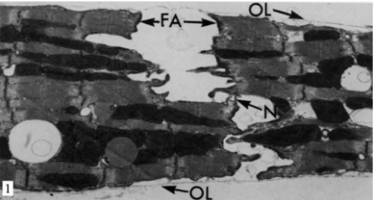
Fig. 1 - Electron micrograph of rat heart after 12 minutes of perfusion without calcium at 37oC. The intercalated disks are separated in
the regions of the fascia adherens (FA), but are still interconnected by the nexus joints (N). The outer layer of the sarcolemma (OL) or glycocalyx is detached from the plasma membrane of the myocytes. ReproducedfromGanote et al., 1985[23]
The calcium-free medium increases the amount of intra-cellular sodium both in cultured myocytes and in isolated heart. Moving to a medium rich in calcium, this ion rapidly enters cells via antiporter pump Na+/Ca+2, working on a
Lysis at the time the myocyte contacts reperfusion solu-tion rich in calcium after sensitizasolu-tion in medium without this ion occurs because these cells have intermediate disc only connecting to one another, and there is their avulsion during contraction with exposure of the intracellular medium of each of them explaining the massive cell death of them[16].
Another change that occurs during infusion without calci-um is the detachment of glycocalyx, which is usually a focal shift that is not seen until 10 to 15 minutes after infusion of the solution without calcium. Frank et al.[29,30] have shown
that despite the detachment of the external layer of glycoc-alyx, there is an inner membrane that is adhered on the cell membrane.
Ashaf et al.[13] and Frank et al.[29] observed aggregation
and anomalous rearrangement of the constituent molecules of the cell membrane when placed in calcium free medi-um, so that there is irreversible cell damage due to altered membrane permeability. The molecular mechanisms of these changes are not yet known.
Phase of re-entry of calcium into the cell
After 10-15 minutes in medium deprived of calcium, the myocyte is sensitized, and the separation of the intercalated discs between cells is established[23]. The sarcomeres of each
cell condense into a single band of contraction. The fascia adherens remain connected to sarcomeres, but are
complete-ly separate from the membranes of adjacent cells. Regions of
intercalated discs, located between the areas of fascia adher-ens become fragmented and allow the mitochondria to go into the intercellular space [31]. Ganote et al.[19] mention that
hypothermia prevents lysis of the fascia adherens and there-fore cytolysis.
The contractions of the sarcomere and cell necrosis are
identical to those observed in other types of injury as cat
-echolamine necrosis and ischemia/reperfusion injury. How -ever, it should be emphasized that the cellular ultrastructure when in medium without calcium differs from all previous separation of the intermediate disks and the presence of a single central shrink band[23].
Traces of calcium
Rebeyka et al.[32] found in CPB model that dog hearts
per-fused with cold cardioplegic solution without calcium showed a worse recovery of ventricular function and greater area of
necrosis than those in which the solution was only 70 μmol/L
calcium, showing that even small concentrations of calcium
are suficient to protect the heart of calcium paradox.
Glycocalyx
The separation of the external layer of the cell membrane glycocalyx from myocyte occurs after exposure of the cell to calcium-poor medium[23]. Frank et al.[14] believed that this
sep-aration would be responsible for the increase in the membrane
calcium permeability. However, Nayler et al. [17] using 2 mM
calcium instead of magnesium in the private period of calcium showed that despite a detachment of the glycocalyx, there was no increase in membrane permeability to calcium and Slade et al.[33] have also observed that myocytes placed in buffer
medi-um without calcimedi-um also lose glycocalyx, without, however,
observing a change from the inlux of calcium ion.
With the use of neuraminidase there is complete separa-tion of the glycocalyx of myocytes with increased cell per-meability to calcium. To explain this phenomenon, Ganote et al.[23] postulate that in this case the membrane glycoproteins
would also be damaged, losing control of the calcium low.
ATP
Ruigrok et al.[34] showed that the massive release of
en-zymes which occurs during reperfusion of calcium is depen-dent on energy. This conclusion is based on experiments that consumed the intracellular ATP from myocyte with anoxic perfusion or the inclusion of this cell in medium without glu-cose. The non-cardiac cells did not release enzymes during phase of normal calcium after 5 minutes of perfusion without calcium due to depletion of ATP[34].
Calcium channel blockers
Baker & Hearse[35] observed that the effect of calcium
channel blockers is best demonstrated when there is low ex-tracellular calcium concentration during reperfusion. Under these conditions, calcium entry occurs preferentially through the slow channels of the membrane. The limitation of this
en-try enables the recovery from injury of intercalated discs and
sarcolemma. In solutions with physiological concentration of calcium in the reperfusion solution, the calcium channel blockers offer little protection to the paradox, suggesting that more than one route is important for the entry of calcium into the cell[36].
Sodium
Dhalla et al.[37] showed that when the concentration of
sodium is reduced to 35 mm in phase without calcium, the magnitude of tissue damage is reduced during reperfusion with calcium. This is due to the low concentration of sodi-um, which slows the entry of calcium into the membrane by antiport pump Na+/Ca+2, facilitating intracellular ionic
re-balancing and preventing contracture that would lead to cell death[37].
During the period without calcium, low sodium concen-tration reduces the transmembrane gradient and delays the
calcium eflux and sodium inlux. This would slow down the
removal of both intracellular calcium and the cell damage caused by the absence of this ion. In the period with normal
calcium low sodium would also be beneicial because it re
-duces calcium inlux via antiporter pump Na+/Ca+2, working
cell is without it and slower internalization during reperfu-sion provides the cell conditions to reestablish its ionic bal-ance before any structural damage[37].
Hypothermia
Hypothermia protects the myocyte from calcium para-dox[38,39]. It prevents the separation of the intercalated disc
and detachment the glycocalyx[40]. In addition, it reduces the
Na+/Ca+2, and may decrease the loss of Ca+2 ions in
calci-um-free perfusion time[41]. The ideal temperature found for
myocardial protection of calcium paradox was 22oC[42-44]. Mechanical injury
Ganote et al.[20] states that when isolated rat hearts are
placed in anoxia in medium with physiological concentra-tion of calcium, the distension of the volumetric balloon in the left ventricle (LV) occurs with a small increase resulting
from enzymatic cell lysis, but the LV distension is dificult.
When the medium is free of calcium and normoxia, disten-sion is easy and enzyme release is also small. However when we have anoxia and calcium-free medium, there is massive release of cellular enzymes.
This is because anoxic hearts can withstand the wall tension that the balloon prints due to the intercalated disks were incomplete. The cavity is distended by elongation of the sarcomeres, with their lesion. In calcium-free medium and
normoxia, the muscle ibers are now relaxed and the voltage produced by the balloon is not suficient to cause avulsion
of weakened intercalated disks. But when anoxia and me-dium without calcium are superimposed, the cell maintains the stiffness of the sarcomeres to the fragility of the
inter-calated disks. The pressure of the inlated balloon adversely
affects directly the region, causing release of intracellular enzymes[20].
Dinitrophenol
The dinitrophenol (DNP) is a fat-soluble weak acid that acts as protonophore (translocator protons) entering the mito-chondria positively charged and leaves it negatively charged, creating electron transport out of the mitochondria, prevent-ing the conversion of ADP to ATP[45]. It also causes rapid
ventricular contraction both in hearts which underwent cal-cium-free medium as in those with normal concentration of this ion. However, those in medium without calcium causes massive cell lysis[22] (Figure 2).
This observation is consistent with the hypothesis that contraction physically separates the cells, causing cell lysis in those in free-calcium medium. The DNP alone causes contraction of cells, and it is not necessary to add calcium to the medium. Intracellular calcium present in
mitochondria and sarcolemma would not be suficient to
generate environment that simulates medium with normal calcium[22].
Caffeine
Caffeine causes the sarcolemma calcium release but not in mitochondria[46]. Thus, intracellular calcium increases
slightly, but without its overhead[31]. The persistent
contrac-tion produced by caffeine is dependent on calcium and in its absence there is support for only 20 to 30 seconds, followed by relaxation[18].
Hearts perfused with solution containing caffeine, but with-out calcium at 22oC do not manifest increased enzymes, but
those kept at 37oC have similar injury to the calcium paradox[18].
Whereas the increase in intracellular calcium
concentra-tion is not signiicant because there was no reperfusion with
calcium, it is unlikely that the lesion is originally from poi-soning by calcium, but by direct action of ventricular con-traction on sarcolemma[18].
CONCLUSION
We should fear the phenomenon known as “calcium par-adox” because it irreversibly damages the membrane of the myocyte, causing extrusion of cellular contents. However, despite its biomolecular mechanisms are not fully under-stood, measures such as hypothermia, hyponatremia, and the presence of traces of calcium in the perfusion solution
decreases the risk of this injury, enabling the recovery of ven -tricular function after induced cardiac arrest.
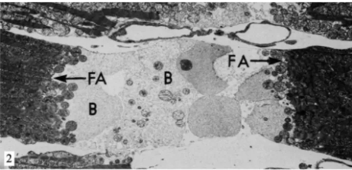
Fig. 2 - Electron micrograph of rat heart after 5 minutes in calcium-free perfusion, followed by 15 minutes with the addition of DNP at the
irst perfusate. The sarcomeres are contracted herein, pulling the fascia
adherens (FA) and causing damage to the cell membrane of the myocyte in the nexus area. The cytosol is exteriorized in the form of blebs (B) in the intercellular region. ReproducedfromGanote et al., 1985[23]
MABO Main Author
ACB Help in bibliographic survey and translated articles CAS Help in literature review and translation of articles PHHB Help in correcting the manuscript
JLLC Help in correcting the manuscript GG Co-supervisor
DMB Advisor
REFERENCES
1. Zimmerman AN, Hulsmann WC. Paradoxical inluence of calcium
ions on the permeability of the cell membranes of the isolated rat heart. Nature. 1966;211(5049):646-7.
2. Zimmerman ANE, Daems W, Hülsmann WC, Snijder J, Wisse
E, Durrer D. Morphological changes of heart muscle caused by successive perfusion with calcium-free and calcium-containing solutions (calcium paradox). Cardiovasc Res. 1967;1(3):201-9.
3. Poole-Wilson PA, Nayler WG. Preface. J Mol Cell Cardiol. 1984;16(2):111.
4. Poole-Wilson PA, Harding DP, Bourdillon PD, Tones MA. Calcium out of control. J Mol Cell Cardiol. 1984;16 (2):175-87
5. Nayler WG, Dresel PE. Ca2+ and the sarcoplasmic reticulum. J Mol Cell Cardiol. 1984;16(2):165-74.
6. Langer GA. Calcium at the sarcolemma. J Mol Cell Cardiol. 1984;16(2):147-53.
7. Piper HM. The calcium paradox revisited: an artefact of great heuristic value. Cardiovasc Res. 2000;45 (1):123-7.
8. Gebhard MM, Bretschneider HJ, Gersing E, Preusse CJ, Schnabel PA, Ulbricht LJ. Calcium-free cardioplegia--pro. Eur Heart J. 1983;4 Suppl H 151-60.
9. Bi SH, Jin ZX, Zhang JY, Chen T, Zhang SL, Yang Y, et al. Calpain inhibitor MDL 28170 protects against the Ca2+ paradox in rat hearts. Clin Exp Pharmacol Physiol. 2012;39(4):385-92.
10. Zhang JY, Tong W, Wu F, Bi SH, Xu M, Jin ZX, et al. Different roles for contracture and calpain in calcium paradox-induced
heart injury. PLoS One. 2012;7(12):e52270.
11. Wenzel S, Tastan I, Abdallah Y, Schreckenberg R, Schluter KD. Aldosterone improves contractile function of adult rat ventricular cardiomyocytes in a non-acute way: potential relationship to the calcium paradox of aldosteronism. Basic Res Cardiol. 2010;105(2):247-56.
12. Kass RS, Lindegger N, Hagen B, Lederer WJ. Another calcium paradox in heart failure. J Mol Cell Cardiol. 2008;45(1):28-31.
13. Ashraf M. Correlative studies on sarcolemmal ultrastructure, permeability, and loss of intracellular enzymes in the isolated heart perfused with calcium-free medium. Am J Pathol. 1979;97(2):411-32.
14. Frank JS, Rich TL, Beydler S, Kreman M. Calcium depletion in rabbit myocardium. Ultrastructure of the sarcolemma and correlation with the calcium paradox. Circ Res. 1982;51(2):117-30.
15. Vander Heide RS, Ganote CE. Caffeine-induced myocardial injury
in calcium-free perfused rat hearts. Am J Pathol. 1985;118(1):55-65.
16. Muir AR. The effects of divalent cations on the ultrastructure of the perfused rat heart. J Anat. 1967;101(Pt 2):239-61.
17. Nayler WG, Elz JS, Perry SE, Daly MJ. The biochemistry of uncontrolled calcium entry. Eur Heart J. 1983;4 Suppl H:29-41.
18. Ganote CE, Sims MA, VanderHeide RS. Mechanism of enzyme release in the calcium paradox. Eur Heart J. 1983;4 Suppl H:63-71.
19. Ganote CE, Sims MA. Parallel temperature dependence of contracture-associated enzyme release due to anoxia, 2,4-dinitrophenol (DNP), or caffeine and the calcium paradox. Am J Pathol. 1984;116(1):94-106.
20. Ganote CE, Sims MA. Physical stress-mediated enzyme release from calcium-deficient hearts. J Mol Cell Cardiol. 1983;15(7):421-9.
21. Ganote CE, Liu SY, Safavi S, Kaltenbach JP. Anoxia, calcium and contracture as mediators of myocardial enzyme release. J Mol Cell Cardiol. 1981;13(1):93-106.
22. Ganote CE, Grinwald PM, Nayler WG. 2,4-Dinitrophenol
(DNP)-induced injury in calcium-free hearts. J Mol Cell Cardiol.
1984;16(6):547-57.
23. Ganote CE, Nayler WG. Contracture and the calcium paradox. J Mol Cell Cardiol. 1985;17 (8 ):733-45.
24. De Mello WC. Intercellular communication in cardiac muscle. Circ Res. 1982;51(1):1-9.
25. Yates JC, Dhalla NS. Structural and functional changes associated with failure and recovery of hearts after perfusion with Ca2+-free medium. J Mol Cell Cardiol. 1975;7(2):91-103.
26. Altschuld R, Gibb L, Ansel A, Hohl C, Kruger FA, Brierley GP. Calcium tolerance of isolated rat heart cells. J Mol Cell Cardiol. 1980;12(12):1383-95.
27. Haworth RA, Hunter DR, Berkoff HA. The isolation of Ca2+-resistant myocytes from the adult rat. J Mol Cell Cardiol. 1980;12(7):715-23.
28. Haworth RA, Hunter DR, Berkoff HA. Mechanism of Ca2+ resistance in adult heart cells isolated with trypsin plus Ca2+. J Mol Cell Cardiol. 1982;14(9):523-30.
29. Frank JS. Ca depletion of the sarcolemma--ultrastructural changes. Eur Heart J. 1983;4 Suppl H:23-7.
30. Frank JS, Langer GA, Nudd LM, Seraydarian K. The myocardial cell surface, its histochemistry, and the effect of sialic acid and calcium removal on its structure and cellular ionic exchange. Circ Res. 1977;41(5):702-14.
32. Rebeyka IM, Axford-Gatley RA, Bush BG, del Nido PJ, Mickle DA, Romaschin AD, et al. Calcium paradox in an in vivo model of multidose cardioplegia and moderate hypothermia. Prevention with diltiazem or trace calcium levels. J Thorac Cardiovasc Surg. 1990;99 (3 ):475-83.
33. Slade AM, Severs NJ, Powell T, Twist VW. Isolated calcium-tolerant myocytes and the calcium paradox: an ultrastructural comparison. Eur Heart J. 1983;4 Suppl H:113-22.
34. Ruigrok TJ, Boink AB, Spies F, Blok FJ, Maas AH, Zimmerman AN. Energy dependence of the calcium paradox. J Mol Cell Cardiol. 1978;10(11):991-1002.
35. Baker JE, Hearse DJ. Slow calcium channel blockers and the calcium paradox: comparative studies in the rat with seven drugs. J Mol Cell Cardiol. 1983;15(7):475-85.
36. Nayler WG, Perry SE, Elz JS, Daly MJ. Calcium, sodium, and the calcium paradox. Circ Res. 1984;55(2):227-37.
37. Dhalla NS, Alto LE, Singal PK. Role of Na+-Ca2+ exchange in the development of cardiac abnormalities due to calcium paradox. Eur Heart J. 1983;4 Suppl H:51-6.
38. Baker JE, Bullock GR, Hearse DJ. The temperature dependence of the calcium paradox: enzymatic, functional and
morphological correlates of cellular injury. J Mol Cell Cardiol.
1983;15(6):393-411.
39. Holland CE, Jr., Olson RE. Prevention by hypothermia of
paradoxical calcium necrosis in cardiac muscle. J Mol Cell Cardiol. 1975;7(12):917-28.
40. Rich TL, Langer GA. Calcium depletion in rabbit myocardium. Calcium paradox protection by hypothermia and cation substitution. Circ Res. 1982;51(2):131-41.
41. Reuter H, Seitz N. The dependence of calcium eflux from cardiac
muscle on temperature and external ion composition. J Physiol. 1968;195(2):451-70.
42. Boink AB, Ruigrok TJ, de Moes D, Maas AH, Zimmerman AN. The effect of hypothermia on the occurrence of the calcium
paradox. Plugers Arch. 1980;385(2):105-9.
43. Hearse DJ, Humphrey SM, Bullock GR. The oxygen paradox and the calcium paradox: two facets of the same problem? J Mol Cell Cardiol. 1978;10(7):641-68.
44. Bulkley BH, Nunnally RL, Hollis DP. “Calcium paradox” and the effect of varied temperature on its development: a phosphorus nuclear magnetic resonance and morphologic study. Lab Invest. 1978;39:133-40.
45. Harper JA, Dickinson K, Brand MD. Mitochondrial uncoupling as a target for drug development for the treatment of obesity. Obes Rev. 2001;2(4):255-65.
46. Blayney L, Thomas H, Muir J, Henderson A. Action of caffeine on
calcium transport by isolated fractions of myoibrils, mitochondria, and