Introduction
Chemically, acebutolol hydrochloride (Fig 1) is (N-[3-Acetyl-4-[2-hydroxy-3[(1-methy-lethyl)amino]propoxy]phenyl]butanamide) hydro-chloride, is a cardio-selective betablocker used in the management of hypertension, angina pectoris and cardiac arrhythmias[1], which normalizes the blood pressure and prevents the occurrence of hypertensive crisis. The Official method[2] for the determination of ABH is non-aqueous titration tech-nique detecting the end point potentiometrically in aqueous medium using 0.1M NaOH titrant. Several methods are reported in the literature for the deter-mination of the beta-blockers including spectropho-tometry [3-7], NMR[8,9], high performance liquid chromatography[10-13], thin layer chromatogra-phy[14], liquid chromatography[15,16], capillary
electrophoresis[17-21], pharmacokinetics[22,23], and fluorescence[24]. The reported spectrophoto-metric methods [4-6] do not discuss the stability of the methods and at the same time have low sensitiv-ity [3-6]. More over, the methods are laborious and the effects of common excipients have not been investigated [3-6]. Sungur and Yurdakul[7] have determined acebutolol by UV Spectrophotometry. When compared to this method, our method is sim-ple and does not involve any tedious/comsim-plex reac-tion condireac-tions. This prompted us to develop simple, sensitive and accurate spectrophotometric methods for the determination of ABH in pure and pharma-ceutical formulations. One of the well-established spectrophotometric methods is through ion-pair complex extraction. In this case, an ion-pair is formed between a basic compound and an anionic dye (e.g., bromophenol blue, bromocresol green,
37 Ecl. Quím., São Paulo, 33(2): 37-40, 2008
www.scielo.br/eq www.ecletica.iq.unesp.br
Volume 33, número 2, 2008
Simple and sensitive spectrophotometric methods for the
determination of acebutolol hydrochloride in bulk sample and
pharmaceutical preparations
D.H. Manjunatha, S.M.T. Shaikh, K. Harikrishna, R. Sudhirkumar, P.B. Kandagal and J. Seetharamappa*
Department of Chemistry, Karnatak University, Dharwad- 580 003, India * [email protected]
Abstract: A direct, extraction-free spectrophotometric method has been developed for the determina-tion of acebutolol hydrochloride (ABH) in pharmaceutical preparadetermina-tions. The method is based on ion-pair complex formation between the drug and two acidic dyes (sulphonaphthalein) namely bromocre-sol green (BCG) and bromothymol blue (BTB). Conformity to Beer’s law enabled the assay of the drug in the range of 0.5-13.8 µg mL-1with BCG and 1.8-15.9 µg mL-1 with BTB. Compared with a
reference method, the results obtained were of equal accuracy and precision. In addition, these meth-ods were also found to be specific for the analysis of acebutolol hydrochloride in the presence of excip-ients, which are co-formulated in the drug.
methyl orange, etc.). At a specific pH, the ion-pair, which is immiscible with water is extracted into an organic solvent and the concentration of it is deter-mined spectrophotometrically[25-28]. The ion-pair extraction technique has some difficulties and inac-curacies arising from incomplete extraction or the formation of emulsions between the organic solvent and the basic compound, containing solution. In response to the problems resulting from extraction of the ion-pair, few articles were published for the analysis of pharmaceutical compounds through ion-pair formation without involving extraction [29-31]. In this paper, we describe the application of acidic dyes to the spectrophotometric determination of ABH without the application of buffers. The ion-pair formed between the drug and sulphonphthalein dye, BCG/BTB requires no extraction and is meas-ured directly in chloroform. The proposed methods are applied successfully for the determination of ABH either pure or in dosage forms with good accuracy and precision. Interference from some commonly co-formulated substances is also studied.
38 Ecl. Quím., São Paulo, 33(2): 37-40, 2008
Figure 1. Structure of Acebutolol hydrochloride.
Experimental
Apparatus
The absorption spectra were recorded on a double beam CARY 50-BIO UV-Visible spec-trophotometer (Varian, Australia) with 1cm matched quartz cells.
Materials and reagents
Pure ABH sample was kindly provided by HIKAL Ltd., India. Commercial dosage forms were purchased from local sources.
Stock solutions of each of 0.1% of bromocresol green (BCG), and bromothymol blue (BTB) were prepared in chloroform. Standard solution of ABH was prepared by
dis-solving 10 mg in 100 mL of chloroform and fur-ther diluted as and when required.
Procedures
Recommended procedure and calibration curve
Suitable aliquot volumes of ABH solution were transferred into a series of 10 mL volumet-ric flasks, so that the final concentration is in the range stated in Table 1. Recommended volume of the dye solution was added (Table 1), mixed well and diluted to volume with chloroform. The absorbances of the resulting colored species were measured at the specified wavelength (Table 1) against chloroform.
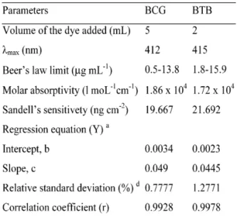
Table 1. Optical characteristics and Statistical Data of the Regression Equations for the Reaction of ABH with BCG and BTB.
a Y = bX + c, where X is the concentration of drug in (µg mL-1)
d Average of six determinations.
Procedure for commercial dosage forms
Results and Discussion
Absorption spectra
The structure of ABH features its basic nature. This structure suggests the possibility of utilizing an anionic dye as chromogenic reagent. In chloroform, ABH is not an absorbing species in the visible region. The dyes employed have almost negligible absorbance (Fig 2). In contrast, when a solution of BCG or BTB in chloroform is added to the drug solution, an intense yellow col-ored product is produced immediately (Fig 2). This is due to the conversion of the dye into an open quinonoidal anionic derivative [32], which forms an ion-pair with ABH.
39 Ecl. Quím., São Paulo, 33(2): 37-40, 2008
Figure 2. Absorption spectrum of reagent blanks-BCG (a) and BTB (b), and ion-pair complexes of ABH (8 µg mL-1) with BCG (c) and BTB (d).
Reaction conditions
The experimental conditions were studied and it was found that 5 mL of BCG or 2 mL of BTB were sufficient to produce maximum and reproducible color. The color products were found to be stable for at least 1.5 h.
Investigation of the ABH-BCG ion-pair complex
The composition of the ion-pair complex formed between ABH and BCG/BTB was inves-tigated by Job’s method of continuous variation. It was found that the composition of drug to dye was 1:1 in both cases.
Analytical performance
Linearity of the method
Under the experimental conditions, linear
correlations were obtained between the absor-bances and ABH concentration over the ranges stated in Table 1 with good correlation coeffi-cients and zero intercepts. The apparent molar absorptivities, Sandell’s sensitivities and detec-tion limits[33] were summarized in Table 1.
Interference studies
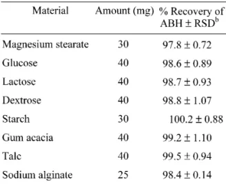
The effects of common excipients and additives were tested for their possible interfer-ences in the assay of ABH. It was also observed that the excipients such as talc, lactose, starch and magnesium stearate did not interfere with the assay, since the formation of an ion-pair complex with anionic dyes needs a basic moiety. The results have been tabulated in Table 2 for a rep-resentative dye, BTB.
Table 2. Determination of ABH a in Presence of Excipients by BTB Method.
a 5 µg mL-1of ABH taken. b Average of five determinations.
Precision and accuracy
Conclusions
The proposed methods are rapid, simple, accurate and in addition, offer advantages in determining ABH, (in pharmaceutical prepara-tions), when extraction difficulties arise with other spectrophotometric methods. Hence, the proposed methods could be adopted routinely for quality control in pharmaceutical industries.
Received 18 December 2007 Accepted 08 May 2008
References
[1] Martindale, The Complete Drug Refernce, 33rd edition, Pharmaceutical Press, London, 2002, p. 825 and 612.
[2] British Pharmacopeial convention, Stationary office, London, 2003, p. 36.
[3] A.F. ElWalily, J Pharm Biomed Anal., 16 (1997) 21. [4] A. Gölcü, C. Yücesoy, S. Serin, IL Farm., 59 (2004) 487. [5] C.S.P. Sastry, R.T. Thirupathi, A. Sailaja, Talanta., 38 (1991) 1057. [6] E.A. Hisham, M.M El-Henawee, H.M. El-Sayed, M.M. Ayad, Spectrochimica Acta Part A., 65 (2006) 1087.
[7] S. Sungur, G. Yurdakul, Scientia Pharmaceutica, 60 (1992) 125. [8] R. Ficarra, P. Ficarra, M.R. Di-Bella, D. Raneri, S. Tommasini, M.L. Calabro, M.C. Gamberin, C. Rustichelli, J Pharm Biomed Anal., 23 (2000) 33.
[9] G.M. Hanna, F.E. Evans, J Pharm Biomed Anal., 24 (2000) 189. [10] B. Alpertunga, S. Sungur, L. Ersoy, S.Y. Manav, Arch Pharm., 323 (1990) 587.
[11] G.W. Caldwell, J.A. Masucci, M. Evangelisto, R. White, J Chromatogr A., 800 (1998) 161.
[12] A.J. Braza, P. Modamio, C.F. Lastra, E.L. Marino, Biomed Chrom., 16 (2002) 517.
[13] I. Lewandowska, J. Tyfczynska, M. Bujak Acta Pol Pharm., 46 (1989) 350.
[14] A.P. Argekar, S.G. Powar, J Pharm Biomed Anal., 21 (2000) 1137. [15] V. Andrisano, R. Gotti, A. Leoni, V. Cavrini, J Pharm Biomed Anal., 21 (1999) 851.
[16] M.E. Abdel-Hamid, Il Farmaco., 55 (2000) 136.
[17] M.I. Maguregui, R.M. Jimenez, R.M. Alonso, J Chromatogr Sci., 36 (1998) 516.
[18] K.A. Assi, B.J. Clark, K.D. Altria, Electrophoresis., 20 (1999) 2723.
[19] A. Jouyban, M. Khoubnasabjafari, H.K. Chan, K.D. Altria, B.J. Clark, Chromatographia., 57 (2003) 191.
[20] A.C. Servais, M. Fillet, P. Chiap, A.M. Abushoffa, P. Hubert, J. Crommen J Sep Sci., 25 (2002) 1087.
[21] W. Lu, R.B. Cole, J Chrom B., 714 (1998) 69.
[22] M. Drozdzik, L. Domanski, J. Wojcicki, A. Pudlo, P. Machoy, J Cli Pharmacol., 43 (2003) 524.
[23] B. Telatynska, J. Wojcicki, M. Drozdzik, B. Gawronska-Szklarz, V. Sulzyc-Bielicka, R. Sterna, Pol J Pharmacol., 55 (2003) 81. [24] J.F. Fernandez-Sanchez, A.S. Carretero, C. Cruces-Blanco, A. Fernandez-Gutierrez, J Pharm Biomed Anal., 31 (2003) 859. [25] J. Milano, G.C. Simone, J Pharm Biomed Anal., 37 (2005) 639. [26] N. Rahman, A. K. Nadeem, H.A.S. Najmul, Il Farmaco., 59 (2004) 47.
[27] A. Safwan, R. Al-Khalil, Il Farmaco., 60 (2005) 771. [28] B.G. Gowda, M.B. Melwanki, J. Seetharamappa, J Pharm Biomed Anal., 25 (2001) 1021.
[29] M.M. Kerdawy, M.A. Moustafa, S.M. Ashry, D.R. El-Waseef, Anal Lett., 26 (1993) 1669.
[30] H.H. Abdine, J Pharm Sci., 14 (2000) 75.
[31] H. Abdine, F. Belal, N. Zoman, Il Farmaco . 57 (2002) 267. [32] T. Higuchi, H.E. Brochmann, Pharmaceutical Analysis., Interscience Publication, New York, pp. 413–418.
[33] J.C. Miller, J.N. Miller, Chapter 4, Wiley, New York, 1984, pp. 83-115.
40 Ecl. Quím., São Paulo, 33(2): 37-40, 2008
Table 3. Evaluation of the Accuracy and Precision of the Proposed Method for ABH Determination.
a Mean of five determinations SD - standard deviation
RSD% - relative standard deviation SAE - standard analytical error.
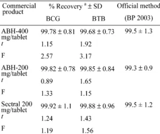
Table 4. Application of the Proposed Spectrophotometric Methods for the Determination of ABH in Dosage Forms.
The theoretical values of t and F at P=0.05 are 2.31 and 6.39, respectively.
a Mean of five determinations.
Pharmaceutical applications