A multicenter validation of an endoscopic
classification with narrow band imaging for
gastric precancerous and cancerous lesions
Authors P. Pimentel-Nunes1, 2, *, M. Dinis-Ribeiro1, 3, *, J. B. Soares2, 4, R. Marcos-Pinto5, C. Santos3, C. Rolanda4, 6, R. P. Bastos7,
M. Areia8, L. Afonso9, J. Bergman10, P. Sharma11, T. Gotoda12, R. Henrique9, 13, L. Moreira-Dias1
Institutions Institutions are listed at the end of article.
submitted 4. March 2011 accepted after revision 14. October 2011 Bibliography DOI http://dx.doi.org/ 10.1055/s-0031-1291537 Published online: 31.1.2012 Endoscopy 2012; 44: 236–246 © Georg Thieme Verlag KG Stuttgart · New York ISSN 0013-726X Corresponding author M. Dinis-Ribeiro, MD Department of Gastroenterology
Portuguese Oncology Institute of Porto
Rua Dr. Bernardino de Almeida 4200-072 Porto Portugal Fax: +351-22-5084055 [email protected]
Introduction
!Gastric adenocarcinoma is the second most lethal cancer worldwide with only a minority of gastric adenocarcinomas diagnosed in a curable and re-sectable form [1, 2]. Helicobacter pylori is consid-ered the most important risk factor for gastric cancer, by promoting a multi-step process of chronic gastritis, atrophy, intestinal metaplasia, dysplasia and, finally, intestinal-type adenocarci-noma [3]. Secondary prevention through diagno-sis of premalignant lesions and early gastric can-cer, and screening or follow-up of individuals at high risk, would probably be the most immediate
strategies for improving survival [4, 5]. Endoscopy examination is therefore of paramount impor-tance. However, endoscopic evaluation of gastric mucosa correlates poorly with histological find-ings [6, 7], and it is not surprising that ancillary techniques such as chromoendoscopy have been used for an accurate diagnosis of precancerous le-sions and/or invasiveness of cancerous lele-sions [8–10]. Even so, for diverse reasons these meth-ods are not very popular among endoscopists, particularly those in Western countries.
Diverse descriptions of new methods of electronic chromoendoscopy, namely high resolution with narrow band imaging (NBI), with or without mag-nification, have been published [11–24]. Good re-sults have been reported for the imaging of intes-tinal metaplasia and cancer; however, reliability
*The authors contributed equally to this study and should
be considered joint first authors.
Background and study aim:The reliability and ex-ternal validity of narrow band imaging (NBI) in the stomach have not been described consistent-ly. The aim of the current study was to describe and estimate the accuracy and reliability of a sim-plified classification system for NBI in the diagno-sis of gastric lesions.
Methods: Consecutive patients undergoing NBI endoscopy at two reference centers (n = 85, 33 % with dysplasia) were included in two studies. In total, 224 different areas were biopsied and re-corded onto video. In the derivation study, pre-viously described NBI features were analyzed in order to develop a simplified classification. In the validation study the accuracy and reliability of this classification were estimated among three groups of endoscopists with different levels of ex-pertise in NBI.
Results:The reliability/accuracy results from the derivation study allowed the creation of a simpli-fied NBI classification. In the validation study, “regular vessels with circular mucosa” (pattern A) was associated with normal histology (accura-cy 83 %; 95 % confidence interval [CI] 75 %–90%);
“tubulo-villous mucosa” (pattern B) was associat-ed with intestinal metaplasia (accuracy 84 %; 95CI 77 %–91%; positive likelihood ratio [LR+]=4.75); and “irregular vessels and mucosa” (pattern C) was associated with dysplasia (accuracy 95 %; 95CI 90 %– 99%; LR+=44.33). The reproducibility of these patterns was high (k = 0.62).“Light-blue crest” was moderately reliable (k=0.49) but specific (87 %) for intestinal metaplasia. A variable vascular density (additional pattern + ) was the best feature for Helicobacter pylori gastritis (accu-racy 70 %; 95CI 59 %–80%) but showed only fair reliability (k = 0.38). Non-experienced endosco-pists presented lower agreement (k = 0.6 vs. k = 0.75) and accuracy (74 % vs. 86 %) than interna-tional experts/experienced endoscopists.
Conclusion:A simplified NBI classification is accu-rate and reliable for the diagnosis of intestinal metaplasia and dysplasia. The classification should be further assessed and validated on a per-patient assessment of NBI, and by comparing NBI with other imaging technologies.
has seldom been evaluated, no study has included the whole spectrum of lesions, and no external validation of any defined features has been reported [25].
Thus, the aims of the current study were: to assess the reliability of previously described NBI features for gastric precancerous and neoplastic lesions; to simplify the features under a new classifica-tion; and to validate this classification on a new sample of pa-tients and observers, and to assess the accuracy of the classifica-tion system in endoscopists with a range of NBI experience.
Methods
!Study design and selection of patients
Patients undergoing routine upper gastrointestinal endoscopy at two hospitals in the North of Portugal (Portuguese Oncology In-stitute of Porto and Braga’s Hospital), between September and December 2009 and between February and April 2010, were con-secutively considered and included in this study after giving in-formed consent. Both hospitals are tertiary centers to which pa-tients with superficial lesions are referred and treated with mini-mally invasive techniques [26]. Patients with chronic liver dis-ease, psychiatric conditions, anticoagulant therapy or coagulati-on disorders, were excluded. The ethical committees of both hos-pitals approved the study.
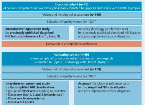
Two studies were planned (see
●
" Fig. e1, available online only):▶the first 45 patients (estimated sample of 100 videos), who were treated between September and December 2009, consti-tuted the“derivation cohort,” which provided data for a study on the reliability of previously described mucosal and vascular features of gastric mucosa using NBI [11– 24]. These data were used to derive a simple NBI classification and were also subject to validity testing using histology as the reference test; ▶ in the second study, a“validation cohort” of 40 new patients
(and a new estimated sample of 100 videos), who were asses-sed between February and April 2010 using endoscopic obser-vations, provided data for the validation of the new NBI classi-fication and assessment of the reliability of the classiclassi-fication within groups of endoscopists with diverse experience.
Endoscopic procedures and selection of videos
Under pharyngeal anesthesia (85 % of the patients) or deep seda-tion (15 % of patients) all patients underwent upper gastrointes-tinal endoscopy using a high resolution (HR) Olympus endoscope with NBI (EVIS EXERA II video system center GIF-180; Olympus, Tokyo, Japan). Detailed observation of esophageal, gastric, and duodenal mucosa was performed and all endoscopic lesions were described accordingly. High resolution videos of low mag-nification ( × 1.5) NBI endoscopy were recorded for further analy-sis from (subject to patient tolerance) five areas of antral, inci-sura, and corpus mucosa. Recordings were also made from those areas with endoscopic changes, either at high resolution white light endoscopy (WLE) or at NBI. A shift between the HR-WLE and HR-NBI was used to ensure the position and precision of the biopsies taken. The total number of histological samples was equal to the total number of videos recorded. From all of the pro-cedures, 140 (derivation study) and 119 (validation study) videos of approximately 10 seconds in duration were recorded consecu-tively and converted into MPEG-4 files of approximately 10 MB each using iMovie (Apple Inc., Cupertino, California, USA). No area was recorded twice. Each video was labeled with a random number and transferred onto a computerized database. Of the
259 videos recorded only those showing highest quality images of mucosal morphology and in which the video observation con-firmed the targeting of the biopsies were selected for use in the respective studies(124 in the derivation study and 100 in the va-lidation study). The quality of the videos was assessed by one ex-perienced endoscopist. The whole potential spectrum of histolo-gical lesions (no intestinal metaplasia or dysplasia [normal mu-cosa], presence of intestinal metaplasia, presence of dysplasia [or carcinoma], and presence of H. pylori irrespective of histology, excluding dysplasia) was considered for video selection, both for the derivation study and for the validation study.
Histopathological procedures
All gastric mucosa specimens were obtained by endoscopic biop-sy at each area selected for video recording, with the exception of videos recorded from superficial lesions where a whole muco-sectomy specimen was obtained. Specimens were fixed in buf-fered formalin, processed for paraffin embedding, sectioned, and stained with hematoxylin and eosin. Gastric specimens were also evaluated for H. pylori infection using modified Giemsa (2 %) stain. Two expert gastrointestinal pathologists, who were blind to the NBI features, made the final histological diagnosis ac-cording to the Sydney–Vienna classification [27,28].
Selection of endoscopists
For the derivation study (reliability assessment of previously de-scribed NBI features), three endoscopists (End1, End2, and End3) who had previous clinical experience of NBI ( > 50NBI gastrosco-pies) each assessed all of the videos; they were blinded to histol-ogy and to the evaluation of the other endoscopists. Endoscopists were instructed to evaluate the videos using previously reported mucosal and vascular features when these features were applic-able to NBI with low magnification [11–24]. In order to over-come the problem of an irregular pattern being assigned differ-ent meanings and being associated with differdiffer-ent pathologies such as H. pylori infection [11], intestinal metaplasia [12], and dysplasia/cancer [16, 18], the endoscopists were instructed to state the pattern as“irregular” only when they observed a com-plete architectural loss of the mucosal or vascular pattern (see below for definition of variables).
In the validation study, nine observers were included:
▶the three experienced endoscopist observers (End1, End2, and End3) who participated in the derivation study;
▶three different gastroenterologists who had not participated in the first assessment, and who had special interest in chro-moendoscopy but with diverse NBI experience ( < 50NBI gas-troscopies); these were designated non-experienced obser-vers;
▶and three international expert NBI endoscopists (from Europe, USA and Japan); these were designated the expert observers. All of the observers classified the second set of videos after re-ceiving a pen drive with a PowerPoint presentation (Microsoft Office 2003, Microsoft Inc., Redmond, Washington, USA), which contained the rationale of the derived classification and example videos.
Variables
In the derivation study the following variables were included [11–24].
▶Mucosal pattern: regular circular (well delineated circular/oval glands), regular tubulo-villous (well delineated tubulo or vil-lous or ridge glandular pattern) or irregular (glandular pattern
is clearly irregular with architecture distortion with absent glandular pattern in some areas possible).
▶Light blue crest (LBC): presence (yes) or not (no) of blue-whit-ish slightly raised areas.
▶White opaque substance (WOS): presence (yes) or not (no) of white material above the mucosa that could be either well defined (regular) or not (irregular).
▶Vascular pattern: regular (vessels well defined in the center or surrounding the glands) or irregular (areas with clearly anomalous vessels associated with architecture distortion of the mucosa). If there were areas where vessels were not seen clearly but without anomalous configurations they were in-cluded in the regular group.
▶Vascular thickness: subjective opinion of normal/thick vessels or somewhat thin or ultrathin.
▶Vascular density: high density (almost all of the glands are surrounded by reddish vessels with some areas with vessel agglomerates possible) or low density (vessels are not seen clearly surrounding all of the glands, pale colored vessels). ▶Variable vascular density (VVD): presence (yes) or not (no) of
alternating areas of high and low density in the same video. To assess the reliability of these features for inclusion in the clas-sification, each video was classified by all observers according to these NBI features and a grade for certainty was assigned. In ad-dition, each observer was asked to make an histological diagnosis based on the NBI features and, again, to assign this diagnosis a grade of certainty. Histopathological assessment was considered to be the gold standard or reference test for accuracy estimates.
Statistical analysis
The Statistical Package for Social Sciences (SPSS 17.0 Package Fa-cility, SPSS Inc., Chicago, Illinois, USA) was used for data support and analysis.
The proportion of overall agreement was the proportion of cases for which two observers agreed. The proportions of specific
agreement relative to each category was an estimation of the probability of, given that one observer makes a rating in a cate-gory, that the other observers will rate the same. The generalized formulae, for more than two observers, for the proportions of overall and specific agreement, were calculated by dividing the total number of actual agreements by the total number of possi-ble agreements. Light’s Kappa (mean of the kappa values obtain-ed from each pair of raters) was also calculatobtain-ed. The nonparamet-ric bootstrap was used to estimate the 95 % confidence intervals (CIs). Strength of agreement was considered as follows: slight 0–0.2; fair 0.2– 0.4; moderate 0.4 –0.6; substantial 0.6– 0.8; al-most perfect 0.8–1.
For estimation of sample size, a target estimate standard error of 0.1 in kappa values was determined. Each video classification was compared with the histological diagnosis of the corresponding specimens (gold standard or reference test). Sensitivity, specifici-ty, and global accuracy were estimated separately and for all nine observers combined, along with the 95 %CIs. Likelihood ratios (LR) were estimated based on mean sensitivity and specificity es-timates.
Results
!Derivation study
Description of participants and videos
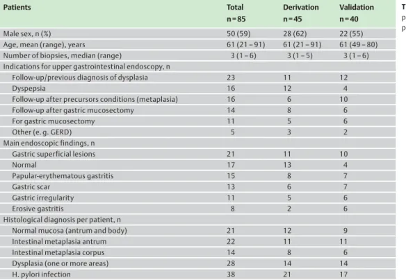
Patient characteristics and a description of endoscopic proce-dures included in the study are shown in
●
" Table 1. In 20 % ofthe patients five or more biopsies were possible and in 28 % four biopsies of different areas were performed. Due to the specialist nature of the institutions, a large number of endoscopic examina-tions were performed for dysplasia or mucosectomy (36 %). This yielded a significant number of histological samples of dysplasia (n = 23) and metaplasia (n = 40). A total of 124 good quality videos (67 normal mucosa, 34 intestinal metaplasia, and 23 dysplasia
Table 1 Description of
participants and endoscopic procedures (n = 85). Patients Total n = 85 Derivation n = 45 Validation n = 40 Male sex, n (%) 50 (59) 28 (62) 22 (55)
Age, mean (range), years 61 (21– 91) 61 (21– 91) 61 (49– 80)
Number of biopsies, median (range) 3 (1– 6) 3 (1– 5) 3 (1– 6)
Indications for upper gastrointestinal endoscopy, n
Follow-up/previous diagnosis of dysplasia 23 11 12
Dyspepsia 16 12 4
Follow-up after precursors conditions (metaplasia) 16 6 10
Follow-up after gastric mucosectomy 14 8 6
For gastric mucosectomy 11 5 6
Other (e. g. GERD) 5 3 2
Main endoscopic findings, n
Gastric superficial lesions 21 11 10
Normal 17 13 4
Papular-erythematous gastritis 15 8 7
Gastric scar 13 6 7
Gastric irregularity 11 5 6
Erosive gastritis 8 2 6
Histological diagnosis per patient, n
Normal mucosa (antrum and body) 21 12 9
Intestinal metaplasia antrum 22 11 11
Intestinal metaplasia corpus 14 8 6
Dysplasia (one or more areas) 28 14 14
H. pylori infection 38 21 17
GERD, gastroesophageal reflux disease.
Ta ble 2 Der iva tio n stud y : co rr e la tio n b e tween fe atu re s o f n a rr o w ba nd im a g in g a nd hi st ol og y ; a cc o rd in g to o b ser ve r c lass ifica ti o n (E n d 1 , E nd 2, a n d E n d 3) rel ia bi li ty measur es a re e st ima ted (p ropor tio n o f a greem ent a n d sp ecifi c p rop or ti on s o f a gr ee ment [P a] a n d k ap p a [k ]) for p re vio u sl y d es cri b ed fe a tur es (n = 1 2 4 ). Histological findings, % O bser v e r classif ication 1, n (%) R eliabilit y [95 %CI] Normal Intestinal metaplasia Dysplasia H. p ylori End1 End2 End3 Pa K Mu co sa lp a ttern 0. 82 [0 .7 7 – 0. 87 ] 0 .7 1 [0 .6 2 – 0. 80] R e gul a r, ci rcu la r 86 9 1 51 5 6 (45 ) 64 (52 ) 62 (5 0) 0. 84 [0 .7 8 – 0. 89 ] R e gul a r, tub u lo -v il lo us 12 89 3 4 5 3 9 (32 ) 3 6 (29 ) 3 9 (3 2 ) 0 .7 3 [0 .6 3 – 0. 81 ] Ir re gu la r 2 2 9 6 4 2 9 (23 ) 24 (19 ) 23 (1 8) 0. 84 [0 .8 2 – 0. 97 ] Ligh t b lu e c res t 0. 89 [0 .8 4, 0. 93] 0 .5 8 [0 .49, 0. 72 ] N o 95 51 97 76 1 0 6 (8 5 ) 104 (8 4) 101 (81) 0. 93 [0 .9 0 – 0. 96 ] Y e s 5 49 3 2 4 1 8 (15 ) 2 0 (16 ) 2 3 (1 9 ) 0 .6 5 [0 .5 1 – 0. 78 ] Wh it e o paq u e su b st an ce 0. 93 [0 .8 9, 0. 97] 0 .5 7 [0 .40, 0. 79 ] N o 100 95 61 10 0 1 17 (9 4) 111 (8 9) 111 (89) 0. 96 [0 .9 4 – 0. 98 ] Y e s 0 5 3 9 0 7 (6) 13 (11 ) 13 (1 1) 0. 64 [0 .4 7 – 0. 77 ] Re g u la ri ty Re g u la r 0 5 1 5 0 4 8 8 Irre gu lar 0 0 1 3 0 3 5 5 Va sc u la r p a tt e rn 0. 96 [0 .9 3 – 0. 98 ] 0 .8 9 [0 .7 8 – 0. 96] R e gul a r 9 8 9 8 3 96 9 6 (77 ) 100 (8 1) 99 (8 0) 0. 97 [0 .9 6 – 0. 99 ] Ir re gu la r 2 2 9 7 4 2 8 (23 ) 24 (19 ) 25 (2 0) 0. 91 [0 .8 2 – 0. 97 ] Va sc u la r th ic k n e ss 0. 62 [0 .5 6 – 0. 68 ] 0 .1 9 [0 .0 2 – 0. 28] Thi n or ul tr athi n 2 8 2 4 3 4 2 2 6 8 (55 ) 9 (7) 2 8 (2 3 ) 0 .3 2 [0 .2 2 – 0. 41 ] Thi c k (n o rm al ) 7 2 7 6 6 6 7 8 5 6 (45 ) 115 (9 3) 96 (7 7) 0. 74 [0 .6 8 – 0. 79 ] Va sc u la r d e n si ty 0. 65 [0 .6 0 – 0. 71 ] 0 .2 4 [0 .1 3 – 0. 36] Lo w 3 4 3 8 3 2 4 3 4 9( 3 9 ) 3 6( 2 9 ) 4 4( 3 6 ) 0 .4 9[ 0 .3 9 – 0. 59 ] Hi gh (n or mal ) 66 62 68 57 7 5 (61 ) 88 (71 ) 80 (6 5) 0. 73 [0 .6 7 – 0. 78 ] V a ri ab le va sc ul ar den si ty 0. 61 [0 .5 5 – 0. 67 ] 0 .2 1 [0 .0 9 – 0. 35] N o (no rma l) 61 47 0 3 8 5 5 (57 ) 5 4 (54 ) 5 6 (5 7 ) 0 .6 6 [0 .5 8 – 0. 73 ] Y e s 3 9 5 3 100 62 4 1 (43 ) 46 (46 ) 43 (4 3) 0. 55 [0 .4 6 – 0. 63 ] 1Endosc opists with previous clinical expe rienc e o f NBI (> 50 NBI g as trosc opies).
[20 high grade, 3 low grade]), 50 of which H. pylori positive, were recorded to accompany 140 histological samples.
Accuracy and reproducibility of previously described NBI
features
The correlation of different NBI features with histology, the relia-bility measures for all described NBI features, and the presump-tive histological results are shown in
●
" Table 2 and●
" Table 3.The identification of different mucosal and vascular patterns was associated with high reproducibility (Pa > 80 % with k = 0.71 and k = 0.89, respectively). The identification of LBC or WOS was also associated with substantial reproducibility (Pa > 80 % with k = 0.58 and k = 0.57, respectively). Conversely, other vascular fea-tures such as thickness or density had only weak to moderate re-producibility (Pa = 0.62 and Pa = 0.65 with k = 0.19 and k = 0.24, respectively).
Variable vascular density was the most accurate parameter for identification of H. pylori gastritis, although inter-observer agree-ment was only fair (Pa = 0.61 and k = 0.21).
Mucosal and vascular patterns derived from the histology results were highly valid for metaplasia and dysplasia (
●
" Table3). Thehistological diagnosis proposed by the observers showed a high agreement with histological diagnosis (Pa = 0.82, k = 0.71) but only a weak to moderate agreement for H. pylori infection (Pa = 0.61, k = 0.21).
Development of a simplified classification
Using the results from the derivation study, a simplified classifi-cation of gastric lesions was established (
●
" Table 4 and●
" Fig.2).The presence of LBC contributed but was not essential to the agnosis of intestinal metaplasia. Also, WOS contributed to the di-agnosis of dysplasia or cancer; however, it could only be consid-ered when an irregular mucosal/vascular pattern was seen.
Validation study
Description of participants and videos
The characteristics of patients and endoscopic procedures inclu-ded in the validation study are shown in
●
" Table 1, and weresim-ilar to those of the derivation study. Dysplasia in previous biop-sies and mucosectomy were important indications for endoscopy (45 %). A total of 100 good quality videos (40 normal mucosa, 38 intestinal metaplasia, 22 dysplasia [19 high grade, 3 low grade], and 39 of which had H. pylori) were recorded to accompany 119 histological samples.
Accuracy and reproducibility of the new NBI classification
●
" Table5 shows the correlation between NBI patterns andhisto-logical findings, inter-observer agreement, and accuracy for the different NBI patterns proposed. WOS was not evaluated because only one cancer lesion presented this feature in the validation study. The identification of the different patterns was associated with substantial reproducibility (Pa = 0.76, k = 0.62). There were no differences in the reproducibility between the experts and the experienced observers. However, the agreement between the group of experienced observers was higher when compared with the non-experienced group (k = 0.75 vs. 0.60). The identifi-cation of the LBC was associated with a moderate agreement (Pa = 0.77, k = 0.49); again, however, this agreement was better be-tween the experienced than bebe-tween the non-experienced ob-servers (k = 0.77 vs. k = 0.40, respectively). The reproducibility of the variable vascular density NBI pattern for H. pylori was fair to moderate (Pa = 0.71, k = 0.38) with no differences between the groups.
The new NBI patterns derived and proposed were highly accurate for metaplasia and, in particular, for dysplasia (
●
" Table 5). Theac-curacy of the patterns (A– C) was higher in the experts and ex-perienced observers compared with the non-exex-perienced
(pat-Table 3 Derivation study: features on narrow band imaging with accuracy estimates for the diagnosis of the several gastric lesions.
Mucosal/vascular pattern
Outcome Mean sensitivity (range)
[95 %CI]
Mean specificity (range) [95 %CI]
Mean accuracy (range) [95 %CI]
Regular, tubulo-villous Intestinal metaplasia 0.89 (0.85– 0.94)
[0.78– 1.00] 0.90 (0.88– 0.95) [0.84– 0.96] 0.90 (0.87– 0.94) [0.85– 0.95] Irregular Dysplasia 0.96 (0.92– 1.00) [0.82– 1.00] 0.98 (0.96– 0.99) [0.92– 1.00] 0.97 (0.97– 0.98) [0.95– 1.00]
Light blue crest Intestinal metaplasia 0.48 (0.42– 0.55)
[0.31– 0.66] 0.96 (0.95– 0.97) [0.89– 0.99] 0.83 (0.81– 0.85) [0.77– 0.90] Variable vascular density H. pylori infection 0.62 (0.45– 0.69) [0.46– 0.75] 0.70 (0.58– 0.78) [0.56– 0.82] 0.66 (0.52– 0.73) [0.56– 0.75]
Table 4 Proposed classification for gastric lesions on narrow band imaging. Regular mucosal and vascular patterns favor the absence of dysplasia, ridge or
tubulo-villous being found in areas with intestinal metaplasia. The light blue crest should be considered specific for intestinal metaplasia but its absence does
not exclude intestinal metaplasia. A variable vascular density may favor the presence ofH. pylori infection.
Proposed classification
A B Hp + C
Mucosal pattern Regular
circular
Regular
ridge/tubulo-villous
Light blue crest Regular Irregular/absent
White opaque substance
Vascular pattern Regular
Thin/peripheric (body (b) or thick/central (a) vessels
Regular Regular
with variable vascular density
Irregular
Expected outcome Normal Intestinal metaplasia H. pylori infection Dysplasia
tern A: accuracy 87 % vs. 74 %; pattern B: 88 % vs. 76 %; pattern C: 97 % vs. 91 %) as well as the accuracy of LBC (87 % vs. 81 %). There were no differences between the groups with regard to H. pylori pattern accuracy.
The predictive values for the classification were as follows (
●
"Ta-ble 6). For the diagnosis of intestinal metaplasia using pattern B, a likelihood ratio for a positive test (presence of a certain mucosal pattern) was estimated to be 4.75 and the likelihood ratio of its absence (LR–) was 0.13.The diagnosis of intestinal metaplasia using LBC showed LR + = 5.13 and LR– =0.37.Variable vascular density produced predictive values of LR + = 2.53 and LR–=0.48. Importantly, pattern C presented LR + = 44.33 and LR–=0.16 for dysplasia.
Discussion
!To the best of our knowledge this is the first study of reproduci-bility and validity of HR-NBI endoscopy for the identification of several different gastric lesions that incorporates the entire spec-trum of precancerous and intestinal-type cancer lesions. The study provides significant evidence that NBI endoscopy may be a reproducible and accurate method for the diagnosis of gastric pre-neoplastic and cancer lesions. Indeed, some NBI features were very reproducible and were associated consistently with gastric lesions. Furthermore, it appears that a learning curve for the identification of these NBI features should be allowed for, as
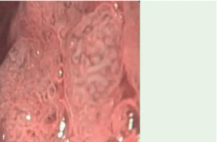
Fig. 2 The simplified classification of gastric lesions using narrow band
imaging. a Pattern Aa–regular circular/oval mucosa surrounding regular
thick vessels in the center of the gland; histology showed normal antrum
mucosa; b Pattern Aa +– variable vascular density with areas of low (right)
and high (top and center) density but with a pattern of a normal antrum;
histology showedH. pylori gastritis. These areas of low or high density may
render the visualization of narrow band imaging (NBI) features difficult, however, they should not be confused with the irregularity seen in
dyspla-sia (f); c Pattern Ab–regular circular mucosa that is surrounded by regular
vessels, not in the center of the gland as in the antrum; histology showed normal body mucosa. It is important to recognize that normal body and
antrum mucosa have a slightly different NBI appearance; d Pattern B–
regular ridge/tubulo-villous mucosa with regular vessels; histology showed
intestinal metaplasia; e Pattern B + with light blue crest–ridge mucosa with
some blue-whitish slightly raised areas and a variable vascular density;
histology showed intestinal metaplasia withH. pylori gastritis; f Pattern C –
irregular mucosa with irregular vessels and a complete architectural loss of the mucosal and vascular pattern; this flat lesion presented high grade dysplasia.
Ta ble 5 V a li d a tion st ud y : co rr e la tion b e tw een fe atur es o n n a rr o w b a n d ima gi ng (N BI) a nd hi st ol og y, a n d rep ro duci b il it y an d d ia gn osi s ac cur a c y of the si m p lifi e d N BI pa tt ern s. Ov erall reproducibilit y M uc osal pat tern V ariable vascular densit y A B C Light blue crest Obs e rv ed outc o me , % N o rm al 76 22 2 1 1 3 9 Inte sti n al m e tapl asi a 9 8 9 2 68 4 9 Dys p lasi a 2 14 8 4 17 1 3 H. py lo ri in fe c tion 3 9 5 9 2 41 6 4 All o bser ve rs [95 % CI] Pa 0. 76 [0. 7 1 – 0. 80 ] 0 .7 5 [0. 67 – 0. 81 ] 0 .7 4 [0. 67 – 0. 79 ] 0 .8 1 [0. 72 – 0. 87] 0. 77 [0 .7 3 – 0 .8 1 ] 0 .7 1 [0. 66 – 0. 75] k 0 .6 2 [0. 55 – 0. 62 ] 0. 49 [0 .3 8 – 0 .5 8 ] 0 .3 8 [0. 29 – 0. 48] M e an se nsi tivi ty (ra ng e) 0. 76 (0 .6 0 – 1. 00 ) [0 .6 3 – 0. 89 ] 0. 90 (0 .7 9 – 1. 00 ) [0. 7 9 – 1. 00] 0. 84 (0 .5 0 – 1. 00) [0 .7 0 – 0. 99 ] 0. 68 (0 .5 6 – 0. 82 ) [0 .5 3 – 0. 84 ] 0. 64 (0 .4 1 – 0. 86) [0 .4 9 – 0. 80] Me a n sp ecif icit y (ran g e) 0 .9 4 (0 .8 3 – 1. 00 ) [0 .8 3 – 0. 97 ] 0. 81 (0 .6 1 – 0. 98 ) [0. 7 2 – 0. 91] 0. 98 (0 .9 5 – 1. 00) [0 .9 5 – 1. 00 ] 0. 87 (0 .6 8 – 0. 98 ) [0 .7 9 – 0. 95 ] 0. 75 (0 .5 6 – 0. 95) [0 .6 1 – 0. 88] Me a n ac cu ra c y (r an ge) 0 .8 2 (0 .6 4 – 0. 95 ) [0. 7 5 – 0. 90] 0. 83 (0 .6 4 – 0. 95 ) [0 .7 5 – 0. 90 ] 0. 84 (0 .6 7 – 0. 95 ) [0. 7 7 – 0. 91] 0. 95 (0 .8 8 – 1. 00) [0 .9 0 – 0. 99 ] 0. 80 (0 .7 0 – 0. 91 ) [0 .7 3 – 0. 88 ] 0. 70 (0 .6 3 – 0. 86) [0 .5 9 – 0. 80] Ex per ts 1[9 5 % C I] Pa 0. 84 [0. 7 8 – 0. 90 ] 0 .8 0 [0. 69 – 0. 88 ] 0 .8 3 [0. 77 – 0. 89 ] 0 .8 8 [0. 78 – 0. 96] 0. 81 [0 .7 5 – 0 .8 6 ] 0 .7 1 [0. 66 – 0. 77] K 0 .7 5 [0. 65 – 0. 83 ] 0. 60 [0 .4 7 – 0 .7 2 ] 0 .4 0 [0. 28 – 0. 53] M e an se nsi tivi ty (ra ng e) 0. 69 (0 .62, 0. 76 ) [0 .5 5 – 0. 83 ] 0. 92 (0 .8 5 – 1. 00 ) [0. 8 3 – 1. 00] 0. 90 (0 .8 3 – 0. 96) [0 .7 8 – 1. 00 ] 0. 72 (0 .5 6 – 0. 82 ) [0 .5 6 – 0. 87 ] 0. 71 (0 .5 7 – 0. 86) [0 .5 7 – 0. 86] Me a n sp ecif icit y (ran g e) 0 .9 5 (0 .9 1 – 1. 00 ) [0 .8 6 – 0. 98 ] 0. 80 (0 .7 9 – 0. 82 ) [0. 7 0 – 0. 89] 0. 98 (0 .9 5 – 1. 00) [0 .9 5 – 1. 00 ] 0. 82 (0 .6 8 – 0. 89 ) [0 .7 3 – 0. 91 ] 0. 73 (0 .6 4 – 0. 85) [0 .5 9 – 0. 87] Me a n ac cu ra c y (r an ge) 0 .8 2 (0 .7 7 – 0. 86 ) [0. 7 4 – 0. 89] 0. 82 (0 .7 7 – 0. 86 ) [0 .7 4 – 0. 90 ] 0. 84 (0 .8 1 – 0. 86 ) [0. 7 7 – 0. 91] 0. 96 (0 .9 4 – 0. 99) [0 .9 2 – 1. 00 ] 0. 78 (0 .7 3 – 0. 94 ) [0 .7 0 – 0. 86 ] 0. 72 (0 .6 3 – 0. 86) [0 .6 2 – 0. 82] Ex peri en ce d o bs er ve rs 2[9 5 % C I] Pa 0. 84 [0. 7 8 – 0. 89 ] 0 .8 5 [0. 77 – 0. 90 ] 0 .7 8 [0. 69 – 0. 86 ] 0 .9 0 [0. 81 – 0. 97] 0. 91 [0 .8 6 – 0 .9 5 ] 0 .7 7 [0. 71 – 0. 83] K 0 .7 5 [0. 65 – 0. 83 ] 0. 77 [0 .6 2 – 0 .8 8 ] 0 .4 6 [0. 32 – 0. 60] M e an se nsi tivi ty (ra ng e) 0. 91 (0 .7 9 – 1. 00 ) [0 .8 3 – 1. 00 ] 0. 89 (0 .8 2 – 0. 94 ) [0. 7 9 – 1. 00] 0. 94 (0 .8 8 – 1. 00) [0 .8 5 – 1. 00 ] 0. 67 (0 .5 9 – 0. 79 ) [0 .5 1 – 0. 83 ] 0. 58 (0 .4 1 – 0. 68) [0 .4 2 – 0. 74] Me a n sp ecif icit y (ran g e) 0 .9 5 (0 .9 1 – 1. 00 ) [0 .8 6 – 0. 98 ] 0. 93 (0 .8 3 – 0. 98 ) [0. 8 7 – 0. 99] 0. 99 (0 .9 7 – 1. 00) [0 .9 6 – 1. 00 ] 0. 98 (0 .9 7 – 0. 98 ) [0 .9 5 – 1. 00 ] 0. 81 (0 .6 4 – 0. 95) [0 .6 9 – 0. 93] Me a n ac cu ra c y (r an ge) 0 .9 1 (0 .8 6 – 0. 95 ) [0. 8 6 – 0. 97] 0. 91 (0 .8 6 – 0. 95 ) [0 .8 6 – 0. 97 ] 0. 92 (0 .8 7 – 0. 95 ) [0. 8 6 – 0. 97] 0. 98 (0 .9 5 – 1. 00) [0 .9 5 – 1. 00 ] 0. 87 (0 .8 5 – 0. 91 ) [0 .8 1 – 0. 94 ] 0. 70 (0 .6 3 – 0. 80) [0 .5 9 – 0. 80] Non-experien ce d o bser ve rs 3[9 5 % C I] Pa 0. 75 [0. 6 8 – 0. 82 ] 0 .7 6 [0. 65 – 0. 84 ] 0 .7 6 [0. 68 – 0. 83 ] 0 .7 0 [0. 56 – 0. 82] 0. 73 [0 .6 6 – 0 .7 7 ] 0 .7 3 [0. 67 – 0. 79] K 0 .6 0 [0. 48 – 0. 71 ] 0. 40 [0 .2 6 – 0 .5 3 ] 0 .4 4 [0. 31 – 0. 57] M e an se nsi tivi ty (ra ng e) 0. 67 (0 .6 0 – 0. 76 ) [0 .5 3 – 0. 82 ] 0. 87 (0 .7 9 – 0. 91 ) [0. 7 6 – 0. 98] 0. 68 (0 .5 0 – 0. 83) [0 .4 9 – 0. 87 ] 0. 67 (0 .6 2 – 0. 71 ) [0 .5 1 – 0. 83 ] 0. 64 (0 .4 9 – 0. 76) [0 .4 8 – 0. 79] Me a n sp ecif icit y (ran g e) 0 .9 2 (0 .8 3 – 0. 97 ) [0 .8 5 – 0. 99 ] 0. 71 (0 .6 1 – 0. 80 ) [0. 6 0 – 0. 82] 0. 98 (0 .9 6 – 1. 00) [0 .9 5 – 1. 00 ] 0. 80 (0 .7 4 – 0. 92 ) [0 .7 1 – 0. 90 ] 0. 70 (0 .5 6 – 0. 77) [0 .5 6 – 0. 84] Me a n ac cu ra c y (r an ge) 0 .7 4 (0 .6 4 – 0. 83 ) [0. 6 6 – 0. 83] 0. 74 (0 .6 4 – 0. 83 ) [0 .6 6 – 0. 83 ] 0. 76 (0 .6 7 – 0. 84 ) [0. 6 8 – 0. 85] 0. 91 (0 .8 8 – 0. 94) [0 .8 5 – 0. 96 ] 0. 76 (0 .7 0 – 0. 84 ) [0 .6 7 – 0. 84 ] 0. 67 (0 .6 6 – 0. 72) [0 .5 7 – 0. 78] 1Three international e xper ts with kno wn interest in NBI. 2The three e xperien ced endos copist (En d1, End2, and End3) who par ticipate d in the derivati o n stud y 3Three dif ferent g as troenterologis ts w ith specia li nterest in chromoe ndosc o p y but with dive rse NBI experienc e (< 5 0 NBI g as trosc opies).
expertise is associated with a more precise identification of the lesions and a more accurate diagnosis.
The main limitations to the study were the fact that NBI features were not compared with HR-WLE and that some gastric lesions that can present dysplasia, such as erosions or ulcers, were not included. Also not included were some gastric pathologies that are associated with increased cancer risk, such as autoimmune gastropathy (pernicious anemia) or Ménétrier disease. Therefore, the results should be regarded as applicable to patients with pre-cancerous conditions and intestinal Lauren-type gastric adeno-carcinoma. In addition, as low grade dysplasia was observed in only six lesions it cannot be accurately stated that the classifica-tion will be applicable equally to low grade dysplasia and high grade dysplasia/intramucosal adenocarcinoma. Also, diffuse-type adenocarcinomas were not included in the current study. Other studies have provided interesting results on the role of NBI for the detection of gastric pre-neoplastic and cancer lesions [11–24]. However, there are several aspects of the previous stud-ies that must be borne in mind. The definitions of the NBI fea-tures were different between the studies, only one study evaluat-ed the reproducibility of some NBI features, and no single study included the whole spectrum of gastric lesions in the same clas-sification or evaluation [25]. Moreover, almost all of the data come from Japan, and therefore applicability to Western coun-tries is uncertain. Another consideration is that almost all of the previous studies used NBI with high magnification (up to × 80), which is not practical in clinical routine as these endoscopes are not available in most centers, at least not in Western countries. To our knowledge, only three other studies have attempted to identify H. pylori gastritis. Alaboudy et al. [23] was the only study that used NBI without magnification; however, the NBI patterns in this study were complex and no reproducibility analysis of these complex patterns was undertaken. Bansal et al. [11] asso-ciated irregularity of mucosal and vascular patterns and a low vascular density to H. pylori gastritis. However, they did not de-fine irregularity, nor was any reproducibility analysis undertak-en, and a low number of patients was included. Tahara et al. [19] associated H. pylori gastritis to different patterns with enlarged pits and increased density of irregular vessels. However, again, no reproducibility analysis was done and in neither of these stud-ies was dysplasia considered. This is important because, in our opinion, the“irregularity” described by those studies is clearly different from the irregularity that is present in cancer lesions. To overcome this problem in the current study, irregularity and the different patterns described in previous studies was defined as“variable vascular density,” which did indeed show a positive association with H. pylori gastritis; however, the reproducibility of this feature on NBI was relatively low and independent of
ex-pertise. The results from all of the studies suggest that, even though NBI may be superior to WLE for the identification of H. pylori gastritis, NBI (at least without magnification) does not re-place other diagnostic tests (e. g. histology) that are clearly more sensitive, specific, and reproducible.
In the case of intestinal metaplasia identification, however, the existing evidence suggests that NBI may be an important tool. In-deed, the two studies by Bansal and Tahara associated a tubulo-villous mucosal pattern to intestinal metaplasia with great accu-racy [11, 19]. These results were confirmed in the present study, with 92 % accuracy of this mucosal pattern for the diagnosis of metaplasia by experienced observers. Uedo et al. [21] suggested that the finding of LBC with NBI is also very accurate for intestinal metaplasia; reproducibility of this finding was not evaluated in the study. In the current study, however, LBC was not very sensi-tive for metaplasia (68 % global sensitivity), though it was specific (87 % global specificity). Nevertheless, at least when using a low magnification and the Olympus EXERA system (Uedo used high magnification and the LUCERA system), it appears that a tubulo-villous mucosal pattern is more consistently associated and in a more reproducible manner with metaplasia.
Evaluation of dysplasia/cancer using NBI has been performed in several studies. Initial studies have also attempted to establish NBI patterns that could help to predict the degree of tumor differ-entiation [13, 18, 20]. Despite some positive results, the authors concluded that NBI was not able to replace histology for tumor differentiation [18]. In these studies, however, the authors did not evaluate which NBI features could help to differentiate be-tween benign and dysplastic lesions. Some authors suggest that “adenoma” may have an NBI pattern resembling pattern B in the current study [20, 22], but the precise histological diagnosis of the lesions was not provided in previous studies and it is likely that they presented foci of low grade dysplasia in the context of extensive intestinal metaplasia. In contrast, Kaise et al. [16] eval-uated NBI criteria for cancer diagnosis in gastric depressed le-sions. They concluded that irregular vascular and mucosal pat-terns were very specific for cancer, although sensitivity was low and reproducibility only moderate. These relatively modest re-sults may be because cancer lesions were compared only with a particular benign lesion – gastric erosions. Indeed, the same group of authors using the same criteria for dysplasia but evalu-ating different suspicious lesions found that the sensitivity and specificity of magnification endoscopy NBI for the diagnosis of dysplasia were > 90 % [29]. Ezoe et al. [14] obtained similar results using similar NBI criteria. In the current study, the accuracy of ir-regular mucosal and vascular patterns, considered as a complete architecture distortion, was evaluated for the diagnosis of gastric dysplasia in the context of all benign lesions and using only low
Table 6 Estimates of predictive values.
Outcome Feature LR + Positive predictive value, % LR– Negative predictive value, %
1 % 10 % 20 % 50 % 1 % 10 % 20 % 50 %
Intestinal metaplasia
B pattern 4.75 5 34 54 83 0.13 0.10 1 3 11
Light blue crest 5.13 5 36 56 84 0.37 0.40 4 8 27
Dysplasia
C pattern 44.33 31 83 92 98 0.16 0.20 2 4 14
LR, likelihood ratio
magnification. The results were very impressive, showing not only that these NBI criteria are very accurate for the diagnosis of dysplasia, similar to the study of Kato et al. [29], but also that they allow dysplasia diagnosis in a very reproducible manner. Taking all of the evidence together, it can be suggested that this pattern irregularity with NBI appears to be a good method for the diag-nosis of gastric dysplasia, at least for high grade dysplasia. What is the clinical utility of all these aspects? The current study is not a comparative study of NBI with WLE, it is not possible to state whether or not NBI is better than WLE for the diagnosis of gastric lesions. However, comparing the current results with those from other studies that used HR-WLE [30–32] NBI appears to be clearly superior to WLE for the diagnosis of these types of gastric lesions. More importantly, Kaise et al. [16], Ezoe et al. [14], and Kato et al. [29] compared WLE with NBI for the diagno-sis of cancer and concluded that the accuracy of NBI is significant-ly superior to WLE. Recentsignificant-ly, Cappelle et al. [12] compared the yield of NBI to WLE in the surveillance of patients with a previous history of intestinal metaplasia or dysplasia. They used the same NBI system as in the current study and similar definitions; how-ever, H. pylori gastritis was not considered and no reproducibility analysis was done. Capelle et al. showed that NBI was better than routine WLE for the diagnosis of intestinal metaplasia and dys-plasia. Taking all of these data together, we can say that NBI may help to select suspicious areas for biopsy and probably replace the random biopsy method with a biopsy strategy directed to the suspicious areas of metaplasia and/or dysplasia. Moreover, NBI can also help to delineate gastric lesions for endoscopic gas-tric resection. Indeed, Kadowaki et al. [15], using similar NBI criteria for dysplasia, have shown that NBI is better than WLE for early cancer demarcation recognition.
Another aspect that is relevant to the current study is the fact that endoscopists with more NBI experience and expertise recog-nize NBI patterns with more agreement and with more diagnos-tic accuracy. This is important as it reflects a learning curve for this new technology and suggests that even highly experienced endoscopists should undergo training before using NBI in clinical routine. Moreover, data are presented as range of expected val-ues for accuracy. These results may therefore help endoscopists to judge the accuracy of their diagnosis when NBI is used for the assessment of gastric lesions in clinical practice.
Using likelihood ratios, it was possible to estimate the predictive values of this classification in different scenarios (
●
" Table 6). Forreference centers in countries with a high prevalence of precan-cerous and canprecan-cerous lesions, as in Portugal and Eastern Europe-an countries, this classification will able to confirm/predict the presence of dysplasia (post-test probability or positive predictive values of 83 %– 98%) and to exclude both intestinal metaplasia and dysplasia within centers of low prevalence countries (nega-tive predic(nega-tive value lower than 1 %). Further studies of a per-pa-tient assessment of utility in such specific settings, namely by comparing NBI with WLE or other technologies are needed. In conclusion, HR-NBI endoscopy is an efficacious technique for the characterization of gastric intestinal metaplasia and, in particular, dysplasia. Irregularity of vascular/mucosal pattern is identified in a reproducible manner and it is consistently asso-ciated with gastric dysplasia. HR-NBI endoscopy may be an im-portant tool for the early diagnosis of gastric pre-neoplastic and neoplastic lesions and for therapeutic procedures.
Competing interests: None
Institutions
1Department of Gastroenterology, Portuguese Oncology Institute of Porto,
Porto, Portugal
2Department of Physiology, Cardiovascular Research and Development Unit,
Porto Faculty of Medicine, Porto University, Porto, Portugal
3CINTESIS/Biostatistics and Medical Informatics, Faculty of Medicine, Porto
University, Porto, Portugal
4Department of Gastroenterology, Hospital de Braga, Braga, Portugal
5Department of Gastroenterology, Centro Hospitalar do Porto, Porto, Portugal
6Surgical Sciences Research Domain, Life and Health Sciences Research
Insti-tute (ICVS), School of Health Sciences, University of Minho, Braga, Portugal
7Department of Gastroenterology, Hospital de São João, Porto, Portugal
8Department of Gastroenterology, Portuguese Oncology Institute of Coimbra,
Coimbra, Portugal
9Department of Pathology, Portuguese Oncology Institute of Porto, Porto,
Portugal
10Department of Gastroenterology, Academic Medical Center, Amsterdam,
The Netherland
11Division of Gastroenterology and Hepatology, Veterans Affairs Medical
Center, Kansas City, Missouri, USA
12Department of Gastroenterology and Hepatology, National Center for
Global Health and Medicine, Tokyo, Japan
13Department of Pathology and Molecular Immunology, Institute of
Biomedical Sciences Abel Salazar, University of Porto, Porto, Portugal
Acknowledgments
!
This study was supported by a grant for medical investigation from the Portuguese Digestive Endoscopy Society (SPED 2009 In-vestigation Grant).
This study was in part presented as an oral communication at the 18th United European Gastroenterology Week, 23–27 October 2010; Barcelona, Spain.
References
1 Parkin DM, Bray F, Ferlay J et al. Estimating the world cancer burden: Globocan 2000. Int J Cancer 2001; 94: 153–156
2 Hundahl SA, Menck HR, Mansour EG et al. The National Cancer Data
Base report on gastric carcinoma. Cancer 1997; 80: 2333–2341
3 Correa P. Human gastric carcinogenesis: a multistep and multifactorial
process– First American Cancer Society Award Lecture on Cancer
Epi-demiology and Prevention. Cancer Res 1992; 52: 6735–6740
4 deVries AC, van Grieken NC, Looman CW et al. Gastric cancer risk in pa-tients with premalignant gastric lesions: a nationwide cohort study in the Netherlands. Gastroenterology 2008; 134: 945–952
5 Stemmermann GN, Fenoglio-Preiser C. Gastric carcinoma distal to the cardia: a review of the epidemiological pathology of the precusors to
a preventable cancer. Pathology 2002; 34: 494–503
6 Lin BR, Shun CT, Wang TH et al. Endoscopic diagnosis of intestinal
me-taplasia of stomach– accuracy judged by histology.
Hepatogastroen-terology 1999; 46: 162–166
7 Redeen S, Petersson F, Jonsson KA et al. Relationship of gastroscopic fea-tures to histological findings in gastritis and Helicobacter pylori
infec-tion in a general populainfec-tion sample. Endoscopy 2003; 35: 946–950
8 Areia M, Amaro P, Dinis-Ribeiro M et al. Estimation of the extent of gas-tric intestinal metaplasia by methylene blue chromoendoscopy. Eur J
Gastroenterol Hepatol 2008; 20: 939–940
9 Areia M, Amaro P, Dinis-Ribeiro M et al. External validation of a classifi-cation for methylene blue magnificlassifi-cation chromoendoscopy in
prema-lignant gastric lesions. Gastrointest Endosc 2008; 67: 1011–1018
10 Dinis-Ribeiro M, da Costa-Pereira A, Lopes C et al. Magnification chro-moendoscopy for the diagnosis of gastric intestinal metaplasia and
dysplasia. Gastrointest Endosc 2003; 57: 498–504
11 Bansal A, Ulusarac O, Mathur S et al. Correlation between narrow band imaging and nonneoplastic gastric pathology: a pilot feasibility trial.
Gastrointest Endosc 2008; 67: 210–216
12 Capelle LG, Haringsma J, de Vries AC et al. Narrow band imaging for the detection of gastric intestinal metaplasia and dysplasia during
surveil-lance endoscopy. Dig Dis Sci 2010; 55: 3442–3448
13 Endo T, Nosho K, Arimura Y et al. Study of the tumor vessels in depres-sed-type early gastric cancers using narrow band imaging magnifying endoscopy and cDNA array analysis. Digestive Endoscopy 2005; 17: 210–217
14 Ezoe Y, Muto M, Horimatsu T et al. Magnifying narrow-band imaging versus magnifying white-light imaging for the differential diagnosis of gastric small depressive lesions: a prospective study. Gastrointest Endosc 2010; 71: 477–484
15 Kadowaki S, Tanaka K, Toyoda H et al. Ease of early gastric cancer de-marcation recognition: a comparison of four magnifying endoscopy
methods. J Gastroenterol Hepatol 2009; 24: 1625–1630
16 Kaise M, Kato M, Urashima M et al. Magnifying endoscopy combined with narrow-band imaging for differential diagnosis of superficial de-pressed gastric lesions. Endoscopy 2009; 41: 310–315
17 Kato M, Kaise M, Yonezawa J et al. Trimodal imaging endoscopy may improve diagnostic accuracy of early gastric neoplasia: a feasibility study. Gastrointest Endosc 2009; 70: 899–906
18 Nakayoshi T, Tajiri H, Matsuda K et al. Magnifying endoscopy combined with narrow band imaging system for early gastric cancer: correlation of vascular pattern with histopathology (including video). Endoscopy 2004; 36: 1080–1084
19 Tahara T, Shibata T, Nakamura M et al. Gastric mucosal pattern by using magnifying narrow-band imaging endoscopy clearly distingui-shes histological and serological severity of chronic gastritis.
Gastroin-test Endosc 2009; 70: 246–253
20 Tamai N, Kaise M, Nakayoshi T et al. Clinical and endoscopic
character-ization of depressed gastric adenoma. Endoscopy 2006; 38: 391–394
21 Uedo N, Ishihara R, Iishi H et al. A new method of diagnosing gastric intestinal metaplasia: narrow-band imaging with magnifying
endos-copy. Endoscopy 2006; 38: 819–824
22 Yao K, Iwashita A, Tanabe H et al. White opaque substance within su-perficial elevated gastric neoplasia as visualized by magnification en-doscopy with narrow-band imaging: a new optical sign for differen-tiating between adenoma and carcinoma. Gastrointest Endosc 2008;
68: 574–580
23 Alaboudy AA, Elbahrawy A, Matsumoto S et al. Conventional narrow-band imaging has good correlation with histopathological severity of Helicobacter pylori gastritis. Dig Dis Sci 2011; 56: 1127–1130
24 Okubo M, Tahara T, Shibata T et al. Changes in gastric mucosal patterns seen by magnifying NBI during H. pylori eradication. . J Gastroenterol 2011; 46: 175–182
25 Curvers WL, van den Broek FJ, Reitsma JB et al. Systematic review of nar-row-band imaging for the detection and differentiation of abnormal-ities in the esophagus and stomach (with video). Gastrointest Endosc 2009; 69: 307–317
26 Dinis-Ribeiro M, Pimentel-Nunes P, Afonso M et al. A European case ser-ies of endoscopic submucosal dissection for gastric superficial lesions. Gastrointest Endosc 2009; 69: 350–355
27 Dixon MF, Genta RM, Yardley JH et al. Classification and grading of gas-tritis. The updated Sydney System. International Workshop on the His-topathology of Gastritis, Houston 1994. Am J Surg Pathol 1996; 20: 1161–1181
28 Dixon MF. Gastrointestinal epithelial neoplasia: Vienna revisited. Gut 2002; 51: 130–131
29 Kato M, Kaise M, Yonezawa J et al. Magnifying endoscopy with narrow-band imaging achieves superior accuracy in the differential diagnosis of superficial gastric lesions identified with white-light endoscopy: a prospective study. Gastrointest Endosc 2010; 72: 523–529
30 Eshmuratov A, Nah JC, Kim N et al. The correlation of endoscopic and histological diagnosis of gastric atrophy. Dig Dis Sci 2010; 55: 1364– 1375
31 Gonen C, Simsek I, Sarioglu S et al. Comparison of high resolution mag-nifying endoscopy and standard videoendoscopy for the diagnosis of Helicobacter pylori gastritis in routine clinical practice: a prospective
study. Helicobacter 2009; 14: 12–21
32 Kato M, Kaise M, Yonezawa J et al. Autofluorescence endoscopy versus conventional white light endoscopy for the detection of superficial gastric neoplasia: a prospective comparative study. Endoscopy 2007;
39: 937–941
Fig.e1 is available online:
online content viewable at:www.thieme-connect.de/ejournals/abstract/endoscopy/ doi/10.1055/s-0031-1291537
Inception cohort (n=45)
of consecutive patients in two tertiary hospitals submitted to upper GI endoscopy with HR-NBI Olympus
Vídeos and histological assessment (n=140) Selection of quality videos (n= 124)*
*estimated number of 100 for Pa and kappa confidence intervals estimates with clinical relevance Derivation of a simplified classification
Interobserver agreement study
for previously published described
NBI features (observers End 1, 2 and 3)
Accuracy (histology as reference test) for previously published described NBI features and presumptive endoscopic diagnosis
Validating cohort (n=40)
of new sample of consecutive patients in two tertiary hospitals submitted to upper GI endoscopy with HR-NBI Olympus
Vídeos and histological assessment (n=119) Selection of quality videos (n= 100)*
Interobserver agreement study
for the simplified NBI classification
3 groups of observers according to expertise
▪Observers End 1, 2 and 3 (experienced)
▪Observers Nonexperience
▪Observers Experts
Accuracy (histology as reference test)
for the simplified NBI classification and presumptive endoscopic diagnosis
Fig. e1 Two studies were planned: 1) a
consecu-tive sample of 45 patients (yielding an estimated sample of 100 videos), observed across two tertiary hospitals between September and December 2009,
constituted the“derivation cohort,” which provided
data for a reliability study of previously described mucosal and vascular features using high resolution narrow band imaging (HR-NBI) in gastric mucosa [11– 24]. These features were used to develop a simplified NBI classification, which also considered validity measures (using histology as the reference
test); 2) a“validation cohort” of 40 new patients
(and a new estimated sample of 100 videos) was assessed between February and April 2010; endo-scopic observations were used to validate the new NBI classification and assess the reliability of the classification within groups of endoscopists with di-verse experience.