Jebmh.com
Original Article
J. Evid. Based Med. Healthc., pISSN- 2349-2562, eISSN- 2349-2570/ Vol. 3/Issue 57/July 18, 2016 Page 2988
ROLE OF NARROW BAND IMAGING SYSTEM IN EARLY DIAGNOSIS OF POTENTIALLY
MALIGNANT LESIONS OF ORAL CAVITY
Laveena P. Mehta1, Yessukrishna Prabhakar Shetty2, Sharad B. Bhalekar3, Yogesh G. Dabholkar4, Haritosh K. Velankar5
1Post Graduate Student, Department of ENT, D. Y. Patil University School of Medicine, Navi Mumbai, Maharashtra, India.
2Senior Registrar, Department of ENT, D. Y. Patil University School of Medicine, Navi Mumbai, Maharashtra, India.
3Associate Professor, Department of ENT, D. Y. Patil University School of Medicine, Navi Mumbai, Maharashtra, India.
4Professor & HOD, Department of ENT, D. Y. Patil University School of Medicine, Navi Mumbai, Maharashtra, India.
5Professor, Department of ENT, D. Y. Patil University School of Medicine, Navi Mumbai, Maharashtra, India.
ABSTRACT
INTRODUCTION
Oral squamous cell carcinomas (OSCC) are frequently preceded by or associated with leucoplakia, erythroplakia, oral submucous fibrosis or lichen planus. The standard method of revealing PML potentially malignant lesions of oral mucosa or submucosa is usually done via conventional oral examination (COE), direct visualisation and biopsy with the aid of image-enhanced endoscopy along with high-resolution imaging system. In view of the promising results of using Narrow Band Imaging as a screening aid in cancer screening, a prospective randomised study was performed to evaluate statistically the efficacy of NBIS in diagnosis of potentially malignant lesions in oral cavity using histopathology as gold standard.
METHODOLOGY
236 potentially malignant lesions in oral cavity from 195 patients underwent Broadband white light & NBI-endoscopic evaluation according to the Inoue’s classification. The histopathology results were compared with Broadband white light & Narrow Band light evaluation.
RESULTS
The sensitivity, specificity, positive predictive value, negative predictive value and clinical accuracy for the detection of oral potentially malignant disorders or worse by Narrow Band Imaging were 96 %, 83%, 96%, 83% and 93.22 % respectively. The sensitivity, specificity, positive predictive value, negative predictive value and clinical accuracy with conventional broadband white light oral examination were 72%, 63%, 88%, 36% and 70.33% respectively for the detection of oral potentially malignant disorders.
CONCLUSION
NBI endoscopy has diagnostic utility as ‘Optical Biopsy’ for superficial squamous neoplasms in the oral cavity and hence can be
considered as a screening tool in high risk patients.
KEYWORDS
Narrow Band Imaging, Leucoplakia, Erythroplakia, Lichen Planus, Broadband White light, Oral Premalignant Lesions, Optical Biopsy.
HOW TO CITE THIS ARTICLE: Mehta LP, Shetty YP, Bhalekar SB, et al. Role of narrow band imaging system in early diagnosis of potentially malignant lesions of oral cavity. J. Evid. Based Med. Healthc. 2016; 3(57), 2988-2991.
DOI: 10.18410/jebmh/2016/651
INTRODUCTION: Narrow band imaging (NBI) is an emerging imaging technology for use in detecting dysplasia in potentially malignant lesions by endoscope.1 In contrast
to conventional imaging using white light imaging (WLI), NBI is designed to enhance the viewing of superficial capillaries and neo-angiogenesis in the mucosal surface, based on the principle that light with different bandwidths has different penetration depths.2
In comparison with the normal mucosa, the capillaries and microvessels in a cancerous lesion look enlarged with tortuous, disorganised configuration and high density. Background colouration (BC), a colour change in the area between IPCL, is seen in the neoplastic lesions of oral cavity but not in the benign lesions using NBI with magnifying endoscopy. In combination with IPCL pattern classification, BC can provide additional information on accurate discrimination of squamous cell carcinoma/high grade neoplasia from low grade neoplasia/non-atypia. In view of the promising results of using NBI as a screening aid in cancer screening, a prospective randomised study was performed to evaluate statistically the efficacy of NBIS in diagnosis of potentially malignant lesions in oral cavity using histopathology as gold standard.
Financial or Other, Competing Interest: None. Submission 23-06-2016, Peer Review 01-07-2016, Acceptance 08-07-2016, Published 18-07-2016. Corresponding Author:
Dr. Sharad B. Bhalekar,
Jebmh.com
Original Article
J. Evid. Based Med. Healthc., pISSN- 2349-2562, eISSN- 2349-2570/ Vol. 3/Issue 57/July 18, 2016 Page 2989 METHODOLOGY: Patients with potentially malignant
lesions like homogenous/non-homogenous leucoplakia, erythroplakia, lichen planus were randomly selected for this prospective study. Patients having oral submucous fibrosis (OSMF) were excluded from the study as the submucosal microvasculature cannot be visualised in case of OSMF. 236 potentially malignant lesions in oral cavity from 195 patients underwent Broadband white light & NBI-endoscopic evaluation using ENF-VT2 Visera Rhino-Laryngo Videoscope at a tertiary health care facility with adequate outpatient, inpatient, and diagnostic capabilities during the period from July 2013 to January 2016. All patients were subjected to BWL & NBI evaluation on a back-to-back fashion so that primary BWL was followed by NBI and primary NBI was followed by BWL. To avoid affecting the first imaging results, the two different examinations were done by two different experts & the reporting of the first examination was completed before the second imaging was started. Endoscopic narrow band imaging findings were classified into five types (I to V) according to the features of the mucosal intraepithelial papillary capillary loops assessed according to the Inoue’s classification.3-6 (Figure 1- 4).
Type I: Normal epithelium.
Type II: Inflammatory changes.
Type III: Low-grade intraepithelial neoplasia.
Type IV V-1: High-grade intraepithelial neoplasia carcinoma in situ.
Type V-2, V-3, V-N: invasive carcinoma.
Fig. 1: A. BWL Normal Mucosa;
B. Normal Mucosa on NBI Scopy (Type I IPCL loops)
Fig. 2: A. BWL suggestive of Inflammation; B. Chronic Inflammation on NBI Scopy
(Type II IPCL loops)
Fig. 3: A. BWL suggestive of Inflammation; B. Low Grade Dysplasia on NBI Scopy
(Type III IPCL loops)
Fig. 4: A. BWL suggestive of Chronic Inflammation; B. High Grade Dysplasia on NBI Scopy
(Type IV IPCL loops)
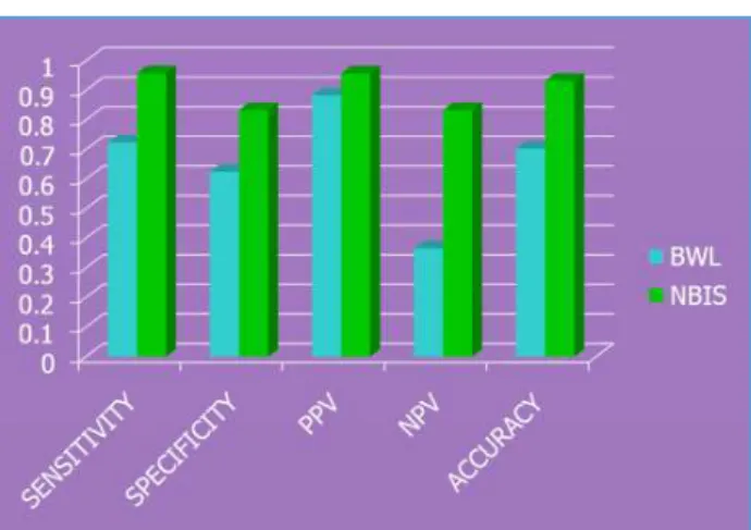
Fig. 5: Comparison between Broadband White Light (BWL) Narrow Band Imaging Scopy (NBIS)
Jebmh.com
Original Article
J. Evid. Based Med. Healthc., pISSN- 2349-2562, eISSN- 2349-2570/ Vol. 3/Issue 57/July 18, 2016 Page 2990 RESULTS: A total of 236 lesions from 195 patients with
mean age of 45.66 years with ± 14.33 years were observed. Out of 236 lesions, 186 lesions were leucoplakia, 46 lesions were erythroplakia and 4 lesions were lichen planus. The sensitivity, specificity, positive predictive value, negative predictive value and clinical accuracy for the detection of oral potentially malignant disorders or worse by Narrow Band Imaging were 96 %, 83%, 96%, 83% and 93.22 % respectively. The sensitivity, specificity, positive predictive value, negative predictive value and clinical accuracy with conventional broadband white light oral examination were 72%, 63%, 88%, 36% and 70.33% respectively for the detection of oral potentially malignant disorders (Figure 5). The negative predictive value of Narrow Band Imaging was far better than the negative predictive value of conventional broadband white light for the detection of oral potentially malignant disorders or worse. Narrow Band Imaging aided the detection of 32 lesions which were undetected by conventional oral examination.
DISCUSSION: Oral cancer is among the 10 most common cancers worldwide and an early detection & prompt treatment offer the best chance for cure. As patient awareness regarding the danger of oral cancer increases, the demand for an appropriate screening tool is bound to increase.6 It has been reported that oral squamous cell
carcinoma (OSCC) are frequently preceded by or associated with leucoplakia, erythroplakia, oral submucous fibrosis or lichen planus.7,8 The standard method of revealing PML
potentially malignant lesions of oral mucosa or sub-mucosa is usually done via Conventional Oral Examination (COE), direct visualisation and biopsy with the aid of image-enhanced endoscopy along with high-resolution imaging system. NBI allows to display the vascular micro-architecture in oral mucosa & hence the disorganised, immature neo-vascularisation in malignancy can be visualised in vivo. The short wavelength blue light (415 nm) penetrates into the mucosa superficially and highlights the superficial vasculature in brown against the blue-green mucosa background. The larger wavelength green band (540 nm) passes through to the sub-mucosal layer and identifies prominent vessels. In contrast to the conventional white light system using broadband RGB technology, the NBI endoscopy system provides images with better contrast and higher resolution on the superficial structures.9 The basic
unit of the vascular micro-architecture of oral mucosa is the intra-papillary capillary loop (IPCL).The normal IPCL loop is
seen as characteristic brown dots with “hair pin” pattern.
Immature angiogenesis plays a critical role in the transition from premalignant to malignant lesions. IPCL analysis demonstrates characteristic changes like dilatation, twisting, elongation, meandering, calibre change and non-uniformity in the appearance of each IPCL which is closely related to structural atypia of the squamous epithelium.10 Background
colouration (BC), a colour change in the area between IPCL, is seen in the neoplastic lesions of oral cavity but not in the benign lesions using NBI with magnifying endoscopy. BC positivity seems to be a useful and reliable finding in the oral cavity malignant lesions to discriminate SCC or high-grade
neoplasia (HGN) from low-grade neoplasia (LGN) or nonatypia.11 In our study, assessment using BC has resulted
in better sensitivity and specificity for distinguishing between HGN/SCC and LGN/nonatypia. A prospective study to elucidate the limitations such as intra- and inter-observer variations is required to acknowledge the significance of BC. Now a days, three established criteria of morphology of IPCL of NBI have been utilised clinically. The characteristics of these criteria was investigated and analysed on oral leucoplakia. Criteria I consisted of brownish spots and demarcation line with irregular microvascular patterns, criteria II consisted of well-demarcated brownish area with thick dark spots and/or winding vessels, and criteria III consisted of elongation, twist, and meandering destruction of IPCL pattern.12 In our study, the clinical accuracy of
Narrow Band Imaging (93.22%) was far better as compared to Broadband white light (70.33%). The odds ratio (95% confidence interval) for detecting dysplasia based on morphologic appearances of BWL, and microvasculature patterns of NBI, were 43.12, and 95.16, respectively, which were significantly better than BWL (p<1×10-15). In case of
oral submucous fibrosis and intramural lesions, clinical palpation is more helpful as Narrow Band Imaging is useful only in case of mucosal/submucosal lesions like leucoplakia, erythroplakia or carcinoma in situ.
CONCLUSION: NBI is a promising optical technology which can be utilised in the early detection of potentially malignant lesions of oral cavity. NBI endoscopy has diagnostic utility as
‘Optical Biopsy’ for superficial squamous neoplasms in the
oral cavity and hence can be considered as a screening tool in high risk patients. Further prospective studies are required to interpret clinical significance of Background coloration in Narrow Band Imaging System.
REFERENCES
1. Gono K, Yamazaki K, Doguchi N, et al. Endoscopic observation of tissue by narrowband illumination. Opt Rev 2003;10(4):211-215.
2. Li ZH, Gao W, Lei WB, et al. The clinical utility of narrow band imaging in the surveillance of mucosa and submucosa lesions in head and neck regions. Head Neck Oncol 2013;5(3):29.
3. Inoue H. Endoscopic diagnosis of tissue atypism (EA) in the pharyngeal and esophageal squamous epithelium; IPCL pattern classification and ECA classification. Kyobu Geka 2007;60(Suppl 8):768-775.
4. Piazza C, Dessouky O, Peretti G, et al. Narrow-band imaging: a new tool for evaluation of head and neck squamous cell carcinomas. Review of the literature. Acta Otorhinolaryngol Ital 2008;28(2):49-54. 5. Chai NL, Ling-Hu EQ, Morita Y, et al. Magnifying
Jebmh.com
Original Article
J. Evid. Based Med. Healthc., pISSN- 2349-2562, eISSN- 2349-2570/ Vol. 3/Issue 57/July 18, 2016 Page 2991
6. Scully C, Bagan JV, Hopper C, et al. Oral cancer: current and future diagnostic techniques. American Journal of Dentistry 2008;21(4):199-209.
7. Villa A, Villa C, Abati S. Oral cancer and oral erythroplakia: an update and implication for clinicians. Aust Dent J 2011;56(3):253-256.
8. Lapthanasupkul P, Poomsawat S, Punyasingh J. A clinicopathologic study of oral leukoplakia and erythroplakia in a Thai population. Quintessence Int 2007;38(8):e448–e455.
9. Yang SW, Lee YS, Chang LC, et al. Light sources used in evaluating oral leukoplakia: broadband white light versus narrowband imaging. Int J Oral Maxillofac Surg 2013;42(6):693-701.
10. Kikuchi O, Ezoe Y, Morita S, et al. Narrow-band imaging for the head and neck region and the upper gastrointestinal tract. Japanese Journal of Clinical Oncology 2013;43(5)458-465.
11. Minami H, Inoue H, Ikeda H, et al. Usefulness of background colouration in detection of esophago-pharyngeal lesions using NBI magnification. Gastroenterology Research and Practice Article ID 529782, 2012;p. 6.