Susana Gomes Rodrigues Martins
[Nome completo do autor]
[Nome completo do autor]
[Nome completo do autor]
[Nome completo do autor]
[Nome completo do autor]
[Nome completo do autor]
[Nome completo do autor]
Licenciada em Biologia Celular e Molecular
[Habilitações Académicas]
[Habilitações Académicas]
[Habilitações Académicas]
[Habilitações Académicas]
[Habilitações Académicas]
[Habilitações Académicas]
[Habilitações Académicas]
Setembro, 2019
A protective role for ataxia-telangiectasia mutated
in hemolytic conditions
[Título da Tese]
Dissertação para obtenção do Grau de Mestre em
Genética Molecular e Biomedicina
Dissertação para obtenção do Grau de Mestre em
[Engenharia Informática]
Orientadora: Doutora Ana Rita Carlos, Investigadora Pós-doutorada
Instituto Gulbenkian de Ciência, Portugal
Júri:
Presidente: Doutora Paula Gonçalves, Professora Associada, Faculdade de Ciências e Tecnologia da Universidade Nova de Lisboa
Arguente: Doutora Birte Blankenhaus, Investigadora Pós-doutorada da Faculdade de Medicina da Universidade de Lisboa
Susana Gomes Rodrigues Martins
[Nome completo do autor]
[Nome completo do autor]
[Nome completo do autor]
[Nome completo do autor]
[Nome completo do autor]
[Nome completo do autor]
[Nome completo do autor]
Licenciada em Biologia Celular e Molecular
[Habilitações Académicas]
[Habilitações Académicas]
[Habilitações Académicas]
[Habilitações Académicas]
[Habilitações Académicas]
[Habilitações Académicas]
[Habilitações Académicas]
Setembro, 2019
Dissertação para obtenção do Grau de Mestre em
Genética Molecular e Biomedicina
Dissertação para obtenção do Grau de Mestre em
[Engenharia Informática]
Orientadora: Doutora Ana Rita Carlos, Investigadora Pós-doutorada
Instituto Gulbenkian de Ciência, Portugal
A protective role for ataxia-telangiectasia mutated
in hemolytic conditions
A protective role for ataxia-telangiectasia mutated in hemolytic conditions
Copyright © Susana Gomes Rodrigues Martins, Faculdade de Ciências e Tecnologia, Universidade Nova de Lisboa.
A Faculdade de Ciências e Tecnologia e a Universidade Nova de Lisboa têm o direito, perpétuo e sem limites geográficos, de arquivar e publicar esta dissertação através de exemplares impressos reproduzidos em papel ou de forma digital, ou por qualquer outro meio conhecido ou que venha a ser inventado, e de a divulgar através de repositórios científicos e de admitir a sua cópia e distribuição com objetivos educacionais ou de investigação, não comerciais, desde que seja dado crédito ao autor e editor.
v
Acknowledgments
This was a long but grateful year. I had the opportunity to meet and work with fantastic people throughout this year that contributed to make my year happier and successful in the laboratory. First, I would to thank my supervisor, Rita, that for one year helped me to become a scientist. Thank you for all the patience and support and for being an excellent teacher. You treated me like a scientist and tried to make me as independent as possible. I learned a lot with you. I hope everything goes well in your new laboratory and that our pathways one day will cross again. Next, I would like to thank Sílvia for helping me and teach me almost everything I know about mice. I said almost everything, because Rita also helped. You helped me overcome my fear of being bitten by a mouse and to feel comfortable with them. Now, we will work closer and I hope to learn even more with you.
Then, I would like to thank Miguel Soares, the principal investigator of my host laboratory, for giving me the opportunity to work in this amazing laboratory, with these amazing people. This is a very special laboratory where people do excellence science.
I also would like thank Sofia, a person that helped me from day one. You did a tour of IGC with me on day one and helped me to become a member of this community. Every time I have a problem, I go see you and you are always willing to help with a big smile.
I also would like to thank Susana for the help with the malaria model. Thanks to you, now I know how to recognize a parasite and you made my work easier in the microscope. I want to thank you also for all the help and suggestions revising my thesis and for saying yes promptly when Rita asked you to help. Regarding this matter, I also want to thank Jess that also promptly accepted to read my thesis and help.
Thanks to all the members of Inflammation Laboratory, including former members, for all the fun lunches and all the conversation and moments we shared together. Thank you Faouzi, Gil, Jess, Patricia, Qian, Rita, Rui, Sílvia, Sofia, Sumnima, Susana, Vital and Wilson.
I also want to thank the Instituto Gulbenkian de Ciência for receiving me and to thank the flow cytometry facility and the animal house facility that also helped to make this project possible. Finally, but not less important, I would like to thank my family and friends. I am lucky to have a special and wonderful family that always supports me and contribute to make my life happier and fulfilled. I want to make a special thank you to my parents for all the patience they have with me, for making all the efforts to make my life easier and for always being there for me. And a thank you to all my friends that I had the pleasure to meet across the years who accompanied my journey and always support me. They contribute to make life happier and funnier.
vi
Thanks to all of you,“Sometimes people come into your life for a moment, a day, or a lifetime. It matters not the time they spent with you but how they impacted your life in that time” – Unknown
vii
Resumo
Ataxia-telangiectasia mutada (ATM) é uma cinase que desempenha funções chave na sinalização da ativação de vias de reparação do DNA e de resposta ao stress oxidativo, entre outras. Mutações em ATM levam ao desenvolvimento de ataxia-telangiectasia (A-T), uma doença caracterizada por exemplo por ataxia, imunodeficiências e desregulação do potencial redox.
Malária é caracterizada por hemólise excessiva com acumulação de hemoglobina e heme no plasma, causando danos nos tecidos. Dados não publicados do laboratório anfitrião demonstraram que: i) ratinhos sem Atm são mais suscetíveis a malária do que ratinhos controlo, revelando uma nova doença onde Atm é essencial; ii) ratinhos controlo infetados com malária apresentam aumento da ativação das vias de reparação do DNA no baço, um órgão hematopoiético. Considerando estes dados, o principal objetivo do presente estudo é estudar a contribuição de Atm no compartimento hematopoiético para proteção contra a malária. Dados obtidos durante o presente estudo revelaram que ratinhos controlo tratados com heme, libertado no contexto da malária, mostram aumento da ativação das vias de reparação do DNA no baço, suportando uma possível função protetora de Atm no compartimento hematopoiético no desenvolvimento da malária. Para testar esta hipótese foram gerados e caracterizados ratinhos que apresentam uma deleção em Atm apenas neste compartimento, Vavicre/wtAtmΔ/Δ. Estes
ratinhos são férteis e têm peso normal, ao contrário do observado em ratinhos Atm ko. Ratinhos
Vavicre/wtAtmΔ/Δ infetados com malária revelaram ser mais suscetíveis que os ratinhos controlo,
contudo, apresentam níveis de parasitas semelhantes, sugerindo que Atm tem um papel que não está associado à resistência contra o parasita causador de malária, mas protege de potenciais danos causados por este, um mecanismo designado de tolerância à doença. Este estudo representa um importante passo para a compreensão do papel de Atm no contexto da malária, podendo também ter implicações noutras doenças hemolíticas.
Palavras-chave: Ataxia-telangiectasia mutada (ATM), heme, danos no DNA, compartimento hematopoiético, malária e tolerância à doença.
ix
Abstract
Ataxia-telangiectasia mutated (ATM) is a kinase that plays key roles in DNA repair signaling pathways and oxidative stress response, among others. ATM mutations lead to ataxia-telangiectasia (A-T), a disease characterized for example by ataxia, immunodeficiencies and redox potential dysregulation.
Malaria is characterized by excessive hemolysis with hemoglobin and heme being accumulated in plasma, causing tissue damage. Unpublished data from the host laboratory showed that: i) mice without Atm are more susceptible to malaria than controls, revealing a new disease where the presence of Atm is essential; ii) control mice infected with malaria show increased activation of DNA repair pathways in the spleen, a hematopoietic organ. Considering these data, the main objective of the present study is to address the contribution of Atm in the hematopoietic compartment in the protection against malaria. Data obtained during the present study showed that control mice treated with heme, released from cells in the context of malaria, show increased activation of DNA repair pathways in the spleen, supporting a possible protective function of Atm in the hematopoietic compartment in respect to malaria. To test this hypothesis, mice with Atm deletion exclusively in this compartment, Vavicre/wtAtmΔ/Δ, were generated and
characterized. These mice are fertile and have normal weight, in contrast to Atm ko mice. Malaria-infected Vavicre/wtAtmΔ/Δ mice were found to be more susceptible than control mice, but had similar
levels of parasites, suggesting that Atm plays a role that is not associated with resistance against the malaria parasite, but protects against potential damage caused by it, a mechanism designated disease tolerance. This study represents an important step in understanding the role of Atm in the context of malaria and may also have implications for other hemolytic diseases.
Key-words: Ataxia-telangiectasia mutated (ATM), heme, DNA damage, hematopoietic compartment, malaria and disease tolerance
xi
Content
Acknowledgments ... v
Resumo ... vii
Abstract ... ix
List of Figures ... xiii
List of Tables ... xv
List of Abbreviations and Symbols ... xvii
Chapter 1. Introduction ... 1
1.1 Ataxia-telangiectasia mutated and disease in humans and mice ... 2
1.2 ATM protein ... 3
1.3 ATM as a DNA damage sensor and its targets ... 5
1.4 ATM as an oxidative stress sensor and its targets ... 7
1.5 ATM and the hematopoietic compartment ... 9
1.6 Malaria ... 11
1.6.1 Life cycle of Plasmodium... 11
1.6.2 Mice models of malaria – Plasmodium chabaudi chabaudi ... 13
1.6.3 Malaria infection and heme ... 14
1.6.4 Malaria and disease tolerance ... 15
1.7 Aims of the project ... 16
Chapter 2. Methods and Materials ... 19
Mice ... 19
Mice genotyping ... 19
Deletion check ... 20
Mouse embryonic fibroblasts ... 21
Heme treatment ... 21
Hemolysis models ... 22
Flow cytometry analysis ... 22
SDS-PAGE and western blot ... 23
Chapter 3. Results ... 29
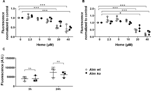
Response of Atm-deficient cells to heme treatment ... 29
Protein expression after heme treatment in vitro ... 32
Heme treatment in vivo ... 33
xii
Atm role in the context of malaria ... 42
Atm role in the context of sterile hemolysis ... 43
Chapter 4. Discussion ... 47
Chapter 5. Conclusion and Future Perspectives ... 53
xiii
List of Figures
Figure 1.1 Disease tolerance in the context of infection. Figure 1.2 Ataxia-telangiectasia mutated structure. Figure 1.3 Examples of ATM targets.
Figure 1.4 Plasmodium life cycle.
Figure 1.5 Atm-deficient mice are more susceptible to Plasmodium chabaudi chabaudi (AR.
Carlos, unpublished data).
Figure 3.1 Atm-deficient MEFs are equally susceptible to heme as wild type MEFs. Figure 3.2 Crystal Violet measurement of MEFs viability.
Figure 3.3 Atm-deficiency alters γ-H2AX and p53 protein expression upon heme treatment. Figure 3.4 Heme treatment induces DNA damage and oxidative stress in vivo.
Figure 3.5 Genotyping of mice from the breeding pair Vavicre/wtAtmlox/wt vs Atmlox/wt.
Figure 3.6 Comparison of Atm expression in the spleen, bone marrow, thymus and liver in different mouse genotypes.
Figure 3.7 Atm expression is significantly reduced in Vavicre/wtAtmΔ/Δ mice in the hematopoietic
compartment.
Figure 3.8 Vavicre/wtAtmΔ/Δ have normal weight.
Figure 3.9 Vavicre/wtAtmΔ/Δ mice are more susceptible to Pcc infection than controls.
Figure 3.10 Sterile hemolysis induced by phenylhydrazine has no effect on Vavicre/wtAtmΔ/Δ mice
survival.
xv
List of Tables
Table 2.1 PCR program.
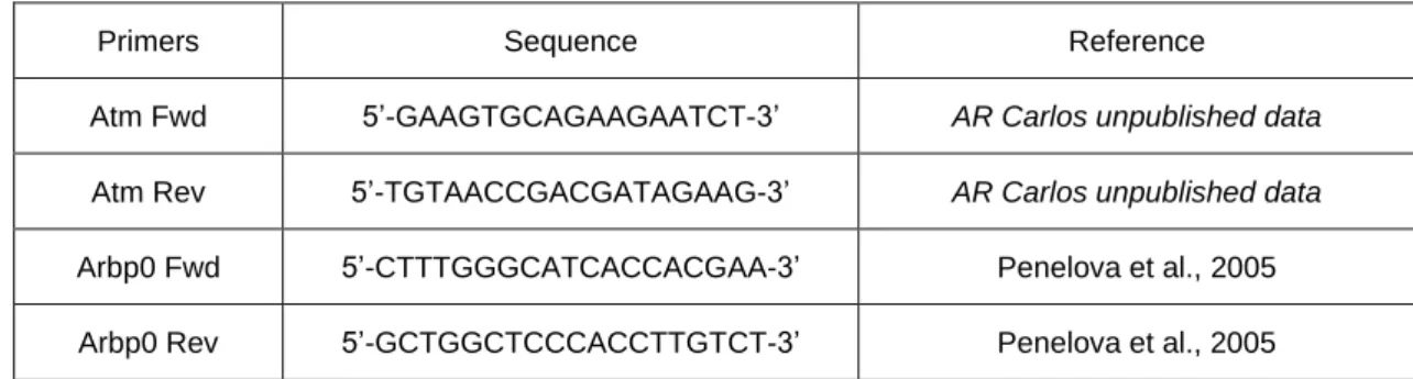
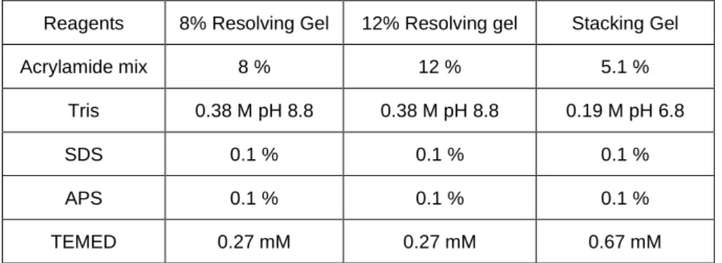
Table 2.2 Primers for genotyping. Table 2.3 Primers for Real-Time qPCR. Table 2.4 Composition of polyacrylamide gel.
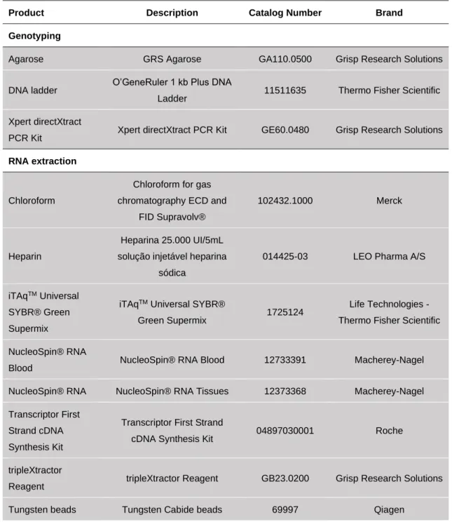
Table 2.5 Running conditions of SDS-PAGE and transfer conditions of western blot Table 2.6 Detailed list of all the reagents, antibodies and media.
Table 3.1 Fertility of Vavicre/wtAtmΔ/Δ mice.
Table 3.2 Number of littermates from each genotype and gender born from the breeding pair Atm
het vs Atm het.
Table 3.3 Number of littermates from each genotype and gender born from the breeding pair
xvii
List of Abbreviations and Symbols
AMPK – AMP-activated protein kinase Arbp0 – acidic ribosomal phosphoprotein PO A-T – ataxia telangiectasia
ATM – ataxia telangiectasia mutated ATR – ATM and Rad 3 related
BID – BH3-interacting domain death agonist bp – base pair
cDNA – complementary DNA Chk2 – checkpoint kinase 2 Cys – cysteine residue
DNA-PK – DNA dependent protein kinase DSB – double strand breaks
FAT – FRAP-ATM-TRRAP FATC – FAT C-terminal motif FBS – fetal bovine serum Fwd – forward
h – hours
H2AX – histone H2A Hb – hemoglobin Het – heterozygous
HR – homologous recombination HRP – horseradish peroxidase HSC – hematopoietic stem cells Hz – hemozoin
IgG – immunoglobulin G i.p. – intraperitoneal
iRBC – infected red blood cell kb – kilobase pair
kDa – kilodalton
kg – kilogram
ko – knock out Lys – lysine residue
MDM2 – murine double minute 2 MEFs – mouse embryonic fibroblasts mg – milligram
min - minutes ml – milliliter
MRN – Mre11-Rad50-NBS1 complex mRNA – messenger RNA
mTOR – mechanistic target of rapamycin NADH -nicotinamide adenine dinucleotide NADPH – reduced nicotinamide adenine dinucleotide phosphate
NHEJ – non-homologous end joining nm – nanometer
Pcc – Plasmodium chabaudi chabaudi PCR – polymerase chain reaction PHZ – phenylhydrazine
PI3K – Phosphoinositide 3-kinase PIKK – PI3K like protein kinase
PIRS – Plasmodium interspersed repeat family PPM1D – type 2C phosphatase WIP1
RBC – red blood cells Rev – reverse
ROS – reactive oxygen species RT – room temperature
Ser – serine residue Thr – threonine residue
xviii
TSC2 - tuberous sclerosis complex 2wt – wild type
μl – microliter μM – micromolar °C – degrees celsius
1
Chapter 1. Introduction
Resistance, a mechanism characterized by its impact on pathogen burden, is the most well-known defense mechanism against infection (McCarville and Ayres, 2018). However, in the recent years, researchers around the world have shown the importance of disease tolerance in the context of infection (McCarville and Ayres, 2018). Disease tolerance includes various mechanisms that have as a primary function the protection of the host against the deleterious effects caused by the presence of the pathogen without targeting directly the pathogen (Martins et al., 2019; McCarville and Ayres, 2018). In this case, the deleterious impact on the host is often a side effect of an attempt of the host to produce a hostile environment for the pathogen and cause its death. These extreme measures sometimes cause damage to the host itself (Chovatiya and Medzhitov, 2014; Martins et al., 2019; McCarville and Ayres, 2018; Soares et al., 2014). One example is the production of ROS as a result of the activation of pathogen killing mechanisms, which leads to DNA, protein and lipid oxidation (McCarville and Ayres, 2018; Medzhitov et al., 2012; Paiva and Bozza, 2014). On the other hand, the pathogen can cause stress and tissue damage, for example through the release of toxins or by inducing the lysis of host’s cells. Thus, disease tolerance mechanisms play a central role in protecting the host from the direct action of the pathogen (Medzhitov et al., 2012).
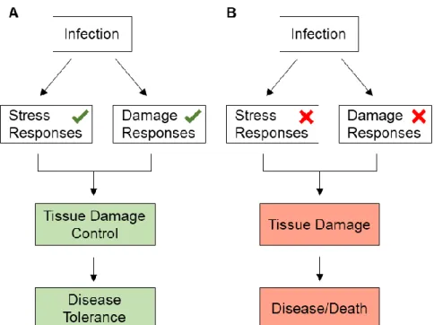
Disease tolerance mechanisms include stress responses, such as responses to oxidative stress, or damage responses, such as DNA damage responses (Martins et al., 2019; Soares et al., 2014). Independently of the response, the goal is to limit the extent of damage as well as prevent and repair it to consequently control tissue damage, ensuring disease tolerance (Figure 1.1 A). When these mechanisms and responses fail, the damage persists and disease develops, compromising the survival of the host (Figure 1.1 B) (Chovatiya and Medzhitov, 2014; Soares et al., 2014). Many proteins drive and regulate stress and/or DNA damage responses, and an example that is required in both types of responses is ATM (ataxia telangiectasia mutated) (Shiloh and Ziv, 2013).
2
Figure 1.1 Disease tolerance in the context of infection.(A) Upon infection, stress and damage responses are activated to counter the stress and damage caused by the pathogen and/or immune-driven mechanisms against the pathogen. When the repair is successful the tissue damage is controlled leading to disease tolerance. (B) When the stress and damage responses are not activated upon infection or repair fails, the damage caused by the parasite persists, leading to disease and possibly compromising host survival.
1.1. Ataxia-telangiectasia mutated and disease in humans and mice
ATM is a protein kinase involved in many pathways related to DNA damage repair (Ambrose and Gatti, 2013; Blackford and Jackson, 2017). Mutations in this protein are associated with the recessive autosomal disease ataxia-telangiectasia (A-T)(Blackford and Jackson, 2017). The most commonly observed features of this disease are neuronal degeneration, such as lack of coordination in voluntary movement (ataxia), and dilation of the blood vessels (telangiectasia) (Blackford and Jackson, 2017). A-T patients also present genomic instability, sensitivity to DNA-damaging agents, such as radiation, and problems with lymphocyte development (e.g. V(D)J recombination and immunoglobulin class switching), leading to immunodeficiency (Balestrini et al., 2016; Shiloh and Ziv, 2013). A-T patients are more susceptible to cancer, specially cancers types associated with the hematopoietic compartment (see section 1.6 ATM and the
hematopoietic compartment), which is a direct consequence of all the changes at the molecular
level in the immune cells, (Shiloh and Ziv, 2013). A-T patients also show elevated levels of oxidative stress (Kamsler et al., 2001; Reichenbach et al., 2002).
The phenotype observed in humans with A-T is similar to the one described for mice lacking functional Atm, allowing for mouse models of the disease to be used to study ATM function and the pathways where it is involved(Barlow et al., 1996). The mouse Atm gene is localized in chromosome 9C, while the human homolog ATM is localized in the long arm of chromosome 11.
3
At nucleotide sequence level, human and mouse gene show an 85 % identity and at amino acid level 84 % identity (Pecker et al., 1996).To study A-T and ATM protein, mouse models of A-T have been generated. An Atm-deficient mice was generated by disrupting the Atm gene with a PGKneo gene (Barlow et al., 1996). Introducing this antisense construct at nucleotide position 5790 of Atm gene causes a frameshift mutation, resulting in a truncated and non-functional protein(Barlow et al., 1996).
Fibroblasts isolated from Atm-deficient mice show cell cycle checkpoint abnormalities, namely in G1 phase, and poor growth (Barlow et al., 1996). These deficiencies in cellular growth, means that Atm-deficient mice have a smaller size at birth and weigh less than wild type (wt) and
heterozygous mice during their lifetime. They also have neurologic abnormalities, although mice
do not show signs of neurodegeneration or gross ataxia, as observed in A-T patients, most likely because they succumb to tumors before reaching the phase of neurodegeneration(Barlow et al., 1996). Even though mice do not seem to have cerebellar degeneration, they have malfunction of the nigrostriatal pathways, age-dependent reduction in dopaminergic neurons, reduced synaptic function in hippocampal neurons and defective neural network(Shiloh and Ziv, 2013). Both male and female Atm-deficient mice are infertile, since they are not able to produce mature gametes due to problems in repairing double strand breaks (DSB) that occur during normal meiotic recombination to form both female and male gametes (Barlow et al., 1996; Coticchio et al., 2014; Rotman and Shiloh, 1998). Immunologic abnormalities and a higher probability of developing malignant thymic lymphomas are also characteristics of these Atm-deficient mice. Besides that, they have a much greater sensitivity to radiation. In terms of molecular effects, the Atm disruption causes chromosomal instability and defects in DSB repair, as observed in A-T patients(Barlow et al., 1996).
1.2 ATM protein
ATM is a kinase that can phosphorylate itself as well as other proteins, contributing directly and indirectly to the regulation of many pathways. Its gene has approximately 150 kb and 66 exons, codifying a 13 kb mRNA transcript and encoding a 350 kDa protein (Barlow et al., 1996) with 3056 amino acid residues(Shiloh and Ziv, 2013). ATM protein localizes predominantly in the nucleus, but it can also be found in the cytoplasm and mitochondria(Ambrose and Gatti, 2013; Shiloh and Ziv, 2013; Valentin-Vega et al., 2012).
ATM is part of the phosphoinositide 3-kinase (PI3K)-like protein kinase (PIKKs) family, which is also constituted by ATR (ATM and Rad3 related) and DNA-PK (DNA dependent protein kinase). The three kinases preferentially phosphorylate serine or threonine residues followed by glutamine (S/T-Q sites). In addition, all three share a similar structure constituted by a kinase domain and a PIKK regulatory domain in the C terminal region, flanked by a FRAP-ATM-TRRAP (FAT) domain and a FAT C-terminal motif (FATC). Between the N-terminal and the FAT domain, there are heat-repeats (HR) domains, with variable length, that mediate protein-protein and DNA-protein interactions (Figure 1.2). ATM also has a leucine zipper region and substrate binding site
4
on the N-terminal region, as well as, a region that functions as an oxidative stress sensor within FATC domain (see section 1.4 ATM as an oxidative stress sensor and its targets) (Bhatti et al., 2011; Blackford and Jackson, 2017).Figure 1.2 Ataxia-telangiectasia mutated structure.
ATM is constituted by a kinase domain (KD, yellow) and a PIKK regulatory domain (PRD, light green) in the C terminal region, flanked by a FRAP-ATM-TRRAP (FAT, red) domain and a FAT C-terminal motif (FATC, blue). At the N-terminal region, ATM has a substrate binding domain (SBD, purple), heat repeats (HR, dark green) and a leucine zipper region (LZR, dark orange). Autophosphorylation site Ser1981 (#) and oxidative stress sensor site Cys2991 (*) are also represented.
The three kinases are responsible for inducing cell cycle arrest and signaling DNA repair mechanisms in response to DNA damage (Blackford and Jackson, 2017). The choice of which pathway is activated depends on the type of damage and phase of the cell cycle.
DNA-PK is recruited and activated in response to DSB and induces DNA repair by classical non-homologous end joining (C-NHEJ) (Blackford and Jackson, 2017). C-NHEJ consist in joining two blunt ends resulting from DSB, without requiring homology, and can occur at any phase of the cell cycle, but predominantly occurs in G1 and G2 (Ceccaldi et al., 2016).
ATM is also recruited in response to DSB and can signal to induce repair through three mechanisms: C-NHEJ, by activating 53BP1 and histone H2AX which are involved in DNA-end protection, processing and bridging; homologous recombination (HR), by activating MRN, CtIP and BRCA1; and alternative end joining (alt-EJ), since MRN activation can also lead to repair via this mechanism (Figure 1.3) (Blackford and Jackson, 2017; Ceccaldi et al., 2016). HR consists in joining two resect ends and, in contrast to C-NHEJ, requires sequence homology, reason why it occurs predominantly in S/G2-phases of the cell cycle (Blackford and Jackson, 2017; Ceccaldi et al., 2016).
ATR is more active in proliferating cells and is activated in response to various DNA damage types, particularly single strand breaks. ATR is involved in preventing replisome fork collapse and is more active in S-phase and G2/M phases, allowing repair by HR (Blackford and Jackson, 2017).
Aside from being activated by DSB, more recent data have shown that ATM can also be activated in response to other stimuli, inducing other pathways unrelated with DNA repair but associated with stress responses (Ambrose and Gatti, 2013; Shiloh and Ziv, 2013). One example that will be discussed below is the activation of ATM by oxidative stress and the pathways induced through this type of activation (Guo et al., 2010a; Kozlov et al., 2016; Shiloh and Ziv, 2013).
5
1.3 ATM as a DNA damage sensor and its targetsATM can act as a DNA damage sensor that promotes DNA repair (Blackford and Jackson, 2017). ATM can phosphorylate substrates involved in many pathways, such as DNA repair, checkpoint activation, apoptosis, senescence, alterations in chromatin structure, transcription and pre-mRNA splicing, which explains its role in the regulation of different cellular mechanisms(Blackford and Jackson, 2017).
DSB are probably the most deleterious form of DNA damage, and can occur due to endogenous insults, such as DNA replication or normal metabolism and development (e.g. V(D)J recombination and immunoglobulin class switching) or due to exogenous insults, such as ionizing radiation or chemical agents (Sancar et al., 2004).
Upon DSB, ATM is recruited to these sites by interacting and binding to the C-terminus of NBS1 (Blackford and Jackson, 2017), which is a component of the MRE11-RAD50-NBS1 complex (MRN complex or MRE11 complex) (Balestrini et al., 2016). RAD50 and MRE11 are responsible for the complex binding to DNA (Bhatti et al., 2011). After recruitment, ATM is catalytically activated so that it can phosphorylate other proteins (Figure 1.3). ATM can also phosphorylate itself in different residues, being some of them important for its activation (Shiloh and Ziv, 2013).
One important step in the process of ATM activation is the transition from dimer to monomer (Bakkenist and Kastan, 2003). At steady state ATM is a noncovalently-associated homodimer, however, during the process of activation, it dissociates into active monomers (Ambrose and Gatti, 2013; Bakkenist and Kastan, 2003). Autophosphorylation on serine 1981 (Ser1981) is the most studied ATM autophosphorylation (Figure 1.2)(Blackford and Jackson, 2017), however its function and importance on ATM activation is still controversial. It was proposed that Ser1981 is important for ATM activation by helping this transition from inactive homodimer to active monomers (Bakkenist and Kastan, 2003). The hypothesis is that monomers of ATM are connected by binding its kinase domains to internal domains of other monomers which contributes to homodimer stabilization. Upon DNA damage, one monomer phosphorylates the Ser1981 of another monomer causing the dissociation of the complex. After the dissociation, the kinase domain is available to phosphorylate other substrates(Bakkenist and Kastan, 2003). Even though some studies support this hypothesis, others show that phosphorylation of Ser1981 is not essential for a normal ATM activation and DNA repair (Blackford and Jackson, 2017; Kozlov et al., 2011).
Other key elements in this process of ATM activation are the sensor protein MDC1 and the histone H2AX. Histone H2AX is phosphorylated by ATM on Ser139 (Burma et al., 2001)and then interacts with MDC1(Shiloh and Ziv, 2013), which binds to DSB sites and to ATM. This enables the recruitment of more ATM molecules, which then phosphorylate more H2AX promoting more MDC1 recruitment to DSB sites and subsequently more binding of ATM molecules. This positive feedback loop accelerates the process of ATM activation and reinforces DNA damage signaling
6
(Shiloh and Ziv, 2013). As mentioned before (see 1.2 ATM protein) H2AX is also important to induce the repair by C-NHEJ.Another important protein required for ATM activation is the phosphorylated form of TIP60 acetyltransferase. TIP60, a tumor suppressor protein, is recruited to DSB sites and activated by interacting with histone H3, when it is trimethylated on lysine 9 (Lys9) (Sun et al., 2009). TIP60 can be phosphorylated by c-Abl tyrosine kinase(Blackford and Jackson, 2017) and is responsible for the acetylation of ATM on Lys3016 that lies on to the FATC domain(Blackford and Jackson, 2017; Shiloh and Ziv, 2013). Without the acetylation, ATM is not correctly activated which can lead to impaired DNA repair(Sun et al., 2009).
Figure 1.3 Examples of ATM targets.
ATM can phosphorylate many proteins in response to oxidative stress and DNA damage. H2AX and 53BP1 are examples of ATM targets involved in DNA repair by non-homologous end joining; MRN, CtIP and BRCA1 are examples of ATM targets involved in DNA repair by homologous recombination. LKB1 is an ATM target involved in metabolic stress regulation pathways; p53 and CHK2 are ATM targets that regulate cell cycle and finally HPS27 is an example of ATM target involved in the antioxidant pathways. These are some of the many proteins phosphorylated by ATM.
After ATM activation by DSB, ATM can induce cell cycle arrest to enable damage repair via the regulation of checkpoint proteins from different phases of the cell cycle (Barzilai et al., 2002; Blackford and Jackson, 2017). ATM can activate and stabilize p53, a tumor suppressor gene involved in the response to DNA damage and a master regulator of the G1/S checkpoint (Barzilai et al., 2002; Blackford and Jackson, 2017). The phosphorylation of p53 by ATM in multiple sites activates and stabilizes p53. As ATM can also phosphorylate other proteins involved in p53 stabilization and activation, it can also regulate p53 indirectly (Blackford and Jackson,
7
2017). Phosphorylation of Ser15 by ATM is the most studied phosphorylation of p53 and contributes to its stabilization by inhibiting the interaction with the ubiquitin ligase MDM2, which is also phosphorylated by ATM (Blackford and Jackson, 2017; Shiloh and Ziv, 2013). MDM2, also known as HDM2 in humans, is a negative regulator of p53 activation and an oncogene (Freedman et al., 1999). The overexpression of this protein was found in many types of cancer, including those on the hematopoietic compartment (Freedman et al., 1999). After activation and stabilization, p53 can influence cell fate, for example by inducing DNA repair and continuation of the cell cycle upon repair or by inducing cell death by apoptosis (Carvajal and Manfredi, 2013).Another important target of ATM is CHK2 (Blackford and Jackson, 2017). CHK2 is a kinase involved in cell cycle arrest. The major targets of CHK2 are p53, that is, as mentioned before, responsible to regulate cell fate, and members of Cdc25 family, responsible for cell cycle progression by dephosphorylating cyclin-dependent kinases (Ahn et al., 2004; Sancar et al., 2004). Thus, phosphorylation of CHK2 on threonine 68 (Thr68) by ATM inhibits Cdc25 function causing cell cycle arrest and also promotes further stabilization of p53(Ahn et al., 2004; Blackford and Jackson, 2017; Sancar et al., 2004).
Aside from being responsible for ATM recruitment, MRN complex can be phosphorylated by ATM to induce DNA repair by HR mostly in S-phase (Ceccaldi et al., 2016). Phosphorylation on Ser343 of NBS1 subunit by ATM is required for the cell cycle arrest, since mutations on Ser343 prevent phosphorylation by ATM and cell cycle arrest (Lim et al., 2000).
Another ATM target is BRCA1, a tumor suppressor that is involved in S-phase and G2/M checkpoints, as well as in DNA repair by HR (Ceccaldi et al., 2016; Xu et al., 2001). Depending on the phosphorylated site, BRCA1 can influence S-phase or G2/M arrest. Phosphorylation of Ser1423 by ATM is required for G2/M checkpoint but not for S-phase(Xu et al., 2001). On the other hand, phosphorylation on Ser1387 is just required for S-phase checkpoint(Xu et al., 2002). After successful DNA repair, it is necessary to inactivate the ATM network so that the cell can return to normal cell cycle (Shiloh and Ziv, 2013). One of the most important proteins involved in this inactivation is the type 2C phosphatase WIP1 (PPM1D). PPM1D reverts several ATM-dependent phosphorylations, including that of p53 (Shiloh and Ziv, 2013). However, this inactivation of ATM network still needs further studies to be fully understood.
1.4 ATM as an oxidative stress sensor and its targets
Apart from its role in DNA repair, ATM can also be involved in responses to oxidative stress (Guo et al., 2010a; Shiloh and Ziv, 2013). Oxidative stress is a type of genotoxic stress that is linked to the formation of reactive oxygen species (ROS). ROS can be endogenous and result from normal metabolism, such as mitochondrial metabolism, or can be exogenous, such as ROS produced by xenobiotics(Yi et al., 1990). At the DNA level, ROS can cause DSB as well as base and/or sugar modifications(Gonzalez-Hunt et al., 2018; Sancar et al., 2004). For the first time, in 1990, the chromosomal instability in A-T patients was associated with oxidative stress and ROS (Yi et al., 1990). Fibroblasts from A-T patients were shown to be more sensitive to treatment with hydrogen
8
peroxide than fibroblasts from healthy individuals and upon treatment revealed chromosomal instability and an increased number of micronuclei, a marker for DNA damage(Yi et al., 1990). This study supported the notion that ATM can have a role in the response to DNA damage caused by oxidative stress, raising the question whether ATM could be activated by the DNA damage that arises from ROS production or directly by oxidative stress.Over the last few years, different studies have shown that ATM can also be activated directly by oxidative stress, and so that it acts as an oxidative stress sensor (Guo et al., 2010a; Shiloh and Ziv, 2013). In this case, ATM activation is independent from the MRN complex and DSB formation and is associated with the cytoplasmatic and mitochondrial form of ATM(Shiloh and Ziv, 2013). When activation is driven by oxidative stress, instead of forming monomers, ATM dimerizes through disulphide bonds (Guo et al., 2010a; Shiloh and Ziv, 2013). Disulphide bonds are covalent ligations between two cysteine residues (Cys) (Wedemeyer et al., 2000). This modification is common in oxidative stress sensing proteins as a response to the environment oxidization and it is important to change the structure, activate and stabilize the protein (Cumming et al., 2004; Storz and Imlayt, 1999). ROS induces the formation of disulphide bonds by oxidizing specific cysteine residues on ATM, leading to changes in protein structure. (Guo et al., 2010a; Shiloh and Ziv, 2013).
In 2010, a study demonstrated the importance of Cys2991 in ATM activation by oxidative stress (Figure 1.2) (Guo et al., 2010a). Using A-T lymphoblastoid cell lines, this study identified that mutation on Cys2991,present in the FATC domain, only compromised the activation of ATM by oxidative stress, but not its DNA repair function. Activation of ATM by oxidative stress can also be impaired in humans, without affecting ATM activation by DSB and the MRN complex, indicative of ROS direct activation of ATM (Guo et al., 2010a). Patients with an ATM variant characterized by the deletion of the last 10 amino acids of the FATC domain (R3047X mutation), a domain involved in the activation by oxidative stress, have less sensitivity to radiation when compared to other A-T patients and do not present with immunodeficiencies (Chessa et al., 1992; Gilad et al., 1998; Guo et al., 2010a; Toyoshima et al., 1998). Thus, depending on the mutation, patients can exhibit features associated with a defective DNA damage response and/or associated with redox regulation(Guo et al., 2010a; Verhagen et al., 2009).
In the case of activation by oxidative stress, autophosphorylation on Ser1981 is observed, but is not essential since mutations in this residue do not affect ATM activation in response to oxidative stress(Guo et al., 2010a).
After the activation of ATM by oxidative stress, ATM phosphorylates other substrates to activate antioxidant pathways and DNA repair mechanisms (Figure 1.3). One important target of ATM in this context is heat-shock protein 27 (HSP27) (Cosentino et al., 2011; Zhang et al., 2018). The phosphorylation of HSP27 by ATM induces glucose-6-phosphate dehydrogenase (G6PD), which is an important protein for the pentose phosphate pathway (PPP). The PPP is a source of ribose-5-phosphate, the sugar present in the structure of nucleotides and essential to produce new nucleotides to consequently repair DNA damage, but also a source of NADPH (Cosentino et
9
al., 2011). NADPH is an endogenous antioxidant that intervenes in the glutathione pathway as a co-factor (Ditch and Paull, 2012). Previous studies associated the activation of PPP by ATM to the production of nucleotides to repair DNA damage as well as NADPH to regulate oxidative stress but without further evaluation regarding the mechanism of ATM activation in this context (Cosentino et al., 2011; Ditch and Paull, 2012). However, a study from 2018 (Zhang et al., 2018) showed a direct link between ATM activation by oxidative stress and the activation of PPP. This study demonstrated that cells with a mutation in the Cys2991 of the ATM gene, which compromises the activation by oxidative stress, have less activation of the PPP, suggesting that HSP27 is phosphorylated by ATM to activate antioxidant pathways, specifically PPP, and that ATM can function as a oxidative stress sensor protein.Another example of ATM phosphorylation following activation by oxidative stress is the phosphorylation of LKB1 tumor suppressor at Thr366 (Ditch and Paull, 2012). LKB1 activates AMP-activated protein kinase (AMPK), which in turn regulates cellular energy homeostasis and, by phosphorylating TSC2 tumor suppressor, inhibits mTORC1 (Ditch and Paull, 2012; Shiloh and Ziv, 2013). Regulation of mTOR by ATM activity, which occurs in response to oxidative stress, is described to regulate metabolic stress, enhance autophagy and to be involved in tumor suppression(Ditch and Paull, 2012; Shiloh and Ziv, 2013).
1.5 ATM and the hematopoietic compartment
A-T patients and Atm-deficient mice have immunodeficiencies and a higher probability of developing hematopoietic related cancers, such as lymphomas (Balestrini et al., 2016; Barlow et al., 1996), supporting the notion that ATM plays an important role in this compartment. In addition, deletion of the long arm of chromosome 11 at the ATM gene region is one of the most common genomic aberration found in lymphomas in general, which suggests that absence of ATM plays a role in the tumorigenesis process(Boultwood, 2001). Hematopoietic compartment includes the blood, spleen, thymus and bone marrow. As it will be discussed below, regulation of DNA repair and oxidative stress responses are essential for hematopoietic function. Thus, ATM activation both by DNA damage and oxidative stress is essential for a functional hematopoietic compartment (Balestrini et al., 2016; Ito et al., 2004; Liyanage et al., 2000; Maryanovich et al., 2012; Pan et al., 2002; Vacchio et al., 2007).
Previous studies have shown that ATM regulates V(D)J recombination and immunoglobulin class switching, two central pathways that allow the generation of immunoglobulin and/or T cell repertoire (Balestrini et al., 2016; Boultwood, 2001).
The Ig (B cell receptors) and TCR (T cell receptors) genes encode different segments (V, D and J) that are cleaved and recombined (V(D)J recombination) to generate the diversity of antibodies and T cell receptors present in our immune system (Vacchio et al., 2007). The generation of B and T-cell receptors relies on inducing DNA breaks followed by recombination and repair (by NHEJ), a process that requires ATM (Vacchio et al., 2007). Analysis of tumors from
10
14, where the T-cell receptor α (Tcrα/δ) locus is located, and translocations involving chromosome 12 at Tcl1 oncogene locus (Liyanage et al., 2000). The same chromosomal aberrations in the homologous genes and chromosomes in humans, and associated B and T-cell development problems, are observed in A-T patients (Liyanage et al., 2000; Vacchio et al., 2007). Atm-deficient mice show reduction in mature CD4 and CD8 single-positive T cells and increased immature double-positive T cells when compared to wild type mice (Barlow et al., 1996; Vacchio et al., 2007) and reduction in Tcr rearrangements (Vacchio et al., 2007), supporting the notion that absence of Atm affects T cell maturation.In addition to V(D)J recombination, B cells can also undergo class switch recombination (CSR). CSR is characterized by genetic alterations in the loci that codify the antibodies’ constant region, allowing the expression of antibodies that recognize the same antigen but with different constant regions and effector functions (Dudley et al., 2005). As in V(D)J recombination, in CSR the genetic alterations are related with DSB that can be repaired by ATM (Pan et al., 2002). Analysis of CSR using spleens from mice that lack Atm in the hematopoietic compartment revealed defects in repair of DSB necessary for immunoglobulin class switch recombination, and lower levels of IgG when compared to those in wild type mice (Balestrini et al., 2016). Analysis of CSR in A-T patients revealed that these patients show aberrant switch junctions (Pan et al., 2002) It was proposed that ATM also has a role on self-renewal of hematopoietic stem cells (HSC) by controlling the redox homeostasis (Ito et al., 2004). By transplanting bone marrow cells from
Atm-deficient mice into congenic recipients, an in vivo study showed that Atm-deficient bone
marrow cells have impaired long-term repopulation capacity, but not short-term, which affects lymphoid and myeloid cell lineages (Ito et al., 2004). These defects were caused by increased levels of ROS observed in Atm-deficient mice which lead to upregulation of p16, an inhibitor of retinoblastoma pathway, and consequently blocking cell cycle progression and HCS self-renewal. Treatment with N-acetyl cysteine (NAC), an antioxidant, rescued the phenotype (Ito et al., 2004), supporting the notion that oxidative stress regulation by ATM is essential to HSC self-renewal.
Previous studies showed that BH3-interacting domain death agonist (BID), a proapoptotic factor, can be phosphorylated by ATM in response to different stimuli (Kamer et al., 2005; Maryanovich et al., 2012). It was shown that BID is phosphorylated by ATM on Ser61 and Ser78 to induce cell cycle arrest in response to DNA damage (Kamer et al., 2005). Furthermore, another study also showed that phosphorylation of Bid by Atm in response to oxidative stress is important for HSC modulation between quiescence and proliferation, being this modulation essential to prevent HSC exhaustion (Maryanovich et al., 2012). Loss of Bid phosphorylation results in i) increase of cell cycle progression and inhibition of self-renewal by upregulating genes involved in cell cycle regulation and ii) decrease in the percentage of HSC in G0/quiescence phase. Both phenomena lead to the exhaustion of HSC pool (Maryanovich et al., 2012). The absence of phosphorylated Bid results in Bid accumulation in the mitochondria and deregulation of ROS balance in this organelle (Maryanovich et al., 2012). The administration of NAC restored the phenotype (Maryanovich et al., 2012), supporting the notion that, once again, ROS are
11
responsible for the deregulation of self-renewal and quiescence states and ATM is essential to restore this balance.In addition, down-regulation of p53 in hematopoietic cells also contributes to the loss of quiescence. p53 up-regulates Necdin (Ndn) and Gfi-1, that are involved in quiescence at steady state. Loss of ATM and CHK2 down-regulates p53 leading to cell cycle progression with DNA errors and loss of quiescence state (Chatterjee et al., 2016).
Thus, DBS repair and oxidative stress regulation by ATM are essential for a healthy hematopoietic compartment and a well-functioning immune response.
1.6 Malaria
Malaria is a disease caused by the protozoan parasite Plasmodium. According to the World Health Organization (WHO) report from 2018, in 2017 the cases of malaria worldwide were about 219 million resulting in approximately 435 000 deaths (61% of them were children under the age of 5 years old). The African region is the most affected with 92% of malaria cases corresponding to approximately 95% of the deaths (World Health Organization, 2018).
There are more than 100 species of Plasmodium and depending on the species, they can infect mammals, birds or reptiles. The vector of the parasite is the female Anopheles mosquito and there are about 60 species of Anopheles capable of transmitting the disease (Tuteja, 2007). Humans can be infected by five species of Plasmodium: P. falciparum, P. vivax, P. ovale,
P. knowlesi and P. malariae (Garrido-Cardenas et al., 2019). According to WHO data, P. falciparum is the most prevalent (World Health Organization, 2018).
Malaria can be classified as asymptomatic, uncomplicated or severe. In asymptomatic cases the patients show no symptoms and therefore no treatment is prescribed. In uncomplicated, the patients show symptoms, such as fever, shaking chills, cough, respiratory distress and diarrhea, but no major organ dysfunction. In severe malaria, the patients show the same symptoms as uncomplicated malaria plus severe anemia and organ damage (specially brain, kidneys and lungs) (Garrido-Cardenas et al., 2019; Tuteja, 2007).
Malaria is a curable disease if treated, and the usual treatment is based on anti-malaria drugs, with the most common being artemisinin-based combination therapy, Fansidar and chloroquine (Tuteja, 2007; World Health Organization, 2018). Nowadays, the focus is on controlling the vector, developing more efficient drugs to overcome parasite resistance and developing a functional vaccine to prevent the transmission and evolution of the disease (Tuteja, 2007).
1.6.1 Life cycle of Plasmodium
Plasmodium infection in mammals can be divided into two stages: liver stage and blood stage.
Upon the mosquito bite, the sporozoites, localized in mosquito’s salivary glands, are injected into the bloodstream of the host. After reaching the liver, they penetrate the hepatocytes and start
12
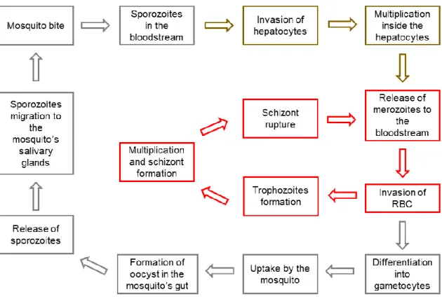
asexual reproduction, which represents the liver stage and is also known as exoerythrocytic schizogony (Figure 1.4. brown). The invasion occurs due to the binding of thrombospondin domains on the circumsporozoite protein (CSP) and thrombospondin-related adhesive protein (TRAP) to the heparin sulfate proteoglycans present on hepatocytes (Miller et al., 2002; Tuteja, 2007). Both CSP and TRAP are sporozoite surface antigens that can be recognized by the immune cells and the major target for malaria vaccination. TRAP is also involved in sporozoites motility (Weedall et al., 2007). In the hepatocytes, the sporozoites replicate into thousands of merozoites that are released to the bloodstream, giving rise to the blood stage of the disease (Miller et al., 2002; Tuteja, 2007).Figure 1.4. Plasmodium life cycle.
Upon mosquito bite, the sporozoites are injected into the bloodstream of the host where they travel until they reach the liver. In the liver, sporozoites penetrate hepatocytes and multiply into merozoites that are posteriorly released into the bloodstream. Once in the blood stream, merozoites invade RBC, where they can undergo asexual reproduction or sexual reproduction. In asexual reproduction pathway, after invading RBC, the merozoite forms a trophozoite. The trophozoite undergo multiple nuclear division without cytokinesis forming the schizont. After the lysis of RBC and rupture of the schizont, the merozoites that where inside the schizont are released into the bloodstream, where they can invade non-infected RBC again. On the sexual reproduction pathway, merozoites differentiate into gametocytes which are ingested by the female mosquito once it bites an infected host. On the mosquito’s gut, the gametocytes fuse and form a zygote, which develops into an oocyst. After the maturation and rupture of the oocyst, the sporozoites migrate to mosquito’s salivary glands and start the cycle again. In red is represented the blood stage of infection and the asexual reproduction pathway in RBC; in brown the liver stage and asexual reproduction in hepatocytes; and in grey the mosquito stage and sexual reproduction pathway.
13
Once in the bloodstream, merozoites invade red blood cells (RBC), where they can undergo asexual or sexual reproduction (Miller et al., 2002; Tuteja, 2007). In the asexual reproduction, also known as erythrocytic schizogony (Figure 1.4 red), merozoites invade RBC. After invasion, the merozoite forms a trophozoite, also known as the ring form. At this phase, the parasite takes advantages of the host’s resources to obtain glucose, performs proteolysis of hemoglobin (Hb) to obtain amino acids and ingests host’s cytoplasm to obtain other essential nutrients. The trophozoites undergo multiple nuclear divisions without cytokinesis in this way forming the schizont, a structure with merozoites inside. After the lysis of RBC and rupture of the schizont, between 8 and 32 merozoites are released into the bloodstream, where they can invade non-infected RBC, rapidly increasing the number of parasites in each cycle. Another feature of the disease is the sequestration and accumulation of infected RBC (iRBC), i. e. RBC infected with the parasite at any given moment of the infection, in different organs caused by their adhesive properties(Miller et al., 2002; Tuteja, 2007).As mentioned, merozoites can also undergo sexual reproduction pathway. In this case merozoites differentiate into female or male gametocytes which are ingested by the female mosquito once it bites an infected host. In the mosquito’s gut, the gametocytes fuse and form a zygote. The zygote penetrates the midgut wall and develops into an oocyst. After the maturation and rupture of the oocyst, the sporozoites, that were generated inside, migrate to the mosquito’s salivary glands where they can be transmitted to the next host through a blood meal and start the cycle again. The process of sexual reproduction is also known as sporogony (Figure 1.4 grey) (Miller et al., 2002; Tuteja, 2007).
The liver stage is asymptomatic and usually the symptoms start during the blood stage, after lysis of iRBC and consequent release of cellular contents that trigger the immune response. The duration of the cycle varies according to the species of parasite(Tuteja, 2007).
1.6.2 Mouse models of malaria – Plasmodium chabaudi chabaudi
Rodent models can be used to study human malaria. However, because none of the human parasites infect rodents, researchers use different species of Plasmodium but with genetic and phenotypic similarities to the human malaria parasites. The Plasmodium species that infect rodents are P. chabaudi, P. berghei, P. yoelii and P. vinckei.
One of the most used species in laboratory is P. chabaudi and the specific cloned line is P.
chabaudi chabaudi (P. chabaudi chabaudi AS or Pcc). This clone causes synchronous non-lethal
infection in C57BL/6 mice, where the schizont can adhere to vascular endothelium. The parasite genome encodes for Plasmodium interspersed repeat family (PIRS) which are expressed on iRBC surface and are important for its adhesion and sequestration. The symptoms of the disease start at the peak of infection, which is between days 7 and 10 post infection (with parasitemias ranging between 20-50% iRBC). Contrary to the fever observed during human malaria, mice develop hypothermiain response to malaria infection. To study blood stage malaria, the parasite
14
can be transmitted from mouse to mouse using blood from infected mice, and in this case no vector is needed (Stephens et al., 2012).Mice also develop anemia due to lysis of iRBC, suppression of hematopoiesis, clearance of uninfected RBC by phagocytosis and dyserythropoeisis. All these phenomena are also seen in human malaria. Thus, P. chabaudi chabaudi AS is a good model to study blood stage malaria. In C57BL/6 mice, the infection is resolved as the host clears the parasite(Stephens et al., 2012).
1.6.3 Malaria infection and heme
To obtain the necessary amino acids for their growth, parasites digest around 80% of the Hb present in RBC. During Hb proteolysis, besides the amino acids, there is also the release of heme (Moore et al., 2006). Heme is the prosthetic group of hemoproteins, such as hemoglobin and myoglobin, and is composed by a tetrapyrrole protoporphyrin IX ring and an iron atom (Gozzelino et al., 2010). Heme is catabolized by heme oxygenase 1 (Ho-1), leading to the production of carbon monoxide, biliverdin and labile iron (Gozzelino et al., 2010). Although essential to life, iron can be toxic to cells, accumulating into labile iron pool (Muckenthaler et al., 2017). To counter this, iron can be up taken by ferritin, a multimeric complex, composed by a light and an heavy chain, that detoxifies the iron by converting Fe2+ into Fe3+ and then store it (Gozzelino et al., 2010; Harrison and Arosio, 1996).
Plasmodium’s HO-1(PfHO) amino acids sequence is not similar to the one observed in mammalian HO-1 and in all the heme-degrading enzymes, and its function is not analogous (Okada, 2009; Sigala et al., 2012). While a study reports no activity of PfHO regarding heme degradation (Sigala et al., 2012), another study shows that PfHO is involved in the conversion of heme into bilirubin supporting the notion that PfHO plays a role in heme catabolism (Okada, 2009).
The major pathway of heme detoxification by Plasmodium parasite is the conversion into hemozoin (Hz). In order to prevent heme toxicity for the parasite, upon digestion of Hb by
Plasmodium, heme is immediately converted into Hz. Hz is an insoluble crystal formed in the
digestive vacuole of the parasite(Moore et al., 2006). When the schizont rupture occurs, Hz is released, together with merozoites, and is immediately removed by phagocytosis. Both lysed RBC and Hz are mainly phagocytosed in the liver in mice(Deroost et al., 2014). The amount of Hz present in the iRBC depends on the stage of the life cycle of the parasite. In the trophozoite stage the levels of Hz are low and in the schizont the levels are high(Moore et al., 2006). Upon the lysis of the iRBC, the non-digested Hb is also released to the bloodstream.
Hb at steady state is a tetramer in the reduced form (Fe2+), however cell-free Hb, when in contact with free radicals, dissociates into dimers and is readily oxidized into methemoglobin (Fe3+) with the consequent release of heme (Gozzelino et al., 2010).
Even though heme is an essential molecule for life, in higher concentrations in the plasma can be prooxidant, cytotoxic and pro-inflammatory (Larsen et al., 2012). Because of the presence of an iron atom, it can be involved in the production of ROS by the Fenton chemistry (Larsen et
15
al., 2012). Fenton chemistry refers to the conversion of hydrogen peroxide into hydroxyl radicals, i.e. powerful oxidants, using the iron atom to catalyze the reaction. It can also induce programmed cell death in non-hematopoietic cells, compromising the integrity and function of tissues (Larsen et al., 2012). Since heme has a lipophilic structure, it can intercalate into the cell membranes, thus allowing heme to reach the nucleus and cause DNA damage through ROS production (Aft and Mueller, 1983). In addition, heme leads to lipid and protein peroxidation (Aft and Mueller, 1984; Vincent et al., 1988).Under normal conditions, cell-free Hb is recognized and bound by haptoglobin, preventing its oxidization (Gozzelino et al., 2010). When the oxidization of Hb occurs, with the consequent release of heme, there are mechanisms to avoid accumulation of heme in the plasma, as well as the production of ROS, and prevent the damage they may cause (Gozzelino et al., 2010; Larsen et al., 2012). For example, apart from the predominant role of HO-1 in heme catabolism, some hemoproteins, like albumin and hemopexin, can bind and sequester heme (Gozzelino et al., 2010). However, in cases like malaria, where the host defense mechanisms are overwhelmed by the excessive hemolysis, heme and Hb accumulate in the plasma leading to oxidative stress and tissue damage (Larsen et al., 2012).
1.6.4 Malaria and disease tolerance
In the last decades, disease tolerance mechanisms have been associated with survival and protection to malaria (Cumnock et al., 2018; Gozzelino et al., 2012; Martins et al., 2019; Ramos et al., 2019). The excessive hemolysis and consequent accumulation of heme and Hb in the plasma are the major features of malaria disease and the main potentiators of tissue damage (Larsen et al., 2012). By Fenton chemistry, iron atom released from heme contributes to ROS production, leading to general tissue damage, through the oxidation of different macromolecules including proteins and DNA (Aft and Mueller, 1983, 1984; Candeias and Wardman, 2013; Larsen et al., 2012). Thus, mechanisms to counter these deleterious effects of heme and ROS are critical to establish disease tolerance and survival to infection. During infection, mice present sickness behavior characterized, for example, by loss of appetite and changes in body temperature. Infection impacts on host metabolism and induces changes in energy source (Cumnock et al., 2018; Vandermosten et al., 2018). Initially, mice use glycogen storage as primary energy source, however, together with anorexia of infection, leads to glycogen exhaustion and severe hypoglycemia (Cumnock et al., 2018; Vandermosten et al., 2018). In order to prevent severe hypoglycemia, fatty acids and ketone bodies are then used as sources of energy (Cumnock et al., 2018). Thus, even though this sickness behavior is part of the host defense mechanisms against the parasite, it can induce damage and organ dysfunction by decreasing the amount of glucose available (Cumnock et al., 2018; Vandermosten et al., 2018). Treatment with glucose ameliorated the disease without interfering with pathogen load which shows that glucose confers protection against infection through a mechanism of disease tolerance (Cumnock et al., 2018). Therefore, both stress and damage responses are important to confer protection against
16
Plasmodium infection, by preventing and repairing tissue damage, redox imbalance and
hypoglycemia state. Understanding disease tolerance mechanisms may allow to identify potential targets for therapeutic approaches.
The host laboratory had made several contributions to our understanding of disease tolerance mechanisms in the context of malaria (Gozzelino et al., 2012; Jeney et al., 2014; Ramos et al., 2019; Seixas et al., 2009). Gozzelino and co-workers showed that induction of ferritin, the multimeric complex that stores iron, particularly in the liver, allows metabolic adaptation to iron overload that is essential to confer tolerance to malaria (Gozzelino et al., 2012). Seixas and co-workers showed that expression of Ho-1, the heme catabolizing enzyme, in the liver also confers protection by preventing hepatic failure (Seixas et al., 2009). Recent work from the host laboratory showed that expression of Ho-1 and ferritin in the kidney, more specifically in renal proximal tubule epithelial cells, is essential to confer protection against malaria and prevent tissue damage in the kidney (Ramos et al., 2019). These studies reveal the importance of controlling heme/iron metabolism in order to prevent tissue damage, supporting the notion that stress and damage responses are essential to confer disease tolerance. In addition, these studies also support the notion that stress and damage sensor proteins in specific organs contribute to protect against malaria by promoting organismal homeostasis.
1.7 Aims of the project
Since heme and ROS induce both oxidative stress and DNA damage, and ATM can act as a DNA damage and oxidative stress sensor, it is possible that ATM plays a role in the activation of disease tolerance mechanisms, in particular in the context of diseases characterized by excessive heme accumulation, such as malaria. Unpublished data from the host laboratory showed that
Atm-deficient mice are more susceptible to blood stage malaria than wild type controls (Figure
1.5 A), indicating that ATM plays a role in the response to malaria infection. The percentage of iRBC, i. e. parasitemia, is similar comparing Atm-deficient (Atm ko) and Atm wild type (Atm wt) control mice (Figure 1.5 B) suggesting that Atm does not impact directly on the pathogen itself but confers protection against malaria through a mechanism that confers disease tolerance.
Knowing that Atm-deficient mice are more susceptible to malaria (Figure 1.5 A) and that one of the characteristics of this disease is the elevated levels of heme (Larsen et al., 2012), the first hypothesis to be tested in this project is that Atm-deficient cells are more susceptible to heme treatment. To test this hypothesis the following specific aims will be addressed:
- Evaluate the susceptibility of Atm-deficient and wild type mouse embryonic fibroblasts to heme treatment;
- Analyse the expression of DNA damage and oxidative stress marker proteins upon heme treatment.
17
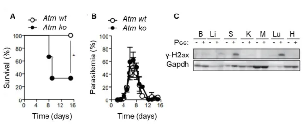
Figure 1.5 Atm-deficient mice are more susceptible to Plasmodium chabaudi chabaudi(AR. Carlos unpublished data).
(A) Atm wt (n=5) and Atm ko (n=6) mice were injected with Pcc and survival was monitored over 15 days.
Statistical significance was determined using Mantel-Cox test. * - p≤0.05. (B) Parasitemia of mice treated as in A. (C) γ-H2ax and Gapdh protein expression detected by western blot in the brain (B), liver (Li), spleen(S), kidney (K), muscle (M), lung (Lu), and heart (H) of C57BL/6 mice, not infected (-) or 7 days after intraperitoneally (i.p.) injection with the malaria-causing agent Plasmodium chabaudi chabaudi (Pcc) (+). Data are representative of four mice per group in one experiment.
Since ATM has an important role in the hematopoietic compartment (see 1.5 ATM and the
hematopoietic compartment) and Plasmodium infection causes DNA damage in the spleen, which
is part of this compartment (Figure 1.5 C), the second hypothesis to be tested is that mice lacking functional Atm in the hematopoietic compartment are more susceptible to the hemolysis caused by malaria. To test this hypothesis the following specific aims will be analyzed:
- Evaluate which organs are more affected by heme, in terms of DNA damage and oxidative stress;
- Characterize mice that lack Atm in the hematopoietic compartment, Vavicre/wtAtmΔ/Δ
mice;
- Analyse the susceptibility of Vavicre/wtAtmΔ/Δ mice to Plasmodium chabaudi chabaudi
infection;
19
Chapter 2. Methods and Materials
For more details about reagents, kits, antibodies and media, see Table 2.6 at the end of this section.
Mice
Atmlox/- mice, kindly provided by F. Alt (Howard Hughes Medical Institute, The Children's Hospital,
Boston) and Vavicre/wt mice, originally obtained from Jacksons Laboratory (B6.Cg-Commd 10Tg(Vav1-icre)A2Kio/J, #008610) were intercrossed and bred until the final breeding format Atmlox/wt vs
Vavicre/wtAtmlox/wt was obtained. Atmlox/wt and Vavicre/wtAtmlox/wt mice were bred at the Instituto
Gulbenkian de Ciência, Lisboa, Portugal in specific pathogen-free (SPF) conditions to generate experimental Vavicre/wtAtmΔ/Δ and control Atmlox/lox and Vavicre/wt mice. All the experiments were
done using mice, females and males, between 7 and 12 weeks of age.
Mice genotyping
Genotyping was performed using the tail tip of mice with 3-5 weeks of age. Tail lysis and genotyping was conducted using Xpert directXtract PCR Kit, according to the manufacturer’s instructions. Details of the PCR program are described on Table 2.1, and the primers described on Table 2.2. After PCR, electrophoresis in 1,5 % (w/v) agarose gel was performed.
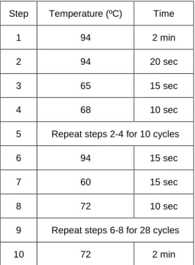
Table 2.1 PCR program.
Description of PCR steps for genotyping protocol.
Step Temperature (ºC) Time
1 94 2 min
2 94 20 sec
3 65 15 sec
4 68 10 sec
5 Repeat steps 2-4 for 10 cycles
6 94 15 sec
7 60 15 sec
8 72 10 sec
9 Repeat steps 6-8 for 28 cycles
20
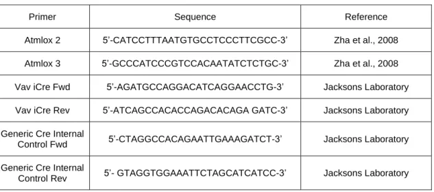
Table 2.2 Primers for genotyping.Primers used for genotyping protocol.
Primer Sequence Reference
Atmlox 2 5’-CATCCTTTAATGTGCCTCCCTTCGCC-3’ Zha et al., 2008 Atmlox 3 5’-GCCCATCCCGTCCACAATATCTCTGC-3’ Zha et al., 2008 Vav iCre Fwd 5’-AGATGCCAGGACATCAGGAACCTG-3’ Jacksons Laboratory Vav iCre Rev 5’-ATCAGCCACACCAGACACAGA GATC-3’ Jacksons Laboratory Generic Cre Internal
Control Fwd 5’-CTAGGCCACAGAATTGAAAGATCT-3’ Jacksons Laboratory Generic Cre Internal
Control Rev 5’- GTAGGTGGAAATTCTAGCATCATCC-3’ Jacksons Laboratory
Deletion check
Deletion of the Atm allele in the hematopoietic compartment of Vavicre/wtAtmΔ/Δ mice was confirmed
using blood, spleen, bone marrow and thymus samples. Liver samples were used as negative control for deletion. Vavicre/wtAtmΔ/Δ mice have two loxP sites on exons 57 and 58 of Atm gene and
iCre recombinase expression under the control of the Vav promoter, which results in deletion of this fragment of the Atm gene only in the hematopoietic compartment. A volume of approximately 200 μL of blood was collected in heparin and RNA was extracted using the kit NucleoSpin® RNA Blood, according to the manufacturer’s instructions. Spleen, bone marrow, thymus and liver were harvested, and RNA was extracted after tissue homogenization. For that, tissues were lysed in 1 mL of tripleXtractor Reagent for 5 min at room temperature (RT) and disrupted mechanically with Tissue Lyser (Tissue Lyser II, Qiagen) using 1 tungsten bead per tube (conditions: two times - frequency 30/s and time 3 min) (in the case of bone marrow, disruption was achieved only by pipetting). After lysis, 200 μL of chloroform were added to each sample, and then samples were vortexed, and incubated 3 min at RT. Following incubation, samples were centrifuged at 12,000 x g for 15 min at 4ºC to separate the different phases and enable the recovery of the aqueous phase, which contains the RNA fraction. The aqueous phase was recovered and transferred into a fresh tube. RNA isolation proceeded using the kit NucleoSpin® RNA protocol, according to the manufacturer’s instructions. Upon RNA extraction from blood and tissues, RNA was converted into cDNA using Transcriptor First Strand cDNA Synthesis Kit or Xpert cDNA Synthesis kit and Real-Time qPCR was then performed using iTAqTM Universal SYBR® Green Supermix using the primers described in Table 2.3.